Lymedin2010
Frequent Contributor (1K+ posts)
Member # 34322
posted
Eight Legs Bad, thanks for the info. His postings have been floating around on Facebook & I have done my part
Thanks for the info & others should help out as well. Dr. Alan MacDonald is one of my new idols post Lyme! His perseverance
Posts: 2087 | From NY | Registered: Oct 2011
| IP: Logged |
Lymedin2010
Frequent Contributor (1K+ posts)
Member # 34322
posted
TNT, great video & it looks as you said it would & that pic serves as a good example. As I said before anything coccoid would be more difficult for anyone to confirm, as I have asked near & far in the past.
But yours has flagella, as you said it does, & it should be easier. I have never seen or heard of such an organisms & most of the babs & bart organisms I have seen are from STAINED RBC slides & never any live ones.
I will do some research on it in the oncoming days & hopefully we can ID this one.
How often do you find this in your blood & about how many can you find on ONE slide? Are they all pretty much the same size or do they come in slightly smaller or larger diameter?
All my past Youtube videos I did not have any conversion & display issues with, except for my last one. I will have to upload another video just to determine if Youtube might have changed any specifications. Perhaps they have moved on & they are expecting most videos nowadays to be in the WIDE screen format (HD) & are no longer 3x4 format friendly?
Posts: 2087 | From NY | Registered: Oct 2011
| IP: Logged |
TNT
Frequent Contributor (1K+ posts)
Member # 42349
posted
quote:Originally posted by Lymedin2010: How often do you find this in your blood & about how many can you find on ONE slide? Are they all pretty much the same size or do they come in slightly smaller or larger diameter?
This is the first time I have seen cocci with flagellum. There may have been one or two cocci in previous samples, but it was very rare and never before with flagellum.
The bart-like bacteria I HAVE seen before... maybe 3 other times/samples (one organism per sample those other times). The one that was pummeling the kete was identical to this one. Actually, all the previous bart-like ones were identical to this one.
Would anyone be interested in the video where the bart-like bacteria was attacking the kete? It may have resolution issues, too, since I zoom in most of the time, and, since I may lose some clarity in the upload.(I will be more careful with the zoom from now on-at least until I get a better camera).
Thanks for your help Lymedin!
Posts: 1308 | From Eastern USA | Registered: Oct 2013
| IP: Logged |
Lymedin2010
Frequent Contributor (1K+ posts)
Member # 34322
posted
Was the bart like organism a rod shape in any form, or strictly round?
How large is original video, as it may not pass the max file size requirement?
Posts: 2087 | From NY | Registered: Oct 2011
| IP: Logged |
TNT
Frequent Contributor (1K+ posts)
Member # 42349
posted
The bart-like organism that attacks the kete is identical to the one in the second video I posted. It is rod-shaped. The bart-like organisms I refer to are all rod-shaped.
The kete-attacker video is a few minutes long, depending on which one I select.
Posts: 1308 | From Eastern USA | Registered: Oct 2013
| IP: Logged |
TNT
Frequent Contributor (1K+ posts)
Member # 42349
posted
The bart-like organism (rod-shaped one) I am learning could easily be E. coli bacteria. According to Wiki, cells are typically rod-shaped, and are about 2.0 micrometers (μm) long and 0.25–1.0 μm in diameter, with a cell volume of 0.6–0.7 μm. That would fit the bill for the "bart-like" (rod-shaped) organisms.
Here are some pics of E. coli from Dennis Kunkel Microscopy Inc.:
Here is an interesting one in which it looks like there could be a cocci form, but I think it is just on end:
Some E. coli strains do possess flagella, but my rods did not. Only the cocci looking organisms had flagella.
So, this only gives me a possible answer for the rods, but not for the round flagellate organisms. And a quick search did not reveal a "cocci" morphology for E. coli. So, I don't think the round flagellates were E. coli.
So, I'm still looking for suggestions on what the round flagellates could be.
Posts: 1308 | From Eastern USA | Registered: Oct 2013
| IP: Logged |
Lymedin2010
Frequent Contributor (1K+ posts)
Member # 34322
posted
Here is a great video of E. Coli & it shows both rod & rounded shaped forms, I would imagine it being dependent on where in its growth cycle it is at. Also, the rounded ones appear to have flagella & is more noticeable when one pauses the video & reviews it frame by frame.
Another one of my colleagues suggest that it might be a platelet (aka thrombocyte), which are 2-3µm in diameter. As you make more microscopic observations see if any appear without flagella. Or do they ALL appear with flagella all the time? This is a Giemsa-stained peripheral blood smear. https://upload.wikimedia.org/wikipedia/commons/thumb/5/51/Platelets2.JPG/1280px-Platelets2.JPG
[ 08-01-2015, 08:15 PM: Message edited by: Lymedin2010 ]
Posts: 2087 | From NY | Registered: Oct 2011
| IP: Logged |
TNT
Frequent Contributor (1K+ posts)
Member # 42349
posted
Hey, thanks for the video of the E. coli! I, too, think it appears as though the round ones have flagella. The rod shaped ones, too. Though, I wish the video was of a higher magnification to tell for certain.
I don't think those round flagellate organisms in my blood are platelets.... I can see platelets very well, and are fairly numerous. The round and rod-shaped "bacteria" are very rare, and are quite different from the platelets. To name just one major difference, platelets look dark in my dark phase view. When I upload more videos, I can try to get ones with some platelets in the field.
I would tend to think my organisms are E. coli, especially if there is round-form stages with flagella. It seems to me that it would be very easy for a predominant organism (E. coli bacteria) of the GI tract to get into the blood stream in few numbers, especially if the mucosal membrane is not completely healthy and there is an imbalance of gut flora.
The scary thought is that the gut could seed other parts of the body via the bloodstream in a situation as that...or just as scary, lead to sepsis.
Posts: 1308 | From Eastern USA | Registered: Oct 2013
| IP: Logged |
Lymedin2010
Frequent Contributor (1K+ posts)
Member # 34322
posted
One thing that I have learned, especially from watching the Animal Planet "Monsters Inside Me" series, is that we are susceptible to many different varieties of tertiary infections with the immuno suppression of Lyme.
If you watch the series you will become all too familiar with the wording "most people can fight off the infection, however the young, elderly, or immuno compromised individuals are at risk."
Posts: 2087 | From NY | Registered: Oct 2011
| IP: Logged |
quote:Originally posted by TNT: I would tend to think my organisms are E. coli, especially if there is round-form stages with flagella. It seems to me that it would be very easy for a predominant organism (E. coli bacteria) of the GI tract to get into the blood stream in few numbers, especially if the mucosal membrane is not completely healthy and there is an imbalance of gut flora.
The scary thought is that the gut could seed other parts of the body via the bloodstream in a situation as that...or just as scary, lead to sepsis.
I think you are very right here! The wild life i have encountered when watching growth in the blood is staggering and the diversity just doesnt seem to match with a single borrelia infection, or even one with a couple of known co infections.
So the idea that gut flora is constantly seeding the body with a wide variety of bacteria is a very good possibility. The growing candida hyphal forms in my blood sort of proves its possible. And the mechanism is leaky gut.
One of the clear observations i have made is that this wild life growth under the microscope is much more abundant when you press the slide with a small object, basically rupturing some of the RBC's and WBC's. So i suspect most of this wildlife is contained in the WBC's. At least an indication that the WBC's are mopping up this gut overflow.
And even without rupturing the WBC's i have pictures showing this wild life just growing from inside whole (but probably dead) WBC's :
Its a bit fuzzy, but it shows 2 WBCs with unknown bacteria and or fungi growing out of them.
So gut flora overspill is probably a very important factor for some of the lyme patients. Continuous overstimulation of the Th1 branch of the immunesystem causes chronic inflammation. Lyme itself with its LPS also adds to this problem of course. Continuous chronic inflammation of the gut causes more bacteria to leak out and possibly causes more SIBO by damaging the ileocecal valve for example. So its a vicious cycle.
quote:Originally posted by Lymedin2010: One thing that I have learned, especially from watching the Animal Planet "Monsters Inside Me" series, is that we are susceptible to many different varieties of tertiary infections with the immuno suppression of Lyme.
So are we immune compromised? Or is it just continuous overstimulation of the immune system that is causing trouble?
Posts: 381 | From The Netherlands | Registered: Nov 2013
| IP: Logged |
Lymedin2010
Frequent Contributor (1K+ posts)
Member # 34322
posted
I have seen a few "normal" & "healthy" blood samples & THEY ALL HAVE FOREIGN ORGANISMS!!!!
The difference is that our Lyme blood is overloaded & overstimulated from many different angles & sources and hence our blood shows the very large loads.
Posts: 2087 | From NY | Registered: Oct 2011
| IP: Logged |
Lymedin2010
Frequent Contributor (1K+ posts)
Member # 34322
posted
Did you guys see Peter Kemp's new centrifuge work with Lyme blood? I almost bought a centrifuge during the first year I bought my scope, since I thought I could sequester & concentrate the spirochetes in a blood layer & have visible masses of spirochetes for time lapse. After my disease spread the spirochetes became more plentiful & I lost the need for a centrifuge.
So in a nutshell this is basically how Advanced Labs & Dr. Burrascano would do a Borreliosis blood culture to prove Lyme Disease in our blood. It is not the exact recipe & but the gist is here. Add a culture medium, add your blood & store in a temp controlled environment for a few days & then check via microscopy.
The only difference here is that Peter was interested in determining which blood fractionation layer would host mot of the spirochetes. I always suspected that the blebs or cysts would be very buoyant & would "float" to the top plasma layer.
Posts: 2087 | From NY | Registered: Oct 2011
| IP: Logged |
TNT
Frequent Contributor (1K+ posts)
Member # 42349
posted
OMG! I watched Peter Kemps video. I thought the buffy coat layer was bad! But, when the video got to the lower RBC layer, I about fell out of my chair! That's BAD! Those RBCs are basically colonies of spirochetes! I do wonder sometimes how we are still alive.
I viewed the blood of a very healthy friend of mine a couple months ago. I was very surprised, yet not surprised, by what I saw. He had one definite spirochete, perhaps 7-8 um long, and two probable shorter ones.
I am certain he has been exposed to lyme one way or another, and probably has been tick-bitten before. So, it was not a complete surprise to me that he did harbor a very small presence of the germ. But, he is as healthy and strong as an ox.
Though, if something would severely stress his immune system, I am sure there is a chance he could "come down" with Lyme disease.... the same way many people "come down" with it (not necessarily right after exposure, but after a STRESSOR on the immune system).
Oh, Lymedin. Do you think there is anyway you could shorten the URL address of that picture of the giemsa stained blood smear to get this page back to normal size?
[ 08-01-2015, 09:10 PM: Message edited by: TNT ]
Posts: 1308 | From Eastern USA | Registered: Oct 2013
| IP: Logged |
Lymedin2010
Frequent Contributor (1K+ posts)
Member # 34322
posted
TNT, the blood sample was CULTURED to get that many spirochetes.
.3ml blood from various layers after the centrifuge + 1ml BSK medium, which was then incubated at 35C for 4 days.
So the few spirochetes in your blood became many by the time he looked at them on the 4th day.
Posts: 2087 | From NY | Registered: Oct 2011
| IP: Logged |
TNT
Frequent Contributor (1K+ posts)
Member # 42349
posted
Phew! That's a relief! I didn't see that it was cultured. NOW that I read the video description, I see that it SAYS it was cultured. Thanks, Lymedin.
If there would have been a snake in that description I could have been bitten.
Thanks also for fixing the page. It's so much easier on my eyes.
Posts: 1308 | From Eastern USA | Registered: Oct 2013
| IP: Logged |
Lymedin2010
Frequent Contributor (1K+ posts)
Member # 34322
posted
TNT, I know a lady who has Lyme so bad & so much circulation issues that she cannot go to bed with her arms & legs crossed , because it hurts/burns so much from the Lyme.
I am willing to bet anything that her blood will look close to that.
There must be some limiting factors that prevent our blood from totally building up with spirochetes though & it must be the utilization of an essential growth component of bb, maybe something like availability of manganese or something along these lines.
Posts: 2087 | From NY | Registered: Oct 2011
| IP: Logged |
Lymedin2010
Frequent Contributor (1K+ posts)
Member # 34322
"In this report we present a method to cultivate Borrelia spirochetes from human serum samples with high efficiency. This method incorporates improved sample collection, optimization of culture media and use of matrix protein. The method was first optimized utilizing Borrelia laboratory strains, and later by demonstrating growth of Borrelia from sera from fifty seropositive Lyme disease patients followed by another cohort of 72 Lyme disease patients, all of whom satisfied the strict CDC surveillance case definition for Lyme disease.
The procedure resulted in positive cultures in 47% at 6 days and 94% at week 16. Negative controls included 48 cases. The positive identification of Borrelia was performed by immunostaining, PCR, and direct DNA sequencing."
Posts: 2087 | From NY | Registered: Oct 2011
| IP: Logged |
Lymedin2010
Frequent Contributor (1K+ posts)
Member # 34322
posted
Just as they mentioned in Under Our Skin 2: Emergence, that they closed down the microscopy work of Prof. Morten Lanne because of what was revealed.
Microscopy visualization of Borreliosis in Chronic Lyme is HUGE.
"Prof. Morten Laane held the presentation: "Easy Detection of Bacteria and Parasites in Infected Human Blood by Microscopy. Some Simple, Low-cost Methods" at the NorVect conference 2014.
Because his and Prof Mysterud's study on Lyme patients were stopped and all results were destroyed by the Norwegian Board of Health Supervision, Prof Laane was not allowed to talk about this particular study. He showed some of his work using two films. One is seen here in this excerpt. His presentation can be watched in full length at http://www.norvect.no
dbpei
Frequent Contributor (1K+ posts)
Member # 33574
posted
I recently came across this thread and saw some pictures that resembled what I have been seeing with my eye floaters for the past few years.
Photos posted by Lymedin2010 7/28/15 at 9:55 a.m. - both pictures look very similar to what I see daily! I could not tell if these photos posted were from H Pylori or Bartonella.
I also have some small cyst shaped floaters, but the ones I see the most, I describe as looking like earwigs with a bunch of long, thin hairs on one end. Do any of you see these same floaters? After seeing these photos, I wonder if it could be bartonella or H Pylori that is keeping me sick.
Lab testing shows that I am making lots of antibodies for brucellosis. Is there a place I could find out what that bacteria looks like under microscope?
I don't own a microscope but am seeing my eye doc this morning. I wonder what she will say when I tell her what my floaters look like. I hope she won't add me to her nutty patient list.
Posts: 2386 | From New England | Registered: Aug 2011
| IP: Logged |
Lymedin2010
Frequent Contributor (1K+ posts)
Member # 34322
posted
Many Lymies have strings as floaters, as I have had for years & continue to have as well, which I suspect are the Borrelia spirochetes.
The pics I posted on that day are of H. Pylori, but your floaters could be Brucella because of your antibodies clue. If you describe them as you do, "like earwigs with a bunch of long, thin hairs on one end," then they could just as easily be H. Pylori escaping from your gut, bart, or another organisms we have not thought of yet.
H. Pylori: Posts: 2087 | From NY | Registered: Oct 2011
| IP: Logged |
dbpei
Frequent Contributor (1K+ posts)
Member # 33574
posted
Thanks so much! It definitely resembles H Pylori missing the bands I see at the end near the hairs, but actually looks more like an earwig than the rod I see here. I saw my eye doc today for my annual exam and told her about them.
She examined my eyes really well and found one of the floaters I described. She told me they are vitrious detachments and not to worry. I hope she is right.
I really appreciate the help in identifying these critters! I will have to follow this thread. It is very interesting.
Posts: 2386 | From New England | Registered: Aug 2011
| IP: Logged |
Lymedin2010
Frequent Contributor (1K+ posts)
Member # 34322
posted
Eye floaters are really hard to prove in either direction, but it is interesting how many Lymies report them.
If anyone is interested in a break down of which cameras are best for pics, video & time lapse, here is a breakdown with focus specifically on microscopy.
Thankfully, it turns out that I did not lose much resolution in the upload.
Posts: 1308 | From Eastern USA | Registered: Oct 2013
| IP: Logged |
Lymedin2010
Frequent Contributor (1K+ posts)
Member # 34322
posted
TNT, the big one sure looks like borrelia!!!! Awesome video & thanks for sharing!
It has the bulbous tips on both ends & it spirals/undulates in that very distinctive way with a bit more aggression than just a stagnant string or anything that may be of human origin & an actual artifact.
BUT, the one you are calling Bart like I also think is Borrelia, just a pre-mature one. It looks like a dumbbell, right?
From the beginning of my thread: "The progression might be DOT--> DOT WITH TAIL-->STUBBY DUMBBELL-->MEDIUM DUMBBELL-->LONG DUMBBELL (STRING)."
At that time I thought it was spirochaeta gallinarum, because it was the only spirochete that I could find where someone discusses the blebs or spore formations. Now I know it is borrelia for sure & the blebs (dots) grow to be tailed blebs at first & then look like very small dumbbells, which are basically growing borrelia organisms.
One of my future wishes is to catch the blebs grow into tailed blebs & then the small spirochetes (small dumbbells).
Posts: 2087 | From NY | Registered: Oct 2011
| IP: Logged |
TNT
Frequent Contributor (1K+ posts)
Member # 42349
posted
Yes, the longer one is definitely borrelia.
Why is the shorter one acting the way it is? It sure appears to me to be attacking the longer one. And the longer one appears to be losing energy.
The shorter one could possibly be borrelia, but I'm not sure. I don't have enough information to go on. I will pay attention to your videos with dumbbell organisms that size.
If you find any dumbbell footage from Sapi or MacDonald, please let me know.
Posts: 1308 | From Eastern USA | Registered: Oct 2013
| IP: Logged |
Lymedin2010
Frequent Contributor (1K+ posts)
Member # 34322
posted
I will be putting out a morphology video soon & it will cover the progression.
The Brownian motion you see often is from surface charges & they end up just bouncing around sporadically. The lighter an object is the more it may be affected by these charges & more they can bounce around.
The blebs & smaller dumbbells bounce around the most from what I see.
I think the phenomena you saw in your video is from surface charge & not really from an attack & it just happens that the two independent spiros had an attraction toward each other or were moving in the path of least resistance in unison. Two spiros attacting each other usually does not happen & is dependent on local concentrations & charges in the plasma.
This is Peter Kemp's video, take a look 13 seconds into the video at the top & center. You will see the small baby spiro or what I called the dumbbell. There is also another one on the lower left side.
9s into this very same video on the lower left, you will see a slightly longer dumbbell that has already gone through "String of Pearl" (SoP) formation & you can be satisfied that these are indeed spiros!!!!
That last spiro has 3 dots or blebs along the entire body, the two that you typically see on either end & it has developed a bleb in the center.
Posts: 2087 | From NY | Registered: Oct 2011
| IP: Logged |
TNT
Frequent Contributor (1K+ posts)
Member # 42349
posted
I am still a little hesitant to say every dumbbell-shaped organism is a juvenile kete. I am definitely open to the strong possibility that some are. But, take a look at this video:
I found this next video fascinating because of the resolution, magnification, and for the sheer awesomeness of phagocytosis! Is this with a DIC microscope?
Lymedin2010
Frequent Contributor (1K+ posts)
Member # 34322
posted
Yea, I have seen this exact Staphylococcus Aureus as one of my first videos when I first started with microscopy.
Proving via time lapse that the blebs turn to tailed blebs & then the dumbbells & then to larger spiros is high on my priority list. But yea it is always possible to confuse them with rods.
Take a look at what Staphylococcus Aureus looks like, since it is spherical, but the bodies can stick to one another & DO NOT HAVE a rod between them!!!! More times over more than one stick together & one would see many, not just two together. All of the dumbbells I see are 2 bulbous tips plus the center body = borrelia I think.
TNT
Frequent Contributor (1K+ posts)
Member # 42349
posted
quote:Originally posted by TNT: Thanks, S13. If you are seeing the pseudohyphae in your blood, does it gently wave around like seaweed in water? The stuff I'm seeing gently waves around.
If so, does it have that delineating cross-section line at the base of the hyphae like what is shown in your pictures above? I'm not seeing any delineation.
I see neutrophils gobbling the balls of "candida" up (if the candida is not too big).
I'm also seeing something very similar to the stuff that is waving around, but it is different. This other stuff very much appears to be thick, faint, spirochetes anchored in what appears to be cysts. The "cysts" are about 1-2 um in diameter and are not necessarily uniform in roundness. The "spirochetes" spin and move just like real spirochetes. I am suspecting these could be l-form borrelia, especially since they appear to be anchored in a cyst. These "ketes" appear to be approx. 3-6 um in lenth (about the same length of many spirochetes).
I have been studying up on L-form (cell-wall deficient) bacteria all morning and am coming to the conclusion that these waving, thick, faint "spirochetes" ARE VERY POSSIBLY a form of cell-wall deficient bacteria.
Here is exactly what I am seeing in my blood and exactly what I am referring to:
I am also seeing "terrain" very much like this (that could look like what has been referred to as candida):
(The above picture is a Phase contrast image of L-form cells from Bacillus subtilis)
Penicillin antibiotics CAUSE cell-wall deficient bacteria!!!!!
I'll probably post my own videos on what is probable CWD bacteria before too long.
Posts: 1308 | From Eastern USA | Registered: Oct 2013
| IP: Logged |
TNT
Frequent Contributor (1K+ posts)
Member # 42349
posted
Here's an interesting page about L-form (CWD) bacteria:
Lymedin2010
Frequent Contributor (1K+ posts)
Member # 34322
posted
Hmmmmm, interesting how they say the coccoids can coalesce into what "appear" as string of pearls. Many different particles can come together to make a pseudo SOP from what I see, but they are never lengthy & do not appear properly aligned to come from a spirochete.
The organisms we did time lapse on don't coalesce into the SOP's, the bodies actually form the SOP as CLEARLY seen in our time lapse videos.
Also, it is stated that they do not easily grow externally to the body & in media. I already know a few people who are culturing the spirochetes externally & when I give you the recipe you will be able to as well. This will also allow you ample time to do time lapse on many of them at once.
Posts: 2087 | From NY | Registered: Oct 2011
| IP: Logged |
posted
I haven't been back here in a long time (I took a break from the Lyme world after being harassed by some nefarious characters a few years ago)-- but I just wanted to thank all of you who contribute to this incredibly important and upbeat thread!!
It is so gratifying to see the explosion of microscopy videos online, showing the spirochete's behavior and habits. At some point, TPTB will no longer be able to suppress the information, and they will be widely viewed by the scientific community as the illegitimate, unscientific liars that they are..
Microscopy is TRUTH---- and the truth will prevail---- thanks to all of you.
Back in 2007/8 there were no videos on Youtube showing Lyme spirochetes, and we were all told the Big Lie--- that spirochetes were impossible to find in blood!!!
I remember stumbling upon a British Lyme guy online who was so pissed off at getting the "Lyme Runaround" from his British doctor that he bought an old microscope. He filmed hungry active spirochetes in the evening, and then loaded several short and poor quality videos of clearly visible live spirochetes in his blood directly onto his webpage. I was amazed that a layman could do this-- and I was so impressed by him. I guess he didn't think to put the videos on youtube, because youtube was fairly new at that time.
I then took the liberty of uploading those amazing, but poor quality videos to youtube-- and his videos-- the first spirochete videos on youtube--- received 10,000 views on my youtube page--- in a short time!!! I was so harassed later that I shut down my youtube page and dropped out of the Lyme world for a while-- because I didn't even know what an ad hominem attack was...and the evil out there directed against vulnerable, chronically ill people was so shocking to me....
This is why I am now so gratified that so many people are now doing microscopy and uploading their amazing videos!!!!
I'm now excited to get my own microscope and camera.
I want to see if promising herbals have any visual effect on diminishing spirochetes in blood over time. I know that only intravenous Rocephin seems to clear the blood completely-- based on one of your findings on this thread... but perhaps we could discover something miraculous working together!!
It sure would be great if there were a "Microscopy Herbal Club"--- we could all agree to take 3 months of a promising herb or herbal cocktail--- while filming befores and afters. (I say 3 months because that is the rough lifespan of a red blood cell-- and microscopy has proven that the spirochetes are intracellular-- i.e., they get safe harbor inside red blood cells) Anyway--- its just a thought.
I would love to see if any of the following herbals have an impact on diminishing spirochetes in blood over a three month period: Olive Leaf Extract; Enzymes like Serrapeptase; Teasle, Monolaurin; Barley Grass; Japanese Knotweed; Smilax; Boron; GSE;Baking Soda; Enula/Houttuynia; Banderol/Samento.... etc, etc.
Posts: 696 | From New York | Registered: Aug 2006
| IP: Logged |
TNT
Frequent Contributor (1K+ posts)
Member # 42349
posted
Welcome back, WakeUp!
Yes, you need to get a microscope and start experimenting!
I think you will find it impossible to get a collaborative study with microscopists who are chronically ill who are looking at their blood who would be able to stop their own protocol to commence one with only herbals. There are just not that many of us.
I am sure you have heard of Sapi's in-vitro studies with Samento and Banderol? This is probably the closest thing you are going to find to your idea. It's a good idea, though, for sure!
Actually, there was one sample I took about 30 minutes after taking approx. 20 drops of Samento that showed MANY ketes. That would have been meaningless except that the previous samples were showing very few ketes. I just assumed that the Samento got intracellular and forced the ketes out into the plasma. But, who knows?
I thought it was interesting because in Sapi's studies the Samento alone caused more round bodies. I don't know if her medium was with whole blood though.
I just chalked it up to a fluke, because the next time I took Samento and then looked at my blood, I found very few ketes again.
My thinking as of late is that the so-called persister bugs are keeping us sick. I think the persister cells are the cell-wall deficient form. But, not just with borrelia, but other bacteria in our blood, too. Cell-wall deficient bacteria are extremely difficult to eradicate. Cysts and spirochetes are easy compared to these.
Perhaps you could do another youtube account and post some of those videos again?? I'd be very interested in seeing them!
Posts: 1308 | From Eastern USA | Registered: Oct 2013
| IP: Logged |
Lymedin2010
Frequent Contributor (1K+ posts)
Member # 34322
posted
Yes, welcome back & welcome HOME!
We can share our experiences with where our abx & herbal protocols take us within our actual course of treatment. Personally I have already done this for some time & I have found that it is lengthy for someone to be on a protocol for 3-4 months in order to fully realize the realities.
But if LLMD's were to implement this by a patient basis, the collective observations & data gathered would be invaluable & would propel the community forward.
It would also be very interesting to see what rife frequencies these spirochetes respond to, since some of us develop additional symptoms to RF, Cellular, & electronic radiation.
I have partly written up my observations based upon abx & herbs and provided theories & insight and I will make that information available soon. My Lyme brain & exhaustion prevents me from systematically gathering all my dozens of ideas into a coherent collection, but as soon as I am done I will post here.
Posts: 2087 | From NY | Registered: Oct 2011
| IP: Logged |
quote:Originally posted by TNT: Welcome back, WakeUp!
Yes, you need to get a microscope and start experimenting!
I think you will find it impossible to get a collaborative study with microscopists who are chronically ill who are looking at their blood who would be able to stop their own protocol to commence one with only herbals. There are just not that many of us.
I am sure you have heard of Sapi's in-vitro studies with Samento and Banderol? This is probably the closest thing you are going to find to your idea. It's a good idea, though, for sure!
Actually, there was one sample I took about 30 minutes after taking approx. 20 drops of Samento that showed MANY ketes. That would have been meaningless except that the previous samples were showing very few ketes. I just assumed that the Samento got intracellular and forced the ketes out into the plasma. But, who knows?
I thought it was interesting because in Sapi's studies the Samento alone caused more round bodies. I don't know if her medium was with whole blood though.
I just chalked it up to a fluke, because the next time I took Samento and then looked at my blood, I found very few ketes again.
My thinking as of late is that the so-called persister bugs are keeping us sick. I think the persister cells are the cell-wall deficient form. But, not just with borrelia, but other bacteria in our blood, too. Cell-wall deficient bacteria are extremely difficult to eradicate. Cysts and spirochetes are easy compared to these.
Perhaps you could do another youtube account and post some of those videos again?? I'd be very interested in seeing them!
Hi.. thanks for the welcome back--- sorry but I'm done with youtube because of google's intrusiveness. I do happen to have one of the original videos (wmf file) from 2007-- its only of historical interest, though-- since what people are making today are just so much better.. I could email it to you if you like, though it might not go thru email. With respect, I don't like to use the word "impossible" as its quite discouraging, and won't help us as a group to find a cure. Of course no one would have to change their own protocol in a Lyme Microscopy collaboration, and I would never in a million years suggest this...
What I am talking about is simply having a list of protocols-- both herbal and antibiotic--- to select on an "online Lyme microscopy club" page, --- kind of like a signup sheet for a potluck dinner, and when and if someone wants to start a new protocol, he or she signs up to grab the slot for that protocol, i.e. "Buhner Protocol" or just "Teasle" alone, or Samento/Banderol. (Or we could have ten slots per protocol) He or she would then just document spirochete behavior in vivo ---and numbers of spirochetes and round bodies observed, say-- once every other week--- for a couple of months, according to a simple, but standardized procedure on the web page. Any side effects could also be documented. All results would be published on an ongoing basis for club members to view and disseminate. I would make it a "Research Microscopy CLUB"---- so that the FDA or other Fed big pharma shills could not claim that it was anything else!!! It might not be hard to do at all... and might provide some valuable anecdotal evidence on treatments. Im just getting tired of spending endless funds on herbs, know-nothing, unproven Doctors, and antibiotics that may not work at all. Your finding on samento (in live blood vivo) is very interesting, combined with Sapi's in vitro finding that Samento, like Doxy increased round bodies!! Perhaps the motile form is stressed by the presence of samento, and flees out of the red blood cells as it senses samento-- and that is what you were observing!! (See this 1912 study on how spirochetes went crazy, shaking like dogs, fleeing in the presence of Salvarsan under the microscope: http://flash.lymenet.org/ubb/ultimatebb.php/topic/1/55039) Wow-- some of us need to follow your anecdotal observation up further!! If you are right, then a combo of samento, banderol and Rocephin might do the trick of clearing both the blood and the red cells. I can't do samento anymore, because I now get a heart palpitation while taking 30 drops a day. I did get an improvement in arthritis recently, while on Olive Leaf Extract with Monolaurin, Boron drops and a daily Green powder drink. My physical fatigue and foot pain, though is still crippling, which I have come to realize is probably Bartonella..(I test negative for everything...so gave up on tests..lol) so Im now making Houttuynia tea (3 grams of leaves and twigs per day) with Enula drops every day-- Sapi's study showed that the Houttuynia/Enula combo is one of the most effective for killing round bodies and biofilm in vitro. Plus, Dr. Schaller believes that Houttuynia kills bartonella. Just think positively and never say "impossible"--- we WILL find a cure or effective treatment, and it won't necessarily be from big pharma.
No one else in government or the AMA cares about us... WE are the ones who must be the cutting edge. A cure or successful treatment will be far more likely if we can create a collaboration with hundreds of Lyme microscopists working together to document this devil's behavior in live blood.. Anecdotes are very valuable things.
Posts: 696 | From New York | Registered: Aug 2006
| IP: Logged |
posted
Sorry--- I think Balfour's Microscopy paper on Salvorsan was from 1911, not 1912, and I don't know whether he had checked the liver juices BEFORE he gave the chicks the Salvorsan--- but the Salvorsan did completely clear the blood of spirochetes!! :
"If a well-infected chick be given a dose of salvarsan, the peripheral blood is soon cleared, or nearly cleared of spirochaetes.
If then a drop of liver juice be examined by the dark-field method, it will be found swarming with spirochaetes and with highly refractile granules. The source of the latter is soon apparent, for attention will be directed to spirochaetes which are not moving in the usual way, but are in a state of violent contortion, or are, so to speak, shaking themselves to and fro.
Indeed, I cannot give a more apt comparison than by likening their movements to those of dogs which have been in water and are shaking themselves vigorously to dry their coats. The object of the spirochaetes, however, is to rid themselves of the bright spherical granules which can be seen within them and which may or may not be aggregations of the so-called chromatin core.
They are forced along the periplastic sheath and suddenly discharged , so that they become free in the medium and dance hither and thither as tiny, solid, spherical, brilliant white particles.
In process of time the spirochaete loses its activity, becomes difficult to see, and eventually all that is left of it is the limp and lifeless sheath drifting aimlessly in the fluid and liable to be caught up and swept away by some still vigorous parasite. Such a sheath may still retain one or two of the granules which it has been unable to discharge. As may be imagined, the process is most fascinating to watch, and my observations have been confirmed by Captain Fry and Mr. Buchanan, of these laboratories and Captain O'Farrell, R.A.M.C. I may also say that the first-named had previously seen a shedding off of granules by trypanosomes in the peripheral blood of experimental animals, a phenomenon which he is now studying.
>b>It is these spirochaete granules in the liver, spleen and lung, and possibly also in other internal organs, which I believe, invade the red cells.
I think I have seen the penetration occur, but require to make futher observations in order to be certain as to the mode of entry.
Such a chain of events fully explains all the puzzling features which this intracorpuscular infection has hitherto presented, and moreover, brings it into line with the infective granules found in the ticks, for these very closely resemble those seen in liver-juice films both when examined by dark-field method and when stained by the Levaditi process."
quote:Originally posted by Lymedin2010: Yes, welcome back & welcome HOME!
We can share our experiences with where our abx & herbal protocols take us within our actual course of treatment. Personally I have already done this for some time & I have found that it is lengthy for someone to be on a protocol for 3-4 months in order to fully realize the realities.
But if LLMD's were to implement this by a patient basis, the collective observations & data gathered would be invaluable & would propel the community forward.
It would also be very interesting to see what rife frequencies these spirochetes respond to, since some of us develop additional symptoms to RF, Cellular, & electronic radiation.
I have partly written up my observations based upon abx & herbs and provided theories & insight and I will make that information available soon. My Lyme brain & exhaustion prevents me from systematically gathering all my dozens of ideas into a coherent collection, but as soon as I am done I will post here.
You are absolutely right.... if LLMDs had a simple web page they could access in order to track live blood observations while their patients are on various therapies--- this would be amazing. I, too am a believer in the possibility of a RIFE treatment. I think Professor Holland was working on RIFE frequencies for Lyme-- he actually "exploded" several blephera organisms live on camera--using only frequency. If we could find a frequency that only worked for Lyme cysts--- that would be a huge advance for all of us!!! It might not be too difficult to do this with an army of microscopists. We can only do all these things with an army of amateur and dedicated microscopists who can spend the time needed to run rife frequencies and or take different herbs, and record the results on film. As for the brain fog you are experiencing-- my sympathies--- I have it too -- it comes and goes. Have you thought to use your iPhone to record all your thoughts and observations so that this community does not lose your precious work? Lastly, you do not have a Lyme brain--- you have your own brilliant brain which is being attacked by a very evil microbe. Never take ownership of this disease----- and never give in to it. I look forward to your results, and wish you strength. We will find a cure.
Posts: 696 | From New York | Registered: Aug 2006
| IP: Logged |
Lymedin2010
Frequent Contributor (1K+ posts)
Member # 34322
posted
I am sure many will be willing to pay for Live blood viewing in a LLMD's office & the MD does not need to waste their time doing it either. Once you nail down the technique the work is very robotic & boring in fact, and they can pay minimum wage workers to simply record any significant findings. One simply hits record when something irregular comes up & the LLMD can then quickly review the video recordings & make an assessment. I am sure they will find a correlation between symptoms & the amount of blood pathogens.
I am sure that many other findings will come along the way & especially if we share & learn from one another.
I have not done any audio recordings on my phone, since then I have to listen to myself for hours & I would rather just read my short-hand note taking.
Posts: 2087 | From NY | Registered: Oct 2011
| IP: Logged |
Lymedin2010
Frequent Contributor (1K+ posts)
Member # 34322
posted
An electron photomicrograph of a white blood cell attacking Borrelia.
Lymedin2010
Frequent Contributor (1K+ posts)
Member # 34322
posted
Electron Cryotomography of Borrelia b.
Yet another clever defense mechanism in the fact that bb keeps its flagellum hidden between the Outer Membrane (OM) & Protoplasmic Cylinder (PC). I was night dreaming of the possibility of putting an end to this organism from the motility standpoint, but this would be a difficult task for bb. Most other organisms have the flagella extending out to the open, where it is more vulnerable, but not bb.
"Figure 1 of Charon et al. Bar, 50 nm. PFs, periplasmic flagella; PS, periplasmic space; PM, plasma (or cytoplasmic) membrane; OM, outer membrane."
Lymedin2010
Frequent Contributor (1K+ posts)
Member # 34322
posted
Guys, this is a very valuable tool to have for microscopy for under $20. Basically it is a mount that you attach either to the trinocular or 1 of the binocular ports & it allows one to attach your Point & Shoot camera (as long as the lens does not stick out too far) to it via the tripod adapter.
Furthermore, if you attach it to the trinocular, you can rest your mobile phone on there & take pics & video, as well as do time lapse video via an app called "Lapse It" or "Lapse It Pro."
If these links should ever expire, then you can search for "Universal Digital Camera Adapter/mount/stand For Scopes/spotting Telescope" on Ebay.
Posts: 2087 | From NY | Registered: Oct 2011
| IP: Logged |
TNT
Frequent Contributor (1K+ posts)
Member # 42349
posted
Someone got an awesome view with Zeiss 100x phase contrast lens of borrelia in at least 3 morphological forms- spirochete, cyst (ketes anchored in cysts), and what I feel is probably l-form (cell-wall-deficient). Oh, yes, I also see "string of pearls."
Notice that this is blood from a "healthy" person.
Notice the frames near the end where it appears like there are many l-form ketes. The "ketes" are ghosted, thicker, and without a distinct outline (cell wall), but definitely resemble ketes.
posted
I think we need to be more careful about claiming whats what under the microscope.
It is an interesting video, absolutely. Apparently this healthy person has a small amount of bacteria in the blood which are mopped up by WBC's quite efficiently. Is that suggestive of borrelia? Im not sure. In fact, borrelia seems to be pretty good at evading WBC's and hides in RBC's instead.
The long wires shown in the video are probably not borrelia imo. They are way longer than whats normal for a spirochete. And they dont seem to be motile, they just wave in the blood stream from brownian motion.
The speed at which this wild grow appears (after just 24 hours) is also strange for borrelia. Borrelia is a slow grower, so the wild grow is imo more suggestive of a different faster growing bacteria or fungal form.
Posts: 381 | From The Netherlands | Registered: Nov 2013
| IP: Logged |
Lymedin2010
Frequent Contributor (1K+ posts)
Member # 34322
posted
So you guys are discrediting Dr. Alan MacDonald, when he says that no other life forms in the blood should present itself as the "String of Pearls?"
You are discrediting a PATHOLOGIST who has graduated Columbia University in 1974 & has made countless blood observations in the lab with actual real life diagnosis?
An individual who has devoted his entire life to Borrelia & who has THE MOST influential connections of people who actually study & care to study this organism. You can bet that when he puts something out to the public that he has done a thorough investigation & thought process & has the support of his colleagues.
A person who has access to some of the best & most current testing & who follows & gets advice from those with cutting-edge contemporary testing (PCR, Culture Tests, DNA Probing/Hybridization, & Immuno staining microscopy) & some incorporating multiple test forms.
Do you think that the scientific Lyme community will allow this video clip in Under Our Skin to be shown without raising questions or doubts? The Lyme community has been VERY conservative even to say that Borreliosis is sexually transmitted & or passed to unborn child & have only hinted at such anecdotes.
Please wait for my full report, which surmises everything that I have learned from microscopy & Lyme thus far.
Posts: 2087 | From NY | Registered: Oct 2011
| IP: Logged |
posted
Im not discrediting anyone. Im just saying we often dont know for sure what we are seeing. Understand that this is all ongoing research. Dr. Alan MacDonald doesnt have all the answers yet, nobody does.
But the scale is smaller than what we often see in the blood under our microscope. So how do we know what we see is the same thing that Macdonald describes?
To say no other organism can make a string of pearls in blood is a daring statement, since we dont know and havent classified all pathogens yet. What about streptococci? They are exactly what we think of as string of pearls. And they are found in massive numbers in the gut, so some of them could very easily wind up in the blood.
So again, im not saying MacDonald is wrong. No, in fact i think he is doing a great job and his research is fascinating and very valuable! But we need to realize there are still more pieces of the puzzle to uncover. And while MacDonald primarily focuses on the borellia bacteria, there are often other infections involved with lyme and other chronic illness.
Remember, good science is to questing everything, not follow the herd. Thats exactly what MacDonald did, and its what we should do to.
Posts: 381 | From The Netherlands | Registered: Nov 2013
| IP: Logged |
TNT
Frequent Contributor (1K+ posts)
Member # 42349
posted
quote:Originally posted by Lymedin2010: So you guys are discrediting Dr. Alan MacDonald...?"
I think his comments were directed towards me, Lymedin... and his caution is well received. We do need to be very careful that we have pretty good evidence to specify what we are seeing under the microscopes.
I feel that since almost no doctors are looking into what we are seeing in our blood (because of the suppressive medical, insurance, and pharmaceutical industries that wants the real nature of disease and cures hidden) that the burden of proof ultimately rests on the ill.
What we are seeing under the scope is REAL. I don't believe in "artifacts." Everything we see is in our blood for a reason. If it is not native, then it is probably pathogenic. Even if we cannot say exactly what things are, I feel we have the liberty to discuss and learn...from valid authoritative sources, and from each other.
When we have laboratory proof of specific infection, we can feel pretty confident that these are the things we are most likely going to see in our blood. But, we need to reference accurate information so that we can correctly identify that we are seeing what we think we are seeing.
I realize that correctly identifying these organisms is not always possible without techniques such as culturing, PCR, or staining. But, hey, even certified labs (think Advanced Labs in PA, and IGENEX in Calif.) are discredited even with verifiable proof.
This has been a very scientific thread, and we need to be careful to keep it that way without making it too narrow that we cannot draw upon our own observations (and each others), and the credible microbiology resources available to us (textbooks, scientific journals and pictures, and our own labwork, etc.), to come to some educated conclusions...
But, sometimes I need reminded that I am not a pathologist....(I missed my life's calling, sigh). I will try to be more careful to use the terms "possible" or "probable" when discussing the organisms I'm not certain about (those that don't have a reasonable level of proof).
Thank you, S13!
Posts: 1308 | From Eastern USA | Registered: Oct 2013
| IP: Logged |
Lymedin2010
Frequent Contributor (1K+ posts)
Member # 34322
posted
It is always a good idea to question, but I just wanted to show you the body that you were up against. I too questioned in the beginning, but the summation of all things that I know about bb now & what I hear & have seen from others is suggestive that it is bb. I WISH that I could say that it is not, but I simply cannot. At least for now you guys accept that they are not "artifacts," which is a huge leap forward for everyone.
At the beginning there were notable biologist that I was sending this information to & they were happy with the "artifact" explanation. When I finally captured the spirochete undergoing cyst formation, spiraling into themselves, undergoing SoP formation & dispersing in the blood, then all felt silent & the matter was taken seriously.
Perhaps for me it hits home, because right after IV Rocephin I could not find a single one of these organisms in my blood. I looked for hours & hours, and days & days, into weeks. Then at one point after my oil immersion technique I started to see them, albeit scarcely. From month to month I saw more & more and eventually an explosive amount in my blood, which followed my increased symptoms DESPITE aggressive combos of oral abs & herbs.
Some of these other guys doing work have cultured their blood, stained them & they have done immuno staining & fluorescent microscopy. They are all confirmed & suggestive of bb. in our blood samples, the very same "STRINGS" that we see in our blood. These guys are far more advanced than I am & when they too tell me that these are definitely bb, I take it seriously & simply pile the evidence on top of everything else.
What I am learning is that INFECTION does not mean DISEASE. Even with my discussions in the veterinary world, where they see a good chunk of dogs with positive borrelia tests, yet they have no symptoms & they do not treat. Carrying the pathogen with no disease is not new, we see it in Malaria infections, Syphilis, TB, herpes...etc. We even see this in HIV+ vs AIDS & I try to see Borreliosis in the light of other known infections where there is a constant battle between immune system & infection. My wife has the same spirochetes that I do, yet she is relatively asymptomatic. I could not find these organisms in the few healthy people that I have checked no matter how hard I looked. Now if you guys are finding this in everyone around you, or in most people then that would be a new finding.
Even from the standpoint of a tick biting a mammal or reptile & picking up the infection is corroborated by this model of spread via the blood. Even the migratory symptoms, shortness of breath, & circulatory issues fits in perfectly in this model. Even the detox issues & sensitivities fall into this, where the body is being overwhelmed by constantly having to filter & contend with toxins (i.e. the massive amounts of bb & toxins in the blood stream).
Do you guys realize the struggle to make blebs & cysts known as part of the borrelia cycle? This despite their known existence in other spirochetal entities. Do you realize the struggle to gain acceptance of the L-forms, the non-aggressive spiraling forms? Reportedly they loose their full spiraling ability & become as we see in our microscopes, as gyrating and worm-like entities.
At 13:42 Dr. Alan MacDonald discusses the non-spiraling form & perhaps why it is non-spiraling. Here I can debate about the REASONS it is non-spiral vs spiral, but I cannot on the existence of a non-aggressive spiraling forms. https://youtu.be/pqKaM_J7KDI?t=822
Even in this video that I made, there is proof that the spirochete still has spiraling abilities, where a very long spirochete forms a hairpin & then spirals into itself. It forms a much thicker double stranded spiral at the point of contact & continues to do so along the length of where it spirals onto itself. https://www.youtube.com/watch?v=kAmEMz-t1dA
When I take a step back & look at all the information collectively, I simply cannot dismiss it & I cannot say this is not bb. There are new & promising tests coming up & it will hopefully allow us all to see the realities.
In my videos I have shown: 1) Cyst formation 2) Spiral activity remains in spiraling onto itself 3) SoP formation of blebs & subsequent release.
The best way to confirm syphilis is still via microscopy. What would be good visual evidence, where we can be more confident in considering this to be bb? For me it is to see an L-form, such as we see in our blood & have that unit give rise to spiraling form within an in vitro culturing environment. Once we have this video, we can go on with more confidence that this is bb.
I am working on it.
Posts: 2087 | From NY | Registered: Oct 2011
| IP: Logged |
posted
Im not questioning the fact that we in fact have lyme disease. I think that is pretty obvious. And yes, those artifacts are not supposed to be in the blood stream. Ive seen that myself countless times on giemsa blood stains. They are bacterial and dont belong in the bloodsteam.
But from a good scientific point of view i want to warn not to contribute everything to borrelia we see in the blood.
For example the neurotoxins by borrelia you mention have never been scientifically shown to be a problem or even exist. So with that im not saying neurotoxins dont exist, we just dont know yet. A lot of other bacteria have been shown to produce potent neurotoxins, so why couldnt they be the source of toxicity for a lot of patients? The idea of neurotoxins fitting in the disease model you have created, doesnt prove the model to be correct. Its a theory that must either be proven right or wrong. And again, dont get me wrong here, its great that you take this approach. Formulating a model of what could happen (the hypothesis) is always the first step in medical research imo. But dont forget to take it to the next level. Find proof that your hypothesis is either right or wrong.
What you mention here is something that puzzles me:
quote:Originally posted by Lymedin2010: At 13:42 Dr. Alan MacDonald discusses the non-spiraling form & perhaps why it is non-spiraling. Here I can debate about the REASONS it is non-spiral vs spiral, but I cannot on the existence of a non-aggressive spiraling forms. https://youtu.be/pqKaM_J7KDI?t=822
MacDonald clearly states in that part of the video that Borrelia becomes elongated and can lose its spiral shape in tissue. Whereas in liquid medium it remains spiral shaped. So why are we seeing the exact opposite in the blood? Ive never seen a true spiral shaped borrelia in my blood. They are all elongated irregular shapes. So again, im not saying those shapes are not borrelia (bacause i actually think they are), but are we truly seeing the same things as MacDonald under our microscopes? MacDonald can back up what he is seeing by using fluorescent molecular DNA probes. We just cannot. For now at least...
So thats why it would be great to get our hands on those dna probes and we can finally start producing some real proof.
There are still lots of shapes and forms of bacteria that i see in my blood and others that i seriously question to be borrelia. For example the medusa heads. Ive never seen fluorescent stained proof from Macdonald or anyone else that those forms are indeed borrelia. For example, stuff like this: https://www.youtube.com/watch?v=IJeVah57y3E
What if these strange shapes are in fact not borrelia? What kind of new co-infection are we perhaps dealing with? Could this be another missing piece of the puzzle why some of the chronic lyme patients just dont get better with standard treatment?
Posts: 381 | From The Netherlands | Registered: Nov 2013
| IP: Logged |
TNT
Frequent Contributor (1K+ posts)
Member # 42349
posted
S13, you have mentioned in the past that some of what you are seeing may be fungal forms of some kind.
I have wondered that as well at times. Especially if the little white round globs the neutrophils are picking up are some kind of fungal form such as candida. That would make it likely that the "l-forms" we are seeing coming out of the WBC are a hyphal form of fungus....like in the video I posted above.
The trouble I see with this possibility is that there is no obvious branching of the "hyphae," as in your CLASSIC skin fungal time lapse. And, the "hyphae" do not seem to be as rigid as would be expected. Unless... these pseudo-hyphae are a fungal l-form.
If these "l-forms" are fungal hyphae, I wonder if the hyphal structures that would "grow" out of these "fungal tubes" would be "as" microscopic as what we are seeing in our blood on the WBC. Approximately what size were those branches in your skin fungal time lapse?
And, lastly, I see the same "l-forms" coming out of RBCs in my blood, and that would be more consistent with an intracellular bacterium. I am not aware that candida can be intracellular in RBCs.
Posts: 1308 | From Eastern USA | Registered: Oct 2013
| IP: Logged |
Candida branches are about 6um in diameter. The medusa stuff is more in the range of ~0.7um diameter. And more important, candida just keeps on growing and growing lengthwise. At least, thats true for the hyphal form. The medusa head never grows that long. So its not candida, but that doesnt rule out all fungal forms of course. Psuedohyphea also dont grow very long either.
What you mention is a good observation. If it also grows out of RBCs then it is probably not a fungal form, but an intracellular bacteria.
If it grows out of WBCs it can be anything, since WBCs will devour anything pathogenic.
Perhaps we need a fungal stain to rule out fungal forms in the blood. I dont think its much more difficult then giemsa stains. However i dont have any specific chemicals for it now. Perhaps some kind of silver stain like GMS: https://en.wikipedia.org/wiki/Grocott%27s_methenamine_silver_stainPosts: 381 | From The Netherlands | Registered: Nov 2013
| IP: Logged |
TNT
Frequent Contributor (1K+ posts)
Member # 42349
posted
Very good, S13, how could I have forgotten that video of yours of the fungal growth in the blood?! Thanks for the clarification-that's very helpful.
Yes, we need to get a fungal stain. You are the man!
For general interest, I just published a couple of my earliest videos of ketes in brightfield (1000x). You can tell I was new at it because of all the zooming in and out, and the jerkiness.
I want to publish a couple videos with the "l-forms" we have been talking about, but I just need to browse through my list and find some good ones.
Posts: 1308 | From Eastern USA | Registered: Oct 2013
| IP: Logged |
TNT
Frequent Contributor (1K+ posts)
Member # 42349
posted
S13, I hope you feel it's alright for me to be posting these videos as "possible" organisms. If you think of a better way I could publish these, please let me know.
I don't exactly know (and can't prove) what some of these organisms are in my blood. But after searching and reading and learning for hours on end (not to mention all I have learned and read over the years besides what has been direct result of microscopy), I feel it is very possible that these organisms could be what I/we refer to them as.
Also, I have strong lab evidence for some of the organisms I refer to.
So, hopefully I am still in line with keeping this thread as scientific as possible. Since we can't absolutely prove some of this stuff as of yet, we are still in the hypothesis stage. But, the correlations we are seeing under our scopes are striking!!! And, I don't believe in "artifacts!"
Ok, that was my disclaimer, lol.
Here are newly published videos. They are not in chronological order. I should eventually go back and post the recording dates.
Here is a couple videos of the possible l-form spirochetes "coming out" of the RBCs.
The next two videos are of possible merozoites in the plasma. (They don't look very alive anymore). I took the one video with my old Canon A540 camera, and the other with my "new" Canon SX160is camera that records in HD. I am comparing the quality of recording between the two, in case you are wondering. My first impression is that the A540 will do just as good of a job and be more presentable because of the 160's "tunnel vision" with it's wide-field lens. What do you all think?
And, a scary one of many possible merozoites on RBCs and in the plasma. These are the triangular-shaped organisms I have referred to before. They definitely resemble apicomplexan merozoites:
Let me know what you guys think.
Posts: 1308 | From Eastern USA | Registered: Oct 2013
| IP: Logged |
Lymedin2010
Frequent Contributor (1K+ posts)
Member # 34322
posted
Hi guys, sorry I have not contributed & responded back earlier. My Lyme symptoms are more crippling at times & brain fog is really high & I am just exhausted with my current treatment & herxing and I did not have the energies to answer concerns to the degree with which it deserves.
No, not all that you see in your blood is due to Lyme & since this is an immunosuppressive disease, as we all now know, you will see additional organisms. The best way for you to determine what is what & to get a better feel for things is to observe "normal" blood as much as you can. With normal blood I also see the rouleaux formation & the spiky protrusions on the rbc's due to crenation. I also see various, although very sparingly, coccoids. Some of which are more rounded, whilst others more oval or cigar shaped.
As I have mentioned in my previous videos that they can be confused with WBC lysosomes, platelets, fat droplets..etc. So because of this I do not report much on smaller entities, since I find it impossible to confirm. I also see what I can more clearly identify as platelets & lipid droplets in normal blood, since they are all rather uniformed in size & there is no presence of other spirochetal morphologies that I mention below.
By now I can pretty much tell when a debri field in the plasma is due to the spirochetes, since there will be many .5-2µm cysts & blebs in the debri field, as I show in My Horrific Lyme blood video when the medusa colony disseminates. The bigger smoking gun &when I can be more confident is when I witness the partially detached or cut up pieces of Borrelia from the terminal stage of SoP formation & extrusion. So you may see the body of borrelia with 3-4 pieces of blebs, all attached in one piece, & free floating in the plasma & along with an unusually high load of what look like blebs & cysts in the surrounding plasma. As stated in my video, this can be replicated in Lyme blood if the blood is left out for at least 24-36 hours. This is when I am more confident that I am looking at spirochetal morphologies & for which I have never seen duplicated in normal blood.
I have also been able to see a single filarial worm & what Lymephotos calls horseshoe mites. The mites I only saw in the very beginning & treatment must have eliminated them, since I have not seen them since. I have never seen the mites in others "normal" blood.
I also see & recorded what might look like candida & even biofilms, but I have not shown this in the past since I cannot confirm what they really are. I was more suspicious of the candida being ghosted & shrunken rbc's. I have since done time lapse & some of candida looking clumps actually grow & bud off, while some are behaving like rbc with no further growth. I have not shown this video either, because I wonder about the possibility of contamination.
I have seen dead & lifeless looking objects that have bends & spiral & that look like spirochetes in my blood, but did I come out to show a video that I found active typical spirochetes? NO! I have found no further evidence to suggest that they are even alive & to this date I have found no TYPICAL AND ALIVE spirochetes in my blood (with deep & aggressive spiraling waves). Just because I have not found them does not mean they do not exist though & perhaps those typical ones are more deeply embedded? Now others that have cultured their blood have observed TYPICAL & text book spiraling ketes IN THE NEW CULTURE, but for some reason this does not happen as often as we expect it to.
Trying to think ahead, the best thing to do is to feed "sterile" ticks with our blood & allow it to fester & grow, where we eventually confirm bb presence via microscopy of xenodiagnosis.
Posts: 2087 | From NY | Registered: Oct 2011
| IP: Logged |
Lymedin2010
Frequent Contributor (1K+ posts)
Member # 34322
posted
THERE ARE objects in the blood that look like spirochetes & that is why I wrote this in the past...
"As far as I am concerned, if one sees the bulbous tips, the undulating motion, and sheer abundance of such objects, then that is a dead giveaway. I think it is hard to logically dismiss that as random artifacts & should force the general scientific/medical community to AT LEAST be interested in further investigation."
So this is where the confusion resides, in that such objects can exist from what I am told, although I have never been able to see them in ANY normal blood slide as of yet. I was able to see one form in normal blood (small dumbbells), which may be another organism & I will discuss this in a bit.
When observing I give higher credibility to when I find the following:
1) BULBOUS OR ROUND TIPS ON BOTH ENDS & MEDIUM LENGTH SPIROCHETES. There are spirochetes that do not have bulbous ends & I have seen video of large quantities of these spirochetes PROPOGATED from the people who have chosen to culture them. So they can exist & I have tons of video on those as well, but I chose to dismiss them. There are also many VERY thin ones which lack the rounded tips, which I also have tons & tons of video but I choose to dismiss.
AND
2) SPIRAL OR UNDULATE WITH SOME AGGRESSION. I prefer to see a bit of life in their mobility. Even Dr. Alan MacDonald makes reference to these relatively docile forms that appear rather lifeless.
AND
3) LARGE QUANTITIES OF ITEM 1 & 2. Large quantities of the 2 items above eliminate the possibility of the chance encounter of any aggregates conforming to this configuration.
If you look at the videos of professionals who release their videos, they fulfill 2-3 out of the aforementioned for a high impact reception.
Furthermore, the morphological transformations of cysts & blebbing is a huge advantage for even more positive identification.
Now go back & watch some of Dr. A. MacDonald's videos & see where this all fits in the big picture. They did not show strings or what may "look like" ketes, they showed actual atypical spirochetes.
Go back & read Morten Laane's paper again... " Figure3. Borrelia from another patient (“EN”) with chronic borreliosis diagnosis (confirmed with both DNA and immune techniques). " http://counsellingme.com/microscopy/MysterudAndLaane.pdf
Now on to the small dumbbells. I have seen these types in normal blood & I am not sure what they are exactly & I don't think they are Staphylococcus aureus as the poster suggests. S. aureus are coccoid & clump together (and usually in multiple quantities & not just two), while these clearly have a body (rod) between the two rounded ends. To this date this remains the bane of my observations until I can show video of a bleb or tailed blow giving rise to an atypical spiro, or with some luck even a typical one.
[ 08-28-2015, 12:16 PM: Message edited by: Lymedin2010 ]
Posts: 2087 | From NY | Registered: Oct 2011
| IP: Logged |
Lymedin2010
Frequent Contributor (1K+ posts)
Member # 34322
posted
I have contributed at a snail's pace because of my symptoms & disease, but imagine how much more can be done to benefit all of us if we all contributed.
I have made my videos available publically to contribute & help us all. I show that they are not artifacts but have morphology & life. They fit the definition of a spirochete & in my videos I show:
1) They burrow out of RBC (as bb is known to do).
2) Form cysts (as bb is known to do).
3) Bleb & SoP formation (as bb is known to do).
4) Disperse blebs effectively throughout the blood during certain conditions(which I don't think has ever been shown before, aside from the bulbous tip blebbing...unless I have just not encountered it?)
5) The appearance of undulating movement is actually spiraling movement, as I show in one of my video.
AGAIN, it fits the mold of a bloodborne disease, whereby it needs to get picked up to propagate infection & it fits into the notion of CHRONIC symptoms & infection. Chronic & continuous migration of spirochetes to & from the blood stream, as they migrate & hit nerve fibers causing pain (migratory symptoms) & setup colonies to cause further damage & pain.
It fits the model of fibromyalgia, shortness of breath occurrences whereby at any given time there may be a greater concentration of spirochetes in a given area (via RBC's partly) such as the lungs, heart, or brain, producing temporary shortness of breath that then resolves as the concentration disperses & evens more out via circulation. As the numbers grow in the blood, this then leads to exercise intolerance & gasping of breath from slight exercise or movement. All this has occurred in me as the load of these organisms has exploded in my blood & I am sure in most of my organs, joints & other tissues.
Peter Kemp has released his fluorescent microscopy videos, which suggest that we are dealing with a LIVE organism & NOT an artifact.
At the end our observations are just that, but take this next reality as an example of what we are doing...we can't prove these are bb with 100% certainty until we do PCR or DNA Probing, but then again neither can the CDC on this next exhibit.
Directly from the CDC on Treponema pallidum (the Syphilis spirochete, which is the cousin to the Lyme Disease spirochete...Borrelia burgdorferi) microscopy. So it is wise to perform microscopy on T. pallidum when antibodies are not present, but because some fail to acknowledge that atypical, cysts & blebs exist, we then forlorn the importance of microscopy observations on Lyme Disease?
A person with Syphilis like symptoms comes in for a blood microscopy sample & they find spirochetes in the samples. Do you want to fight the CDC & medicine on this one & say that their observations are invalid? Any spirochetes visualized might be that of bb, B. Miyamoto, or any of the other 300 spirochetes that they may not know of as disease causing yet.
"A clinical diagnosis of syphilis is confirmed by using DARKFIELD MICROSCOPY to demonstrate Treponema pallidum in material from suspected lesions or regional lymph nodes. A positive darkfield result is an ALMOST CERTAIN diagnosis of primary, secondary, or early congenital syphilis.For patients with early primary syphilis or for patients with syphilitic lesions and advanced acquired immunodeficiency syndrome (AIDS), the darkfield examination may identify the etiologic agent of syphilis and help diagnose the disease EVEN WHEN ANTIBODIES TO T. pallidum CANNOT BE DETECTED."
On microscopy usage & ATYPICAL forms. Atypical- Are the non-aggressive forms are what we see in our blood. Cystic - Are the coiled up & protected B. borrelia forms. Blebs- Smallest unit of bb.
"We characterized these abnormal forms by histochemical, immunohistochemical, DARK FIELD and atomic force microscopy (AFM) methods." "The detection and recognition of ATYPICAL, cystic and granular forms in infected tissues is ESSENTIAL for the diagnosis and the treatment as they can occur in the absence of the typical spiral Borrelia form." http://www.jneuroinflammation.com/content/5/1/40#IDA5STJJ
[ 08-28-2015, 12:20 PM: Message edited by: Lymedin2010 ]
Posts: 2087 | From NY | Registered: Oct 2011
| IP: Logged |
Lymedin2010
Frequent Contributor (1K+ posts)
Member # 34322
posted
TNT, you can do a Giemsa stain to prove merozoites are babs.
TNT
Frequent Contributor (1K+ posts)
Member # 42349
posted
Lymedin, thanks for all your hard work in contributing to this thread.
Yes, I could do a Giemsa stain to help verify. The reasons I have not are a lack of energy and brain clarity to learn and do it, and because of the added expense on top of an already horrible financial situation.
I would like to pursue staining as soon as I can, though.
The trouble with our microscopy is that as we get well enough to do more involved microscopy, there are fewer organisms in our blood to view, ha ha. I have even noticed this with the spirochetes from when I first started viewing.
Thanks for the links. Those tutorials are as clear as I have seen about staining.
Posts: 1308 | From Eastern USA | Registered: Oct 2013
| IP: Logged |
Lymedin2010
Frequent Contributor (1K+ posts)
Member # 34322
posted
I've added these items in the post above:
"Furthermore, the morphological transformations of cysts & blebbing is a huge advantage for even more positive identification."
and
"5) The appearance of undulating movement is actually spiraling movement, as I show in one of my video."
The issues with babs identification, is that it is reported that most times <1% of rbc's are infected, unless it is an acute infection. Now I don't know what is actually going on in my blood with babs either, since I have not done any staining as of yet. It is another one of my future endeavors.
Staining can get expensive & PCR even more expensive. Here is a good virtual pc lab link, just click on "begin" to start your education on PCR techniques to understand how it works at least.
posted
Giemsa staining is not that expensive tbh. You can buy some 100% methanol for fixation for cheap. I paid like $10 for a quart of the stuff. Also Giemsa solution undiluted 500ml set me back about $30.
You can make a lot of giemsa stains with this, probably several thousands.
Other stains are probably more expensive because of less commonly used chemicals, and they are more complex too (involves more steps). Giemsa is just a matter of smearing, fixating, coloring and youre done.
Posts: 381 | From The Netherlands | Registered: Nov 2013
| IP: Logged |
Lymedin2010
Frequent Contributor (1K+ posts)
Member # 34322
posted
TNT is really pressed for monies & understandably so with what we all know Lyme does, both financially & health wise. I have reached that point too.
The microscope was a stretched purchase, but like I said don't worry since you can resell your used microscope & reclaim a large portion of your monies.
S13, can you send us the link where you purchased from?
Posts: 2087 | From NY | Registered: Oct 2011
| IP: Logged |
Lymedin2010
Frequent Contributor (1K+ posts)
Member # 34322
posted
Guys, Morten Laane has embarked on new research that will prove this once & for all.
posted
Since im from the Netherlands, i know only where to get the chemicals here. I use a webshop dedicated for laboratory stuff: http://www.labstuff.nl/contents/nl/d103.html You can find the Giemsa solution in that list, along with a bunch of other staining chemicals. They also sell the methanol, though i originally bought it from a different webshop that sells wood finish restoration products (specifically French polishing).
I would think in the US there would be a similar kind of webshop that sells laboratory equipment?
Posts: 381 | From The Netherlands | Registered: Nov 2013
| IP: Logged |
posted
TNT, your mailbox is full.
Posts: 381 | From The Netherlands | Registered: Nov 2013
| IP: Logged |
TNT
Frequent Contributor (1K+ posts)
Member # 42349
posted
Sorry, I cleared some messages out. You should be able to send a message now. Thanks.
Posts: 1308 | From Eastern USA | Registered: Oct 2013
| IP: Logged |
TNT
Frequent Contributor (1K+ posts)
Member # 42349
posted
quote:Originally posted by Lymedin2010: The microscope was a stretched purchase, but like I said don't worry since you can resell your used microscope & reclaim a large portion of your monies.
There's NO WAY I'm getting rid of my microscope, unless I get a better one!!! Posts: 1308 | From Eastern USA | Registered: Oct 2013
| IP: Logged |
Lymedin2010
Frequent Contributor (1K+ posts)
Member # 34322
posted
I feel the same way about mine
"A common side effect of long-term oral antibiotics is proliferation of yeast, mainly Candida albicans, which is normally a minor fraction of the gut flora but becomes a major part as bacteria - both harmful and beneficial - are killed. The symptoms of candidiasis are usually intestinal bloating, cramps, poor digestion, constipation and/or diarrhea. In advanced cases the normally round yeast can metamorphose into a mycelial form that shoots out filaments that penetrate into the intestinal walls, thus allowing undigested food, yeast and bacteria that normally stay in the gut to pass through into the bloodstream. This is called leaky gut syndrome. "
Candida albicans in normal form
Candida albicans in mycelial form
Candida albicans burrowing into living tissue Posts: 2087 | From NY | Registered: Oct 2011
| IP: Logged |
Lymedin2010
Frequent Contributor (1K+ posts)
Member # 34322
posted
I put up a video such as this one & you guess whether it is from "normal" blood or LD blood. First one in the series.
Lymedin2010
Frequent Contributor (1K+ posts)
Member # 34322
posted
Hot off the press & from Dr. Alan MacDonald's latest research on Alzheimer's Amyloid brain plaques. It appears that the plaques are actually biofilm. See how the blebs, cysts & atypical forms fits into the model he describes in Alzheimer's Disease.
posted
The ghosting is probably the rupturing of an RBC with resulting deflation.
Intracellular bacteria need to keep the cells alive or they dont have a place to hide and feed. So they use specific proteins to carefully open the cell, gain entrance, and seal it back up. So the cell remains alive. That is why you probably wont see ghosting when babesia (or bartonella) enters an RBC.
Posts: 381 | From The Netherlands | Registered: Nov 2013
| IP: Logged |
Lymedin2010
Frequent Contributor (1K+ posts)
Member # 34322
posted
What I meant was that only 1 rbc ghosted out of the few that we see babs go through. There is an outline still of the rbc & it does not look like it ruptured.
This is interesting in the way malaria works, in that it is known to rupture the rbc's. It is also interesting in that every time I have seen a medusa colony out of a rbc, the rbc is ruptured, damaged & ghosted.
Posts: 2087 | From NY | Registered: Oct 2011
| IP: Logged |
Lymedin2010
Frequent Contributor (1K+ posts)
Member # 34322
posted
This is from Fry Labs & looks a lot like what TNT has shown us. I have not found that in anyone's blood yet, or perhaps it does not occur on a large enough scale for me to notice.
They are in essence very short rods that can be mistaken for cocci, hence coccobacilli.
The Lyme Disease Network is a non-profit organization funded by individual donations. If you would like to support the Network and the LymeNet system of Web services, please send your donations to:
The
Lyme Disease Network of New Jersey 907 Pebble Creek Court,
Pennington,
NJ08534USA http://www.lymenet.org/


 UBBFriend: Email this page to someone!


![[Smile]](smile.gif)


![[bonk]](graemlins/bonk.gif)
![[Frown]](frown.gif)
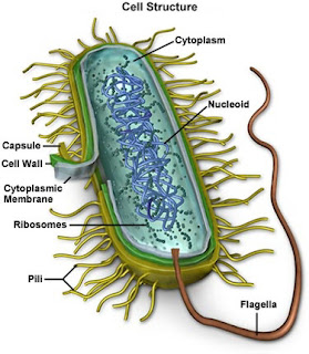

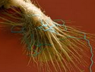
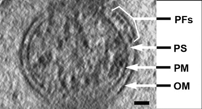
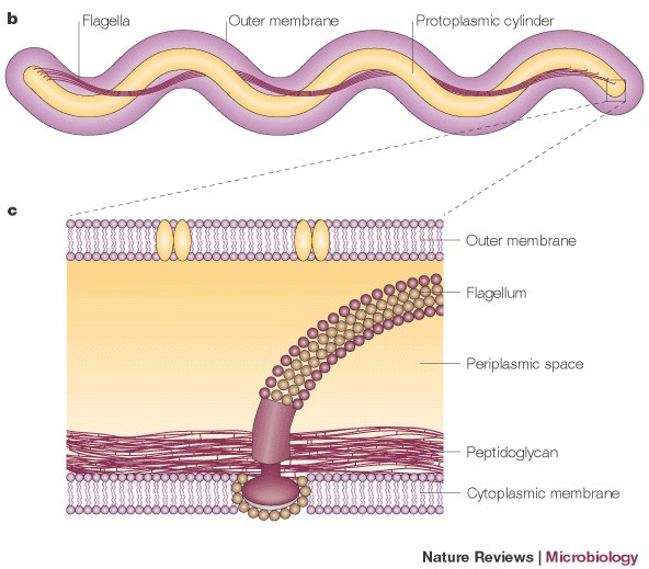

![[Wink]](wink.gif)
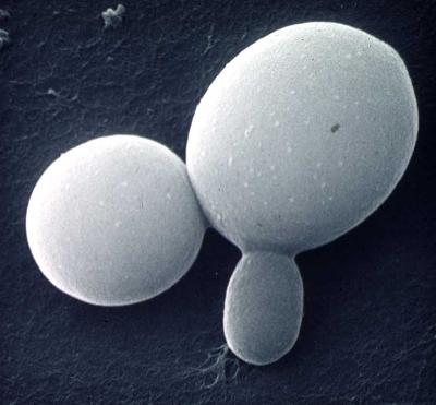
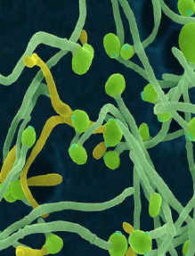
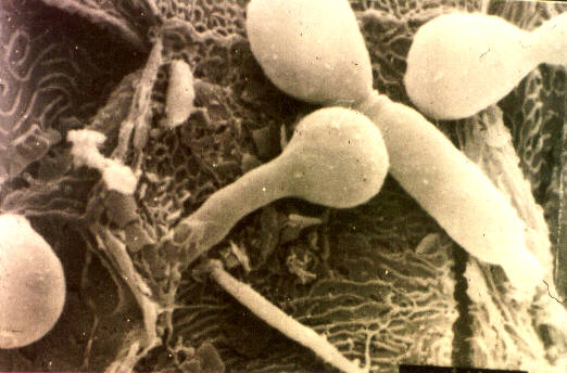


 Printer-friendly view of this topic