TNT
Frequent Contributor (1K+ posts)
Member # 42349
posted
Lymedin, I hope you don't mind me asking if you got your scope yet. I hope it came packaged well and is in good shape.
Posts: 1308 | From Eastern USA | Registered: Oct 2013
| IP: Logged |
Lymedin2010
Frequent Contributor (1K+ posts)
Member # 34322
posted
Emailed ya!
Posts: 2087 | From NY | Registered: Oct 2011
| IP: Logged |
TNT
Frequent Contributor (1K+ posts)
Member # 42349
posted
I INTERRUPT THIS BROADCAST TO BRING YOU BREAKING NEWS!
Bluelyme posted this video on another site and I am watching it for the first time.
This is a video of Eva Sapi's presentation at the NorVect Lyme conference.
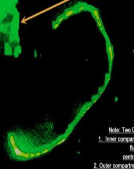
At 6:40 she shows a slide that depicts the response of Bb to minute amounts of Penicillin. Now we all know what Penicillin does to bacteria. It causes the conversion to L-forms. In this case a Spheroplast. Finally my question about what a Bb spheroplast looks like/is has been answered!
Now, for the big news! At 6:55 she settles the debate about what (active) form of Bb it is we are seeing in our blood. The people who don't believe we are seeing Lyme in our blood like to say that since we are not seeing typical (coiled) spirochetes, it must not be Bb, but some other object such as fibrin spicules (which do not move by the way!!!), etc., etc.
But, her slide shows typical spirochetes and it also shows exactly what we are seeing in our Lyme-diseased blood.... the docile "strings." Her slide absolutely shows that these are L-form Bb!!! So, Lymedin, you were right!
Finally, some documentation. You won't get this kind of validation from the CDC or the IDSA, that's for sure. In fact, they'll probably try to get this video taken down. May the truth prevail!
Posts: 1308 | From Eastern USA | Registered: Oct 2013
| IP: Logged |
TF
Frequent Contributor (5K+ posts)
Member # 14183
posted
I like how she said that spirochetes do NOT have to be stressed to go into the round body (cyst) form. Rather, it is a type of backup system that borrelia employs. Everyone with lyme will have them.
Posts: 9931 | From Maryland | Registered: Dec 2007
| IP: Logged |
Lymedin2010
Frequent Contributor (1K+ posts)
Member # 34322
posted
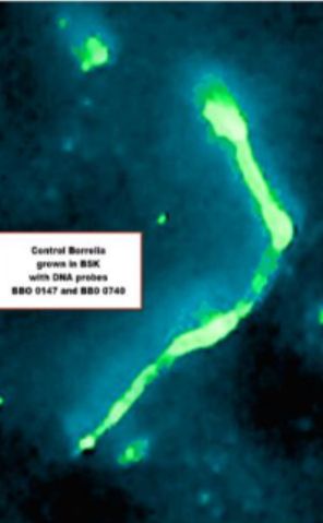
Great video & I had seen that one in the past. I hope you saw Dr. MacDonald's blood stains?
"Results of Research Study on human peripheral blood with Fluorescence In Situ DNA Hybridization(FISH method) and Borrelia burgdorferi family Specific DNA Probes BBO 0147 andBBO 0740"
I also posted TONS of research & materials suggested that Borrelia can & will be in the blood at times & more specifically during later stages of infection. The evidence is overwhelming I think... Join MY MICROSCOPE to see it. https://www.facebook.com/groups/MyMicroscope/permalink/1646490118967316/
I too have seen many of the blebs & cyst in my spiro tick studies. When I add new solution to the mix, the majority of them cyst up & all you see is tons of cysts (tiny ~1 micron dots). What I have noticed also is that when I let the sample dry up, then there are a lot more SOP/Segmented spiros, so I think during the most stressful times the majority will SOP to prepare for maximum dispersion in the open wild (after death of the organism it was in). It is also interesting that they can be kept outside the tick for months & they propagate & grow fine & form biofilm at room temp (73-78 degrees). And they should as they can live in homeothermic mammals & ticks at outdoor & cold temperatures, so room temp becomes a better suite than outdoor tick temps at times.
Also, TNT, I was able to hack the fluorescent lighting & I can get AO to glow with jut the 100W halogen bulb + a UV flashlight under the condenser shining up & even through the front windows of the fluoro light train/condenser. The halogen bulb puts out some UV, but not enough & hence the added supplementation. The images are not crystal clear/sharp, but you get to see what you need & can even make out the nuclear components of WBC's. Trickier to do it with my phone, as I need a better app for longer exposures. I've even been able to get get images to fluoresce with a LED flashlight held at an angle to the slide & w/the UV flashlight under the condenser.
I may put a quick video montage with some pics Fri or Sat. I already have ideas for improved hacks.
Posts: 2087 | From NY | Registered: Oct 2011
| IP: Logged |
Lymedin2010
Frequent Contributor (1K+ posts)
Member # 34322
TNT
Frequent Contributor (1K+ posts)
Member # 42349
posted
I know many of us have tried Artemisia annua at some time or another, but I came across this article and I thought it interesting, so I will share the link.
Lymedin2010
Frequent Contributor (1K+ posts)
Member # 34322
posted
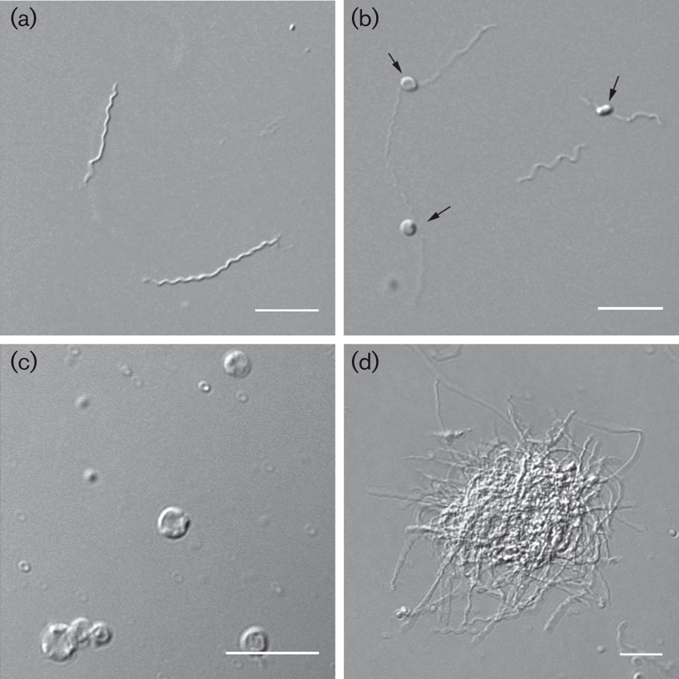
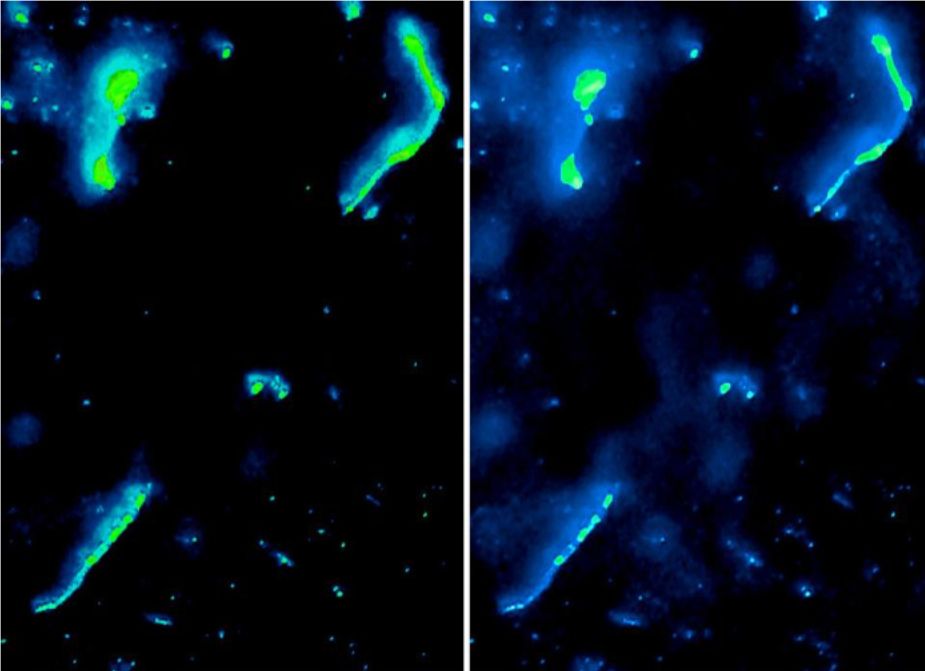
"The spirochaete bacterium Borrelia burgdorferi sensu lato is the causative agent of Lyme disease, the most common tick-borne infection in the northern hemisphere. There is a long-standing debate regarding the role of pleomorphic forms in Lyme disease pathogenesis, while very little is known about the characteristics of these morphological variants. Here, we present a comprehensive analysis of B. burgdorferi pleomorphic formation in different culturing conditions at physiological temperature. Interestingly, human serum induced the bacterium to change its morphology to round bodies (RBs). In addition, biofilm-like colonies in suspension were found to be part of B. burgdorferi’s normal in vitro growth. Further studies provided evidence that spherical RBs had an intact and flexible cell envelope, demonstrating that they are not cell wall deficient, or degenerative as previously implied. However, the RBs displayed lower metabolic activity compared with spirochaetes. Furthermore, our results indicated that the different pleomorphic variants were distinguishable by having unique biochemical signatures. Consequently, pleomorphic B. burgdorferi should be taken into consideration as being clinically relevant and influence the development of novel diagnostics and treatment protocols."
"Fig. 1. Typical pleomorphic forms of B. burgdorferi B31. Live cell DIC images of B. burgdorferi cultures representing (a) spirochaetes, (b) blebs on spirochaetes (black arrows), (c) 10 min H 2O-induced RBs and (d) BFL aggregates. Bars, 10 µm."
________________________________________ Intravital Imaging of Vascular Transmigration by the Lyme Spirochete: Requirement for the Integrin
_________________________________________ This reminds me of this particular spiro I found in my tick spiro video here. This is the same spiros/sample for the majority of the videos in my spiro Lessons video.
posted
Thanks for the links and videos Lymedin2010. Great work as always!
Anyone gotten any good stuff at home lately???
I'm finding some very faint halo's in some of my stains, but nothing convincing enough to post at the moment.
Posts: 71 | From Canada | Registered: May 2016
| IP: Logged |
Lymedin2010
Frequent Contributor (1K+ posts)
Member # 34322
posted
Thanks mustard.
I have not spent a whole lot of time on blood stains in general, but I did some fluorescent blood stains recently without spending too much time on them either. Doing everything at a Lyme snail pace. My first fluoro blood stain I thought I might have been seeing babesia splitting in 2 & tetrads, but I was not sure. I thought I saw signs of them being wbc, but was not sure either & so I made a quick slide show & posted it on my alternate channel. Since this video I have done 1 more stain & this time exposed the stains to full light for comparison and with better blood dispersion & sure enough they end up being wbc nuclei components. On my first slide I was just excited to see something glow, but no energy to investigate it in detail.
The first part of the video shows orange stained wbc's and the orange fluorescence represents DNA in the wbc that is in an acidic environment. But as the wbc ages & some acid escapes it then turns to green. Green fluoro is indicative of DNA in general when spotting bacteria in these stains too.
TNT, are you too noticing that the wbc first stain orange & as time passes, especially 24hrs some finally turn green? The faster the sample is left to dry, the faster the wbc compromise & then turn green.
I have some super exceptional fluoro stains of the tick spiros & it will help us decipher spiros in our blood if/when they stain & pop, but I have to finish putting together the lessons 2 video. I also have a false pathogen video that has some new revelations for us as well & I have to finish that too, the one I spoke of last time and another undetermined video that I am puzzling over.
You can check out this really awesome blood confocal micro live staining video of Bb in the capillary walls of the circulatory sys & there is another really good live microscopy video I will share in the future.
Lymedin2010
Frequent Contributor (1K+ posts)
Member # 34322
posted
EVEN THE MAYO CLINIC NOW KNOWS THAT LYME SPIROCHETES CAN BE IN LYME DISEASED BLOOD IN LARGE QUANTITIES.
They estimate that there can be ~85,000 Borrelia mayonii's per 1ml of human blood using calculations from darkfield microscopy. Given that the human body can have up to 5L of blood, this then means that there can be up to 425,000,000 spirochetes floating in the circulatory system & this is in a DILUTED blood sample, which means they will be even more concentrated in reality.
Given that the spirochetes can be invisible when inside the RBC & there can exist even more massive quantities of granular forms & cysts, then the numbers become even more astronomical & in the BILLIONS!
posted
That's a crap load of bacteria! Scary to think about.
Anyway, I watched part of the Sapi video posted above by TNT and blue, and I have a question that I may have asked earlier, but now can't remember. Any answers would be much appreciated, I'm not sure where else I would ask:
When we're seeing spirochetes with bulbous tips, or string of pearl spirochetes, are those L-forms?
Is that an L-form? Or do L-forms look like something different, and the bulbous tips are just regular spirochetes?
Are L-forms just in between forms of spirochetes and cysts?
And are L-forms killed by spirochete medications (Doxy, Rocephin) or by cyst medications (Flagy, Tini)?
I seem to have a hard time grasping exactly what a cell-wall deficient Borrelia looks like, acts like, and how exactly we kill it.
Posts: 71 | From Canada | Registered: May 2016
| IP: Logged |
Lymedin2010
Frequent Contributor (1K+ posts)
Member # 34322
posted
L-form are cell wall deficient, where they shed their outer membranes & loose their spiral shape. They look like the strings/spiros we see in our blood. They can if needed, revert back to the spiral forms when the conditions are right, such as when they are back in a tick.
BUT I also think that some of the string-like subjects we see are rbc cell wall sheddings & they can look like spiros & sometimes it is hard to tell from one another. In the next few months I am going to try to get better video to try to prove this & show this.
Posts: 2087 | From NY | Registered: Oct 2011
| IP: Logged |
posted
So in the video in my last post, the video title is incorrect, and it's actually an L-form changing into a cyst?
It seems hard to tell the difference between a true spirochete and an L-form if that's the case.
Posts: 71 | From Canada | Registered: May 2016
| IP: Logged |
TNT
Frequent Contributor (1K+ posts)
Member # 42349
posted
quote:Originally posted by Lymedin2010: L-form are cell wall deficient, where they shed their outer membranes & loose their spiral shape. They look like the strings/spiros we see in our blood. They can if needed, revert back to the spiral forms when the conditions are right, such as when they are back in a tick.
BUT I also think that some of the string-like subjects we see are rbc cell wall sheddings & they can look like spiros & sometimes it is hard to tell from one another. In the next few months I am going to try to get better video to try to prove this & show this.
I agree with the first paragraph for sure, and also about the RBC cell wall shedding, but I think most of what we see hanging on the outside of RBCs and even WBCs that are slightly wider than a spirochete, or L-form spirochete, are probably spirochetes in transition to spheroplasts or possibly even cysts. I wouldn't have been able to say that before I saw the mass conversion of the ketes in acridine orange. Man, I've got to get some time-lapse of that transition but I've been too busy. I'm thinking of just using dark phase to document it instead of fluorescence, since I will be able to keep the light on longer with the halogen bulb. I don't want to unecessarily put time on my mercury bulb for something that can just as easily be captured with something other than fluoroscence. So, if I ever get around to doing this don't be surprised if it's in phase. I think it will clear this up for you guys though.
Posts: 1308 | From Eastern USA | Registered: Oct 2013
| IP: Logged |
Lymedin2010
Frequent Contributor (1K+ posts)
Member # 34322
posted
Looking forward to the spheroplast videos.
Guys, you don't need a true fluorescent microscope to do fluorescent microscopy. The fluoro video I put out was all done with a 100W halogen light bulb & no mercury bulb whatsoever. I am on the hunt to replace that with LED as well.
Also, you don't even need a true fluorescent microscope rig either. All you need is the following to do FITC & Acridine Orange microscopy.
2)Short-pass filtered at 495-500nm, able to withstand heat from a 10-20W LED light source.
3)Emission filters, coloured glass long pass filter of 520-525nm. This you stick to the bottom of your objective lens.
They need to be 0 degree filters, not dichroic 45deg type filters like in the cubes.
4)You may need to add a collinator that collects more of the light between your LED light source, under the stage & condenser. Sometimes a flashlight top curved lens might due, or an old condenser, or a collinator from an actual fluoro scope...basically anything to collect the light & concentrate it. You have to see how your condenser & scope do without the collinator & if you need it then add it between the led & condenser.
I put in some questions to a filter place to see if they have them available for you guys.
As with any LED source, never look through the eyepiece as it can burn your eyes, just like a laser pointer & always do it through a camera. And also when your wiring for LED's always watch out for shorts & shocks & good heat dissipation with heatsink & thermal paste between the heatsink & LED unit.
Posts: 2087 | From NY | Registered: Oct 2011
| IP: Logged |
Lymedin2010
Frequent Contributor (1K+ posts)
Member # 34322
posted
So this is the video I was referring to, which I show what can look like more false pathogens. I have seen the rbc spikes out in the plasma & at times the wbc's will pick them up & be loaded with them.
Lymedin2010
Frequent Contributor (1K+ posts)
Member # 34322
posted
So here is a spiro in my blood in 4K and also the subjects I described a while back that fluoresce even without any fluorescent stains & just the filters.
Hard to say what they are for sure, as I need to spend more time on them & I need more refined staining. Before the end of 2017 I will get to the bottom of it with more clues.
They could be: -platelets -rbc crenation spikes (Burr cells) -maybe babs or bart, but no solid evidence of vacuoles or ring or maltese forms. Some evidence yes, but nothing I am totally happy with & I need more staining refinement as I had spent too much time on the tick spiros & neglected staining & improvement for a while now. I have one vacuole video, but I am not totally pleased with it & maybe I will upload that later today.
I will leave this open as to what exactly it is until further investigation & evidence & I do see the same things in my Giemsa/Wright stains & even my freeze trials.
posted
Got any pictures or vidoes of that under Giemsa?
It'd make it easier to determine what they are. Might just be platelets.
Posts: 71 | From Canada | Registered: May 2016
| IP: Logged |
Lymedin2010
Frequent Contributor (1K+ posts)
Member # 34322
posted
I only stained maybe 3-4 slides & did not have a whole lot of energy to look at them with grand detail & the preparations were less than ideal. I made a few scan videos & posted them here.
At one point I will go in stronger with staining & use EDTA to disperse the blood more evenly...same for the fluoro stains as they are too clumped & less than perfect for observations.
I think they might be the spikes from the Burr rbc's, but it can just as easily be platelets. Once I put some normal blood under the fluorescent filters, I will be able to tell & buy some more clues.
If you look at some of the Giemsa videos, there sure are a lot of platelets scattered & some objects appear to be damaging or coming out of the rbc's. So I will just leave everything open & unresolved until further investigation. Some objects almost look like babs ring forms, but I am still not satisfied & prefer to dismiss them.
I am also trying to upgrade my 100W power supply, as it overheats quickly & trying to even get a LED replacement for more pop in the fluorescence.
Posts: 2087 | From NY | Registered: Oct 2011
| IP: Logged |
Lymedin2010
Frequent Contributor (1K+ posts)
Member # 34322
posted
TNT, this one is for you. This seems like a gentler way to spread a drop of blood for fluoro & giemsa/wright stains.
Using EDTA in purple cap tubes or as standalone additions is even better with this method.
Another secret is to use a cover slip to GENTLY place on top of a drop of blood & then run a tissues along the edges of the cover slip. This will siphon blood out of the slide & onto the napkin & leave a more thinner dispersed field of blood on the slide. Then you simply remove the cover slip gently.
___________________________ Those triple stained bovine endothelial cells would be awesome to have for fluoro reference when changing equipment & refining, ouch but the damn price tag!
Lymedin2010
Frequent Contributor (1K+ posts)
Member # 34322
posted
Using this blood prep method provides SUPERB dispersion of rbc's & practically uniformly throughout. I cannot believe how well it disperses the blood!!!!
You basically touch a 2nd glass slide on top of the first one & allow the blood to evenly spread naturally & then you swipe across the length of the glass slide.
_____________________________________________ Here is a lab example of Diff Quick/Dip Quick stain procedure. I would not use tap water as they did here, but RODI or distilled water instead. https://www.youtube.com/watch?v=V9nFq8BvW-QPosts: 2087 | From NY | Registered: Oct 2011
| IP: Logged |
quote:Originally posted by Lymedin2010: Using this blood prep method provides SUPERB dispersion of rbc's & practically uniformly throughout. I cannot believe how well it disperses the blood!!!!
You basically touch a 2nd glass slide on top of the first one & allow the blood to evenly spread naturally & then you swipe across the length of the glass slide.
_____________________________________________ Here is a lab example of Diff Quick/Dip Quick stain procedure. I would not use tap water as they did here, but RODI or distilled water instead. https://www.youtube.com/watch?v=V9nFq8BvW-Q
What method were you using before???
That last video is a great "how to" staining video.
The one thing I do differently is smear a little bit slower. She talks about how her feathered edge is a mono-layer, but I find that if I smear the blood slower, almost my entire slide is a mono layer.
I also use a smaller drop of blood than what she uses, but that's mostly because I can only get so much out of my finger.
Posts: 71 | From Canada | Registered: May 2016
| IP: Logged |
Lymedin2010
Frequent Contributor (1K+ posts)
Member # 34322
posted
For Giemsa/Wright I do it the way she does to get the feathered edge & that sweet spot. Mine don't come out uniformed & I rely heavily on the edge for observations. But doing it the other way, with the 2 glass slides right on top of one another gives great dispersion & I will use this for live blood preps the next few times.
My live smears I do the way I show in the video & when I want less disturbed cells I just gently plop the cover slip on top & I do not smear, at the expense of a lot of clumping but a good amount of whole & undisturbed cells. I also add 0.9% NaCl when I really want good dispersion, but found that this method does not allow many spiros to come out or produce any false spiros.
__________________________ I have checked a few spiders & no spirochetes. "Spirochetes inside the gut of Coptothermes, a termite species, also showing me in my lab with the ECIS-system invented by nobel laureate in Physics, Ivar Giaever."
I wonder if ants have them too, as ants often times consume many other insects?
Lymedin2010
Frequent Contributor (1K+ posts)
Member # 34322
posted
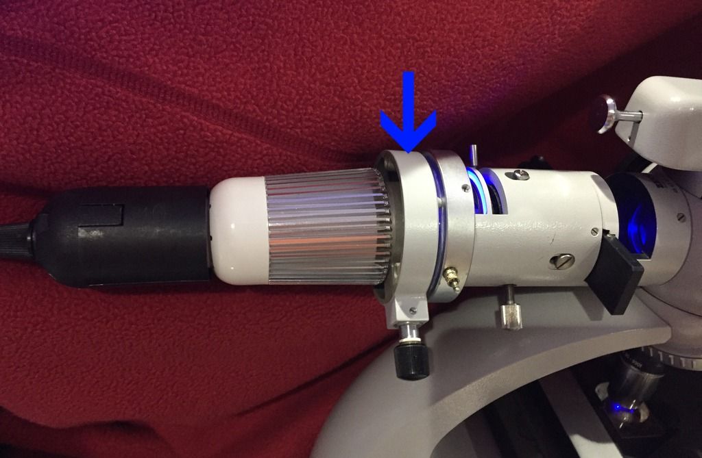
TNT, I did it...fluorescent microscopy mercury hack to the power of 2. Not only does the 100W halogen bulb produce decent images, but I managed to use a $11 27W RGB light bulb w/standard E27 fitting & on a standard lamp socket to produce even better results. I removed the ring from the bulb & then the lens to expose direct light from the LED chip & then I pulled out the collinator from the light house, which conveniently attaches directly onto the light train. Then I place the bulb in front of it & viola!
There are some issues as the bulb in blue light does not use all of the LED's & so I have to angle the bulb slightly & it would have been better to have just a blue light bulb that uses all of the LED's & the whole 27W. Even with this I will have to tone down the intensity 1 or 2 notches, but the unevenness makes me want to get a dedicated blue bulb. At least this is proof of concept & on the road to mercury free, electricity saving, & more pop to the fluoro images. You be the judge....
Pic 1: Image using highest intensity on the 100W halogen lamp:
Pic 2: Using the 27W LED bulb on blue light: Posts: 2087 | From NY | Registered: Oct 2011
| IP: Logged |
TNT
Frequent Contributor (1K+ posts)
Member # 42349
posted
Nice hack! What exactly are we viewing in your picture?
Can you link to the bulb you used, and also post a couple pictures of the actual setup?
Posts: 1308 | From Eastern USA | Registered: Oct 2013
| IP: Logged |
TNT
Frequent Contributor (1K+ posts)
Member # 42349
posted
I wonder if something like this would give you the whole beam you're looking for?
Lymedin2010
Frequent Contributor (1K+ posts)
Member # 34322
posted
No, that will not work as it is too weak with only 3W, but everything else about the bulb fits. Try at least 15W with those smaller bulb, as I think that is the max on those type of bulbs, because of the way the heat sink heat dissipation fans are designed at the base.
You can try this bulb though, as it emits blue in its spectrum as well & should fluoresce. I ordered this one, but it has not come in yet & I could not find the same bulb in RGB with 15W. I don't even know if 15W will be bright enough & it might be just on the verge if we are lucky.
Here is the one I am using now & just for now I simply hold it in the back in front of the light train, with the collinator attached to the light train as it attaches the same way from the lighthouse (but just removed from the light house with the 3 screws on my unit). I am not going to make any final attachments until I test out a few bulbs & LED's.
Also it is important that you observe that with white light all the LED's are used, but with blue only a portion is used & there is unevenness & that is why I have to slightly angle it.
Lymedin2010
Frequent Contributor (1K+ posts)
Member # 34322
posted
Also, just to note that Acridine Orange (AO) will excite in the 440-480 & 502 spectrum range (which is blue light mostly) & so light that is in that peak will produce the best images.
Pure white light LED will work too, since it also emits blue, but you may get some washing out of the fluoro colors & probably depends on the strength & exact position of the blue peak & the bulb overall.
[ 06-21-2017, 08:09 AM: Message edited by: Lymedin2010 ]
Posts: 2087 | From NY | Registered: Oct 2011
| IP: Logged |
Lymedin2010
Frequent Contributor (1K+ posts)
Member # 34322
posted
FYI, word on the street is some have been using Red #40 food coloring to see some of these things in the blood. I have not tried it nor seen any pictures, just heard. It might make for some nice live & undisturbed viewing if anyone wants to try it before me.
Posts: 2087 | From NY | Registered: Oct 2011
| IP: Logged |
Lymedin2010
Frequent Contributor (1K+ posts)
Member # 34322
posted
TNT, last time I did not have a whole lot of stamina to play with the bulb as much as I would have liked & I looked at it again today. It looks like there are 9 LED's (3W*9 = 27W) in the bulb & when the blue lights come on only 3 of the LED's are active & they are not the ones in the middle of the bulb & it looks like that is why I have to angle it to get better penetration.
This means that only 9W of the blue light is used. Also now that I messed with it some more I can see 3 distinctive spots when I hold it a certain way & closer to the light train. Then when I pull away I can tell that only 1 of those LED's becomes centered & can provide good enough penetration...shockingly!
So that means that you might be able to get away with the bulb that you linked. If you look at my setup, the arrow will show you the collinator that I removed from the lamphouse & then screwed back on to the lamp train just as the way it would normally screw on. With the lens ring & lens off the bulb, it fits right nicely onto the collinator & it actually stays on and if I twist it in a specific direction I get very narrow coverage.
Then when I pull out slightly I get even better coverage, but at the risk of the bulb falling. Then when I pull out more & angle the bulb so that the 3 LED's beam towards the middle then even better. And lastly when I pull out even more so that only 1 of the LED's, then I finally get uniformed penetration & great coverage. So now I have it on top of a tall box & a sweater & it stays nicely for observation at the proper angle & position...even this way it beats the mercury lamp! That aluminium housing can easily be screwed off & one can reposition the chip with new taps to center those 3 LED bulbs & I just might do that in the future, but I am good for now.
[ 07-21-2017, 04:23 PM: Message edited by: Lymedin2010 ]
Posts: 2087 | From NY | Registered: Oct 2011
| IP: Logged |
TNT
Frequent Contributor (1K+ posts)
Member # 42349
posted
Great fluoro hack! Very good! I can see the spirochetes very easily in your specimens, even at what appears to be 400x.
It appears like your Zeiss scope is more hack friendly than my American Optical fluoro scope. The dichroic filter/mirror doesn't hinder the operation at all? I guess it is not really doing much (because you already have the proper wavelength with the blue LED) except the essential function of directing the light down through the objectives.
It would be possible to do fluoro work simply by directing your bulb directly on the specimen as long as you have the correct wavelength bulb (Just a FYI for those not acquainted with fluorescent microscopy....).
Posts: 1308 | From Eastern USA | Registered: Oct 2013
| IP: Logged |
TNT
Frequent Contributor (1K+ posts)
Member # 42349
posted
Another idea for your setup would be to try one of these two bulbs (depending on whether you wanted a clear LED lens or not). Both are 5 watt and both of the heat sink flanges could be drilled to provide a mounting surface that you could screw the bulb onto another tube or adapter that could be configured to the exact length so as to be parfocal with the original Zeiss lamp assembly.
The exact distance of your light source from your collimator really matters-- so that you can get a good, strong, focused beam to your dichroic mirror and down through the objectives (just like when you said you pulled your LED further and further out until you only had the light from one LED, and how this made the strongest, most focused beam of light).
So, a customized or machined "tube" adapter that would attach or mount directly to your collimator, and the other end/opening of the adapter would be screwed to the flange of these LED bulb's aluminum heat sinks.
Posts: 1308 | From Eastern USA | Registered: Oct 2013
| IP: Logged |
TNT
Frequent Contributor (1K+ posts)
Member # 42349
posted
Fluorescent microscopy is pretty cool. I knew you would like it, and I know the tick-handler will make full use of it!
Ha Ha, I guess I can take credit for pushing you over the edge Lymedin! How about it?
I'm glad you are brain-storming cheap hacks for your fluorescent microscope. These ideas will come in handy when my mercury bulb blows out. Your idea about the cheap color changing bulbs is a great idea. And, you've proved by your pics above that they will work, even if the exact wavelength of the colors are not known. Much appreciated. I love building upon your ideas. I actually went inspecting my scope this afternoon to see how hard it would be to mount one of those bulbs at the back where my lamp housing presently is. I don't think it would take much customization to be honest.
Posts: 1308 | From Eastern USA | Registered: Oct 2013
| IP: Logged |
Lymedin2010
Frequent Contributor (1K+ posts)
Member # 34322
posted
No, the mirror & filters do not interfere. They are designed to block all the wavelengths except for the desired ones in the range. So a strong peak in only the desired range & with more intensity of that peak will produce more luminosity in the excitation. So even when white light, or green light is passed it can fluoresce, as different shades of green can also have some slight blue in them as well. White light has a broad spectrum of the visible wavelength, including blue.
Yea, in theory you can use a LED chip under the scope & condenser with the specific wavelength needed for the specific dye, in this case blue, & then you will only need a filter between the top glass slide & your objective lens. I am sure if someone tried this, they can hack the entire fluoro microscope & DIY all of it. And if you use a LED under the scope, you should not need a powerful one & could get away with much smaller wattages.
Hard to believe how well this LED bulb fits, almost as if it was made for it. I have ordered the 15w version, as it will have only 5 LED's & I am hoping it will be centralized, but we'll see. I may do a custom PVC to extend out the insert & so the distance can then be perfect for direct attachment of the LED light bulb.
So I tried the various blue spectrums off the bulb & it looks like light blue is even more precise. Then I double checked the Zeiss site on excitation & cross referenced that with other AO lists. I contacted Zeiss about this & they said that It turns out that they are wrong on their website & it is not 430nm and this is the info they gave me about AO with relation to their filters.
Acridine Orange + dsDNA (double stranded) Excite 502, Emit 525 (Emits GREEN) (Orange at pH 8.4 & green at pH 10.4)
So really the excite is more of a range betwen 440-480 & 502 somewhere for BOTH DNA & RNA & can also be dependent on the filters being used & the solution that dye is in, as they can shift these values. Gonna edit my previous post for the true number range.
References to the values here & the Zeiss one is wrong:
[ 06-22-2017, 08:13 AM: Message edited by: Lymedin2010 ]
Posts: 2087 | From NY | Registered: Oct 2011
| IP: Logged |
Lymedin2010
Frequent Contributor (1K+ posts)
Member # 34322
posted
TNT, both bulbs you linked should work. It looks like it may be one led at 5W & should be more powerful than mine I think.
I like the first one & you may have to remove the ring & lens cover to get it to work properly. I like the first one & the 2nd one you definitely have to remove the lens, otherwise you will see the lens rippling patterns.
Lymedin2010
Frequent Contributor (1K+ posts)
Member # 34322
posted
"Detection and Differentiation of Lyme Spirochetes and Other Tick-Borne Pathogens from Blood Using Real-Time PCR with Molecular Beacons Schlachter S, Chan K, Marras SAE, Parveen N. Methods in Molecular Biology, 2017;1616:155-170.
Abstract
Real-time PCR assays have recently been implemented in diagnostics for many bacterial pathogens, allowing rapid and accurate detection, which ultimately results in improved clinical intervention. Here, we describe a sensitive method of detection for three common tick-borne pathogens Borrelia burgdorferi, Anaplasma phagocytophilum, and Babesia microti since coinfections with these pathogens have started occurring with increasing frequency over the last several years in both North America and Europe.
A shared geographic region, the same tick vectors, and similar transmission cycle all favor simultaneous transmission of these three tick-borne pathogens. Furthermore, early symptoms of the diseases are often similar and somewhat nonspecific leading to poor clinical identification.
The multiplex real-time PCR assay we describe here utilizes gene-specific primers, molecular beacon probes tagged with different fluorophores, and optimized PCR conditions to detect even small amounts of specific pathogen DNA without interference. Application of this detection method will offer better diagnostics for acute and persistent infection compared to the two-tier serological tests that are currently approved in North America and Europe, which do not necessarily detect active infection."
Good job! Is that lighting with the RGB LED bulb?
Posts: 1308 | From Eastern USA | Registered: Oct 2013
| IP: Logged |
Lymedin2010
Frequent Contributor (1K+ posts)
Member # 34322
posted
No, that video was taken with the 100w halogen bulb before I got the ideas for the LED's.
I got this one too & this seems to work as well & has 5000 lumens. It also works with the flashlight lens on & the unit can be powered with the provided charger while in use & charged. It seems to be even brighter than the bulb, but it translates to a very narrow view on the actual view & I have to play with that some more when I have energy.
TNT, are you diluting the AO mixture with your blood samples & if so with what?
Posts: 2087 | From NY | Registered: Oct 2011
| IP: Logged |
TNT
Frequent Contributor (1K+ posts)
Member # 42349
posted
So, that headlamp works as an "ultraviolet" light source? Interesting. I definitely think your colored RGB LED bulbs are a better idea.
I'm joking about this next suggestion because it would be extreme overkill, but you could hook this up to a driver and have incredible intensity (it's a laser without the collimator):
I think the RGB LED bulbs we've discussed (like what you are using) are plenty powerful enough though and should work really well as a permanent hack solution. The two 5 watt ones I linked to on Amazon should have plenty of intensity and would be fairly easy to incorporate into a customized light train apparatus.
I didn't dilute the acridine orange. I just added a very minute drop (if you can call it a drop even) to the bottom side of my cover slip before laying the cover slip down on my drop of blood.
Posts: 1308 | From Eastern USA | Registered: Oct 2013
| IP: Logged |
Lymedin2010
Frequent Contributor (1K+ posts)
Member # 34322
posted
The Halogen & Mercury & Xenon bulbs produce UV, but these RGB bulbs do not & the AO & Fitc are stains which do not emit or excite in the UV wavelength range & so it is not needed with these dyes.
Lymedin2010
Frequent Contributor (1K+ posts)
Member # 34322
posted
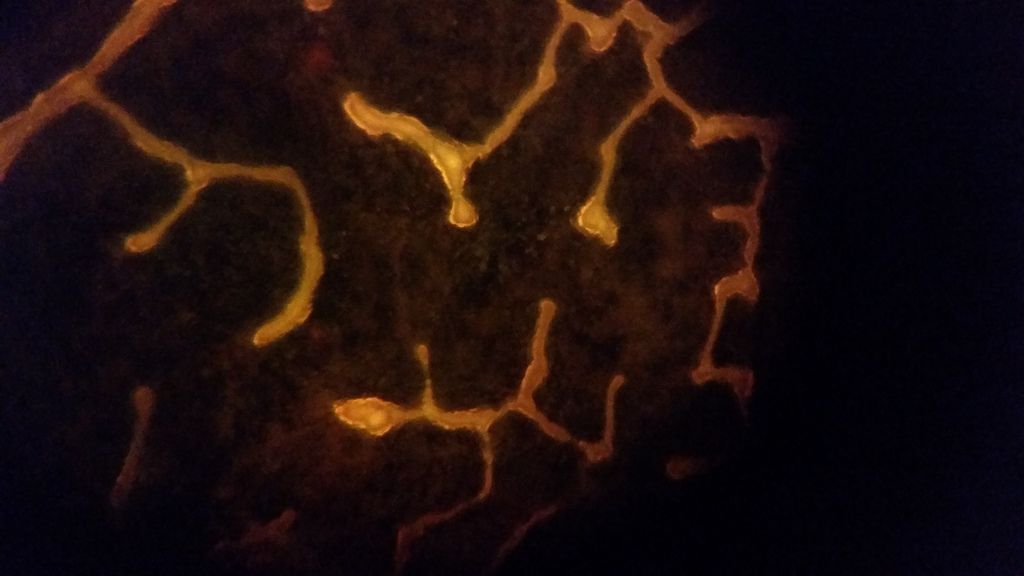
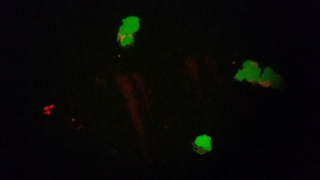
I figured out what those string-like spirochetes are in the pictures I posted. After looking at them for some time, it looks like in certain spots & after the prep has dried out that the rbc's clump together & because some wbc's burst & release some DNA into the plasma, the edges of the rbc's can fluoresce & glow when clumped together. So those string-like subjects are not spirochetes. Sometimes I do also see string-like subjects in the plasma & they are so thin & glow faintly and after their light exposure they grow even dimmer. Hard to say for sure, as I have not caught the perfect specimen yet. I have also seen one subject that looks like what we might call a spiro, but it did not fluoresce though.
On another note, here is what might be either Babs or Bart in my blood. Read the description for more info. TNT, I diluted this prep to get better results & I cross tested one live view with the 100w Halogen bulb. I would have missed all these with the Halogen, as they are too faint & barely visible & it was only with the RGB bulb that I am able to pick them up.
Lymedin2010
Frequent Contributor (1K+ posts)
Member # 34322
posted
Here are some pictures of wbc's with the 27W bulb. I have tried 2 other bulbs with less wattage & it seems like the 27W bulb is the best, the lower wattage ones only have 1 blue led & they still work, but just not as bright. The 3 blue leds on the 27W overlap & if you use the middle led, the light from the other 2 leds overlap into concentric circles. You can actually view the effectiveness of the overlap in the front view window of the light train, as you can see 3 bright spots & they overlap as you pull it further away from the back into a sweet spot.
Some of these pictures are of lightfield of the fluoro view for comparison.
posted
It's a stain ya, but I've never seen a malaria ring look quite like that before so I'm not sure.
Posts: 71 | From Canada | Registered: May 2016
| IP: Logged |
Lymedin2010
Frequent Contributor (1K+ posts)
Member # 34322
posted
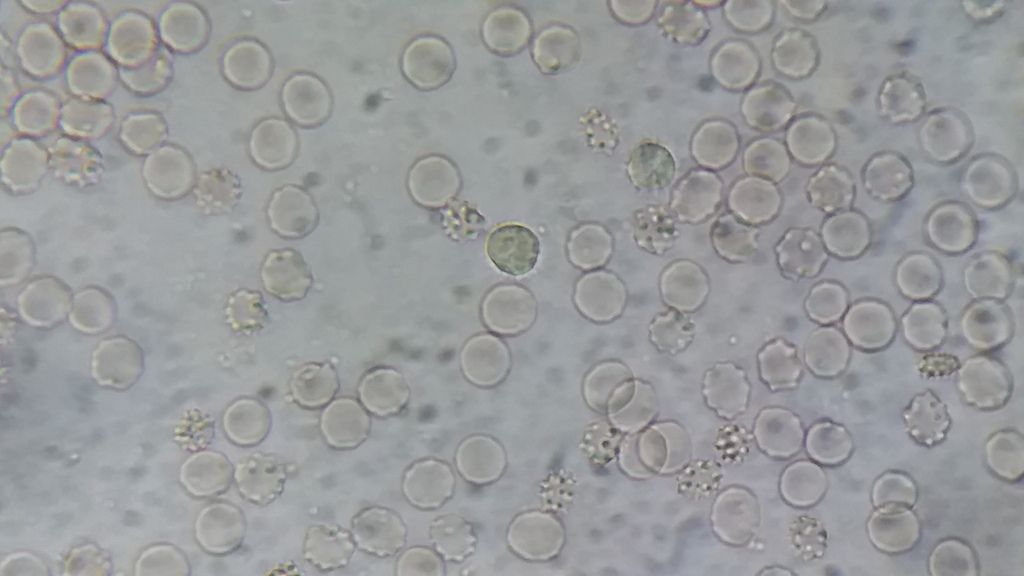
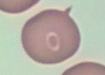
It sure looks like the shape of a ring form, but it did not stain & the circle around it did not stain as well. The center of the circle is pink like hemoglobin and the circle is clear like a halo. As a result I don't think this is Babs ring form. The spike or protrusion of the rbc is just a deformity in the rbc I think.
The rings around stains that do have them stain blue to purple & are clear stained rings AROUND the halo.
Check out these forms here & you can see a halo around them too. I still have not done anything else to figure out if they are platelets or rbc crenation burrs for sure. Maybe even some of the darker ones might be pathogens, around the rbc, but I am still not sure on this video until I do more live viewing with the filters & compare it to normal blood.
I was ready to call it off as a random artifact, but in a stain I did tonight I found two more identical to the one I posted! It's very weird. I'm going to have to dig a little deeper into this...
I tend to think those are platelets, but it's obviously hard to tell. Have you done any work with the quick dip?
Posts: 71 | From Canada | Registered: May 2016
| IP: Logged |
Lymedin2010
Frequent Contributor (1K+ posts)
Member # 34322
posted
Some, but not a whole lot & I never got to refining my technique yet. Before 2017 is over I will get to that.
Posts: 2087 | From NY | Registered: Oct 2011
| IP: Logged |
Lymedin2010
Frequent Contributor (1K+ posts)
Member # 34322
posted
DIRECTLY FROM THE CDC!!!!!
"Although no cases of Lyme disease have been linked to blood transfusion, scientists have found that the Lyme disease bacteria can live in blood that is stored for donation. Individuals being treated for Lyme disease with an antibiotic should not donate blood. Individuals who have completed antibiotic treatment for Lyme disease may be considered as potential blood donors. Information on the current criteria for blood donation is available on the Red Cross website"
If anyone of these anti-Lymers gives you grief, just tell them to put their science where their big anti-Lyme rhetoric mouths are & offer them an infusion of 1 pint of your blood. Then see how quickly they reveal their spineless inconsistencies in the deplorable act of terrorizing the sick.
If they are too worried about receiving blood, then just offer to inject them with 1 CC of Borrelia burgdorferi colonies & tell them don't worry it is easy to treat with 2 weeks of Doxycyline.
Lymedin2010
Frequent Contributor (1K+ posts)
Member # 34322
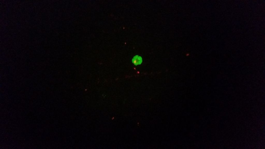
posted
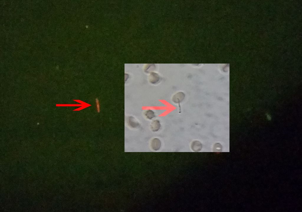
So I have only found 2 string-like subjects in my blood so far out of 7-8 slides, and not all slides were ideally made. One of them did not fluoresce & I have video of it and I will post it up eventually. And this one, which did fluoresce.
I do see a lot of smaller particles fluoresce though, but it is too hard to determine if they are DNA from the WBC's or spiro cysts and/or blebs. Since I am not seeing many string-like objects, be it actual spiros or false spiros, it must be that the AO solution is causing them to get destroyed or cyst up. Once they cyst up, it is hard to tell them from other components in the blood. I have not had the energy to check with much aggression though & the attention/time that it deserves.
Spirochetes are also delicate subjects & can lyse/die easily. If they are not given ample time to cyst up, they can get destroyed too and their delicate nature is known by the people who handle them...hardy but delicate.
In the pic below I also provide a zoomed version of it to the right & one with some light to see the rbc's that it is next to. Also, to the right of the subject in the center is another what might be infected rbc, similar to my last co-infection video.
Lymedin2010
Frequent Contributor (1K+ posts)
Member # 34322
posted
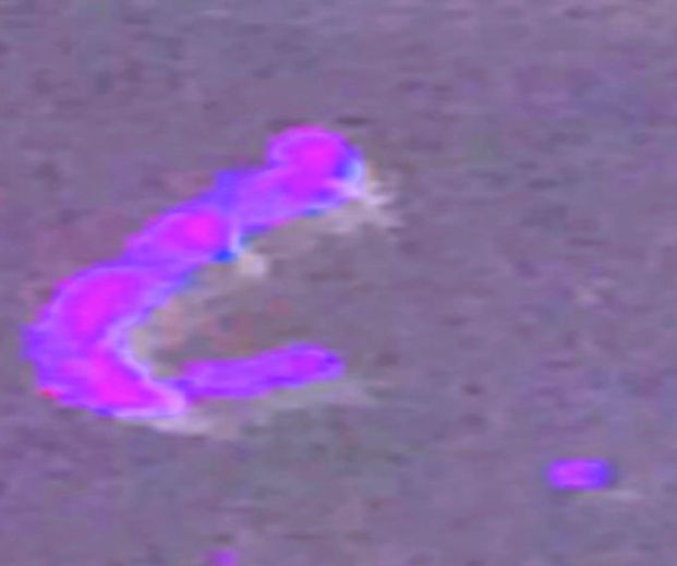
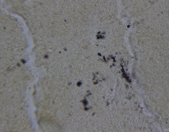
Here are some of Alan MacDonald's PanDNA and DNA Probe stains. The stains are not always ideal & what we would normally expect and they don't look very typical of lab grown spiros.
Lymedin2010
Frequent Contributor (1K+ posts)
Member # 34322
posted
"Figure 3. Spirochetes are present within xenodiagnostic ticks that fed upon saline- or antibiotic-treated mice at 12 months after treatment.
Indirect immunofluorescent staining of B. burgdorferi in the midguts of ticks that fed upon saline-treated (A) or ceftriaxone-treated mice (B) at 12 months after treatment."
posted
Keep up the good work with the fluro lymedin2010. Looks very promising.
Posts: 71 | From Canada | Registered: May 2016
| IP: Logged |
Lymedin2010
Frequent Contributor (1K+ posts)
Member # 34322
posted
Thanks mustard!
A new video from Ann, who grows confirmed laboratory grown Borrelia cultures. Interesting, as at time 3:25 the Borrelia has a slight bend, just like the one that I captured in my lyme blood via fluorescent staining.
I will also attest to the variation in spiro length & size & shapes, as that became obvious with my tick studies. In this one tick I found ridiculously super massive spirochetes of the same unusual type I had been finding. I couldn't believe just how big they were. And it was not just one, ALL OF THEM were big... a DNA lineage of massive spiros.
"Examples of fluorescent antibody staining from one donor showing fluorescence filtered image on the left of each matched pair and normal darkfield image on the right.:"
Lymedin2010
Frequent Contributor (1K+ posts)
Member # 34322
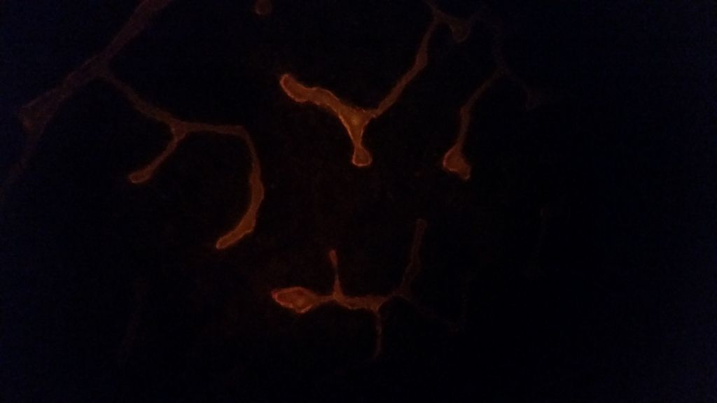
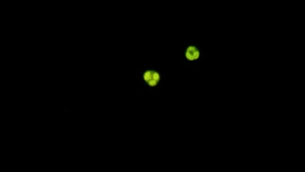
posted
I am finding that a lot of my rbc's have DNA on them at times & they are scattered throughout the sample. The fuller & brighter objects are WBC's & the speckled ones with DNA are rbc's.
________________________________________
This is fluorescence in the plasma. It might be components from WBC's that have lysed, as the WBC's have DNA in them & that is why they fluoresce in AO stains.
Also, this is that vacuole video I was talking about a while back & I finally got to uploading it.Vacuole in the rbc or biconcave lighting tricks. I would probably just dismiss this as lighting tricks until further evidence, although it looks tempting to say it is a vacuole.
Lymedin2010
Frequent Contributor (1K+ posts)
Member # 34322
posted
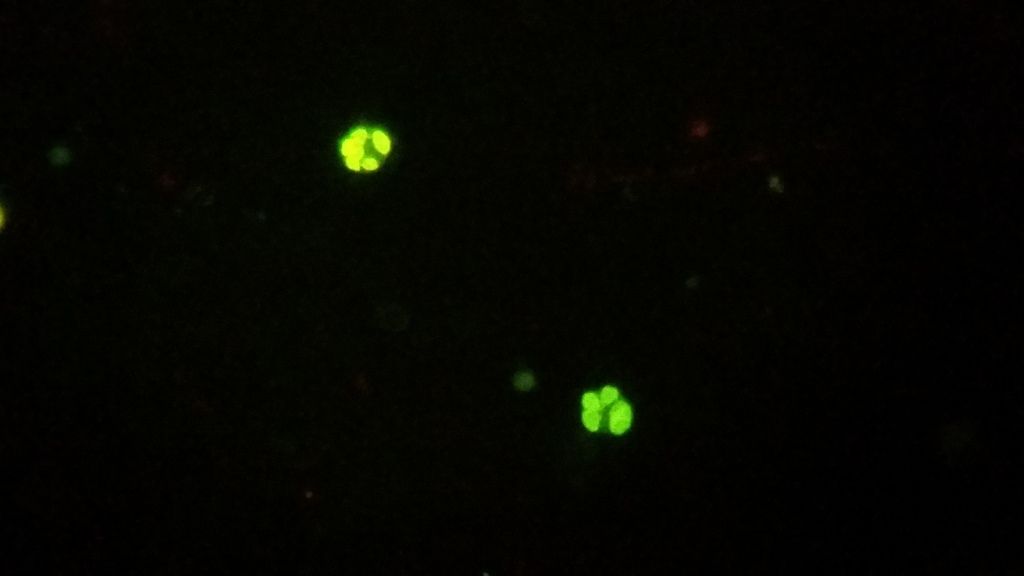
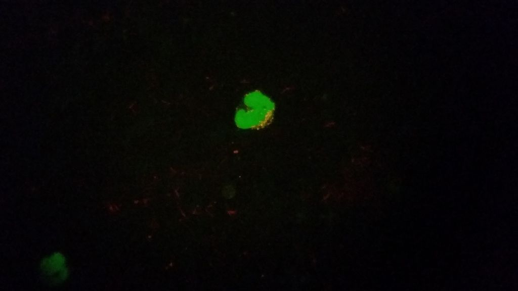
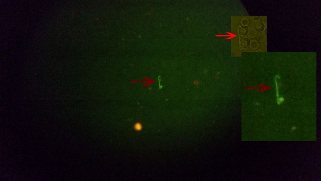
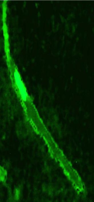
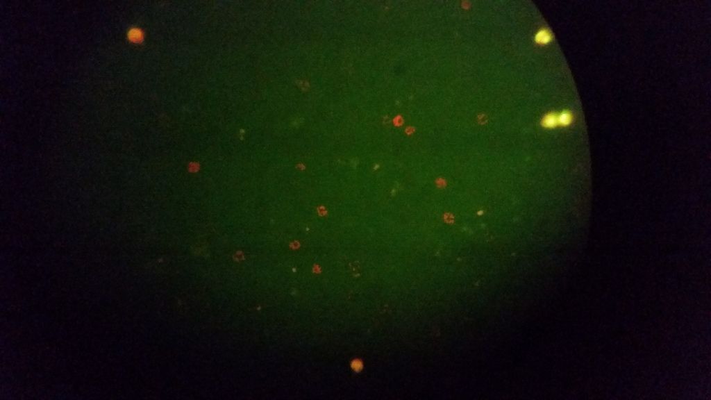
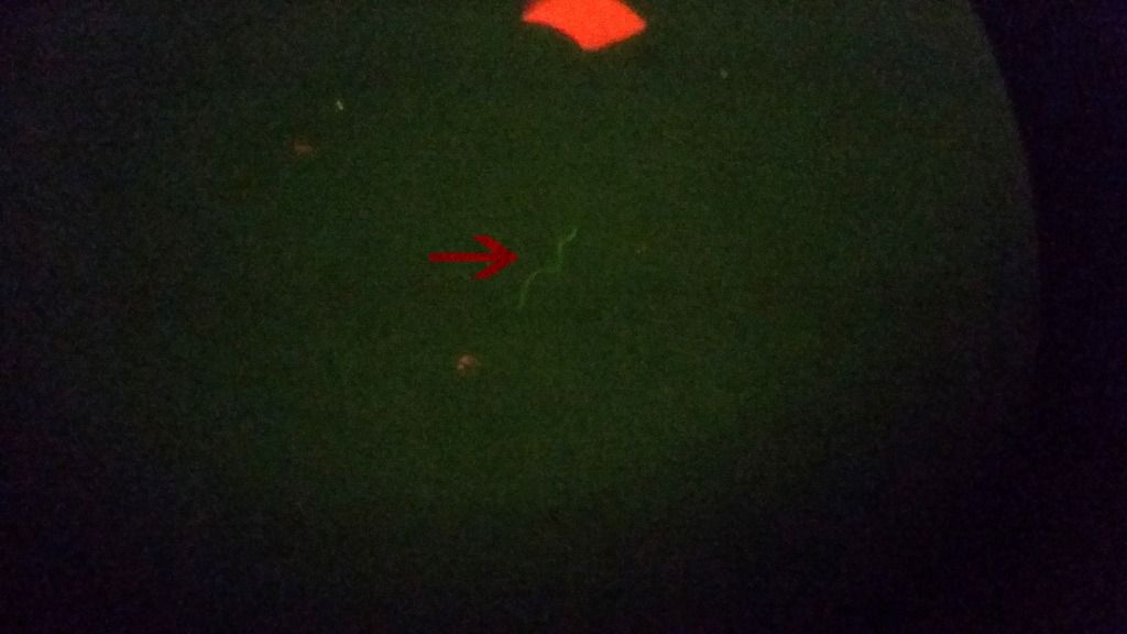
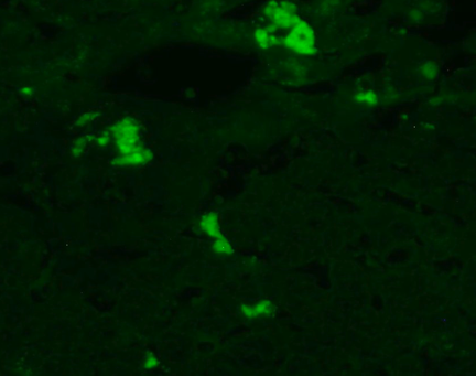
Another spirochete that fluoresced with Acridine Orange in my blood. Green means it is something with DNA, but who knows what species it might be.
Lymedin2010
Frequent Contributor (1K+ posts)
Member # 34322
posted
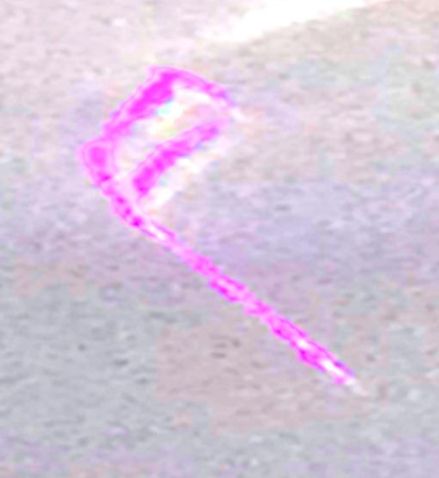
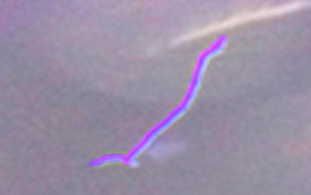
So with refinement of my technique of diluting & spreading the prep thinner I am finding spiros that fluoresce, but they are few & scattered. Another spiro in my blood pic below. I now also have other string-like subjects & even SOP in my preps & they do not fluoresce, which means they do not have DNA/RNA and are probably false spiros. I will make a video of the false spiros eventually.
Lymedin2010
Frequent Contributor (1K+ posts)
Member # 34322
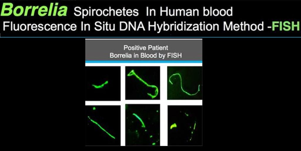
posted
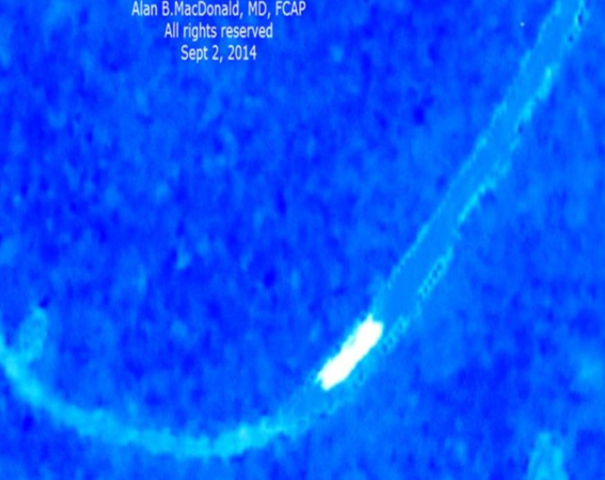
I like to remind myself of the odd & unusual shapes of Borrelia in the human body at times. Alan MacDonald's DNA FISH probes only bind to Borrelia DNA & makes for a good reference. Here is his result from probing in human blood.
bluelyme
Frequent Contributor (1K+ posts)
Member # 47170
posted
Wow what great work ...hard proof that it is indeed very much intercellular ...maybe why dr h loves double and triple intercellular abx .thanks lymed
-------------------- Blue Posts: 1539 | From southwest | Registered: Dec 2015
| IP: Logged |
Lymedin2010
Frequent Contributor (1K+ posts)
Member # 34322
posted
Thanks.
I have found SOP's that fluoresce in my blood now. I also found spiro-like ones that do not fluoresce & segmented ones that do no fluoresce & I will put them in a future false spiro video.
Lymedin2010
Frequent Contributor (1K+ posts)
Member # 34322
posted
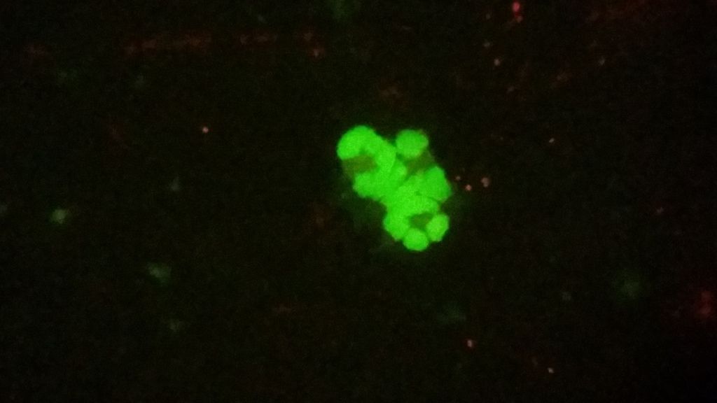
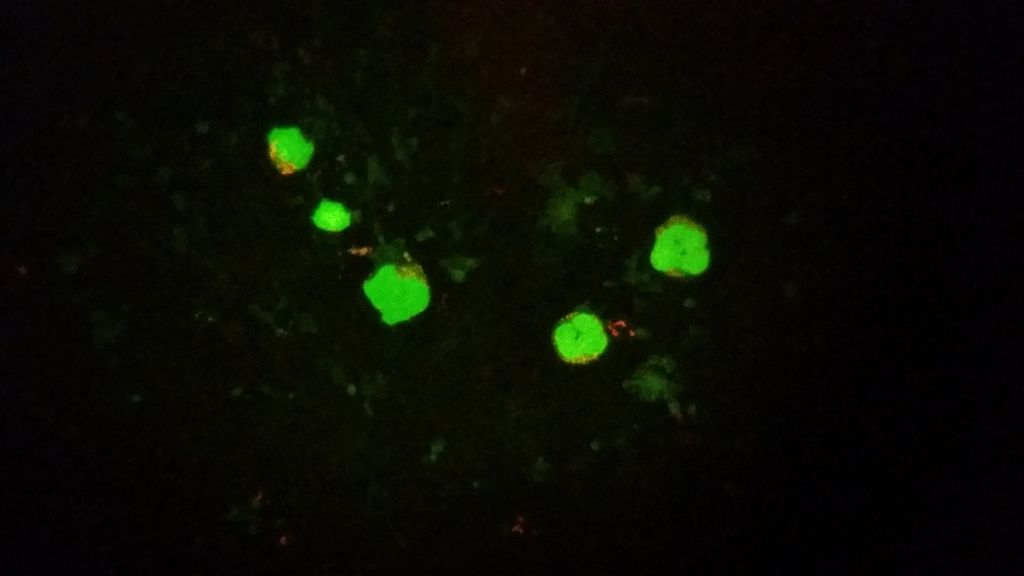
HUGE discovery & further proof for all of us!!!!!!
Spirochetes, SOP's, blebs, 2 daughter splitting in my Lyme blood by fluorescent microscopy...so that explains all the symptoms & the grappling fatigue and shortness of breath...these things are all over my blood just as we see them in regular light microscopy! Only now they pop & stand out more with fluorescent stains and this adds proof that they have DNA & they are not just artifacts!
Plenty of info in the description & since it was too long, I had to add some more info in the comments section as well.
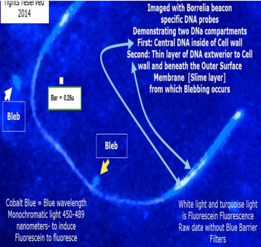
I also figured out what those dumbbells are that we have been seeing since the beginning of our microscopy. On regular lightfield they look like dumbbells & when I switch to fluorescence they reveal as 2 daughter splitting and so the segment between the 2 splitting spiros looks like the handle to the dumbbell and since the dumbbells typically move fast in live blood preps with Brownian motion it distorts it and makes the tips look thicker then what they really are and it gives us the illusion of dumbbells.
You can see the dumbbells & the other many forms from the breakup of SOP in this video I made back in 2014. And now we get to see them really stand out!
Lymedin2010
Frequent Contributor (1K+ posts)
Member # 34322
posted
"Results
Bb Culture Dieterle staining of the blood culture pellet demonstrated both long slender spirochetes ranging from approximately 0.1 to 0.5 μm in width and approximately 2 to 6 μm in length, some with visible helices, and round morphological variants ranging from approximately 0.5 to 2 μm in diameter that looked much like some of the morphological variants seen in the liver section (Figure 1). " " Anti-Bb immunostaining of the blood culture was strongly positive, exhibiting a bright red cherry color (Figure 2). There was some positive staining of cellular debris, possibly because of the antigens released from lysed spirochetes or secreted by Bb. Strongly positive staining was not observed in the culture pellets of control microorganisms. Molecular beacon staining of the culture pellet sections was strongly positive throughout (Figure 3). Culture pellets contained both cultured bacteria and human cellular debris (intact red blood cells and lysed blood cells) indicating that Bb nucleic acid sequences were present throughout the cellular debris and within intact RBCs."
Figure 3. Blood culture beacon stain, Probe Fla B. 400X magnification. Posts: 2087 | From NY | Registered: Oct 2011
| IP: Logged |
Lymedin2010
Frequent Contributor (1K+ posts)
Member # 34322
posted
This one is super special as it is the first stained SOP that I know of from one of our members, Sylvie. Great contrast between the rbc's & the stained SOP to show it is not a false SOP that is generated from the rbc, but a true SOP. So might be from spirochete and others will be adamant that it is from yet another organism.
-------------------- Blue Posts: 1539 | From southwest | Registered: Dec 2015
| IP: Logged |
Lymedin2010
Frequent Contributor (1K+ posts)
Member # 34322
posted
Yea, I have to transfer those pics over to another site one day.
I am not familiar with that scope, but the Nikon's & Zeiss's have some nice turrets that show up from time to time. Some times they show up cheaper & without the filters, and so one can DIY the filters & place them in the turret. It is valuable to have a swivel for the condenser lens, as sometimes a slight angle on the light can make a world of difference, as it does on my Zeiss when I am in the mood for images that stand out. My Reichert Oblique Illumination is outstanding when it comes to impressive views & outperforms my Zeiss in this respect, even with worst optics. The view can appear very 3D & DIC like.
________________________________ After combing through my videos of False Spirochete proofs, I decided to put out a video before I gather more evidence via 4K video & other methods. I think there is enough proof in this video & it shouldn't be too far of a stretch to believe this?
So I think both real & False Spirochetes (FS) can exist in our Lyme blood and sometimes they will be hard to tell apart. There is more info in the video & in the video description.
The Lyme Disease Network is a non-profit organization funded by individual donations. If you would like to support the Network and the LymeNet system of Web services, please send your donations to:
The
Lyme Disease Network of New Jersey 907 Pebble Creek Court,
Pennington,
NJ08534USA http://www.lymenet.org/


 UBBFriend: Email this page to someone!


 Printer-friendly view of this topic