__________________________________________ CYST OR WBC COMPONENTS???
I tried live staining with Parker's blue/black ink to see if I can buy more clues. In this video the objects did stain blue/black/purplish, as spirochetes do stain that color, but the wbc's also stained aquablue, blue, & purplish as well, so it was hard to differentiate whether these were spiro cysts or wbc components. I time lapsed them for 1.5 days here & no sign of uncysting.
That's a really good Bartonella FISH video. Interestingly, my fluorescent "Bart" organisms look almost identical.
It's too bad I cannot get some of this DNA/RNA-specific fluorescent stain. I'd likely have to have a license (a certified lab) to purchase this stuff once they find a distributor.
I'm still having trouble with my AO-stained dry smears. I've done some experimenting--even got some AO from a different company--but my smears keep washing off when I rinse the AO off the specimen. The only other thing I think it could possibly be is perhaps my methanol is not good anymore. I'm not sure why that would be though, unless perhaps it has absorbed some moisture somehow. I can't see how that's possible since I store it in the capped plastic bottle it came in. Just in case, I have some more methanol coming. I hope that's the fix, because I have been really bummed out about microscopy because of this.
Posts: 1308 | From Eastern USA | Registered: Oct 2013
| IP: Logged |
Lymedin2010
Frequent Contributor (1K+ posts)
Member # 34322
posted
Hey TNT. Yea, I would love to get my hands on some DNA Probes.
TNT, it is better to dilute the AO & do live staining, that is how I saw the spirochete SOP's & 2 daughter splitting, blebs, & developing juvenile spiros. Before that I was using pure 0.2% AO & it was too strong & cytotoxic and just destroys the spiros. I spoke to someone in a lab & they said that when they exposed sperm to pure AO it too destroyed the sperm cells & so I confirmed the toxicity. I gave the solution in MY HORRIFIC LYME BLOOD P2 video, it is 2 step dilution to mimic the exact one that I used. You then add a drop of that to a drop of your blood, sometimes a little less than a drop so you can get better dispersion of the rbc's. You seal the cover slip with vaseline or immersion oil so that it does not dry up & you wait for the spiros to take up the dye & fluoresce. Usually some start right away & more will pick it up in 12-24 hrs & sometimes ever more at 36 hrs later.
It seems like when the spiros are not active their double bound cell membranes are hard to penetrate at this low concentration, but if they undergo change, such as SOP formation then the AO is taken up & they light up.
Instead of washing out the AO from your slide, have you tried NOT to wash it out & just simply let it dry out naturally. BUT you must place a smaller amount of blood & disperse them really well so you don't get as much clumping when the AO evaporates. Also, evaporate in a well ventilated areas & better to do it in a garage but enclosed in a box & then open the garage door to vent...any stain fumes are not good for the Lyme body.
Posts: 2087 | From NY | Registered: Oct 2011
| IP: Logged |
TNT
Frequent Contributor (1K+ posts)
Member # 42349
posted
Thanks for the tips Lymedin. I've got to say, you are doing some EXCELLENT work with spirochetes and live blood! I've really been impressed! And, your "schizont" looks more like one than the one in ID-Fish's video. Keep up the great work. I honestly think your channel will eventually go viral as your microscopy work with Borrelia is definitely on the cutting edge.
Personally, I guess I'm just not as interested in doing live blood as much anymore since I've seen the conversion and I've never doubted from the beginning that I was seeing spirochetes. I'm more intent on catching pyriforms/ringforms or morulas in their distinct morphologies under fluorescence because those infections (COMBINED with the Borrelia) are what I believe have made and kept me sick. Trouble is, I have less and less a chance to see this as I keep improving and can't get my game on with the dried smears.
I will probably do more live blood under fluorescence once I have mastered the fluoro dried smears though.
Posts: 1308 | From Eastern USA | Registered: Oct 2013
| IP: Logged |
Lymedin2010
Frequent Contributor (1K+ posts)
Member # 34322
posted
Thanks TNT! I think collectively we did & learned a lot together.
Looking forward to seeing some new stuff from you.
Posts: 2087 | From NY | Registered: Oct 2011
| IP: Logged |
Lymedin2010
Frequent Contributor (1K+ posts)
Member # 34322
posted
Lymedin2010/TNT and all - this is a wonderful thread. Thanks for sharing all of the amazing information. Been reading on and off for a while. First time posting.
I am interested in getting started with some dark field analysis. What do you recommend for a microscope/camera to get started without going with a full pro setup (Olympus/Zeiss etc) in September 2017? More specifically, can I get the job done well with a cheap microscope (Amscope etc)?FWIW - I am good with electronics so I am able to hack/mod anything if necessary if that helps.
Posts: 65 | From minneapolis, san francisco | Registered: Nov 2004
| IP: Logged |
quote:Originally posted by electric: Lymedin2010/TNT and all - this is a wonderful thread. Thanks for sharing all of the amazing information. Been reading on and off for a while. First time posting.
I am interested in getting started with some dark field analysis. What do you recommend for a microscope/camera to get started without going with a full pro setup (Olympus/Zeiss etc) in September 2017? More specifically, can I get the job done well with a cheap microscope (Amscope etc)?FWIW - I am good with electronics so I am able to hack/mod anything if necessary if that helps.
I don't have a specific microscope recommendation, but I just started doing darkfield and here's what I recommend as far as the type of equipment:
Darkfield at 40x isn't all that impressive. I've done some at 63x which is pretty decent, but 100x is the best.
You'll want to make sure that the NA of your objective is always less than the NA of your condenser. For instance, for 100x darkfield, I use a darkfield oil condenser with an NA of 1.25, so my 100x objective (which also has an NA of 1.25) has an adjustable iris to close down the NA (otherwise the image isn't dark enough).
You'll also want a fairly strong light source. My 18 watt incandescent bulb on my microscope is slightly under-powered for 100x darkfield, but it's not terrible either. A high powered LED is probably the way to go.
Check out older microscopes on eBay, or a new Amscope (but make sure it has both an oil condenser and 100x objective with iris; often the Amscopes are sold as "Darkfield" but only come with a dry condenser and 100x objective without adjustable NA, basically limiting darkfield work to 40x).
Posts: 71 | From Canada | Registered: May 2016
| IP: Logged |
Lymedin2010
Frequent Contributor (1K+ posts)
Member # 34322
posted
Personally, I don't own a darkfield & have limited experience. I would def ask & listen to the owners of DF...thanks MS2!
posted
mustardseed2 - awesome information! Thank you, it really helped me understand exactly what I need to look for shopping around.
Also, how valuable is adding phase contrast ability to the setup? Would it be valuable for trying to view lyme/co-infections in blood samples etc?
Lymedin2010 - Thanks for the link to My Microscope group. Will check it out.
Posts: 65 | From minneapolis, san francisco | Registered: Nov 2004
| IP: Logged |
Lymedin2010
Frequent Contributor (1K+ posts)
Member # 34322
posted
Sometimes it helps & other times it can also hinder, depends on how the slides are prepared and how busy the slide is. You don't need it really, but only if you want more impressive images on a clean slide/prep.
Posts: 2087 | From NY | Registered: Oct 2011
| IP: Logged |
Lymedin2010
Frequent Contributor (1K+ posts)
Member # 34322
posted
My new venture on high contrast staining. WBC's & platelets are a dramatic blue contrast to the golden/yellow rbc's. This makes things easier to spot & distinguish.
This is on one of my bad delaminated objectives though, better images in the future.
Even in high clutter, spot things easily. Posts: 2087 | From NY | Registered: Oct 2011
| IP: Logged |
Lymedin2010
Frequent Contributor (1K+ posts)
Member # 34322
posted
Another true spiro in my blood using a proprietary technique other than the freezing method.
posted
Lymedin2010 -Thanks for sharing. What magnification levels are the images above?
Posts: 65 | From minneapolis, san francisco | Registered: Nov 2004
| IP: Logged |
Lymedin2010
Frequent Contributor (1K+ posts)
Member # 34322
posted
They are all w/ 40x objective + 15x eyepiece, so 600x & then I can zoom in & out with my Samsung S5 phone cam.
You can see them better with a 100x oil objective, but I did not use it with the above pics.
Posts: 2087 | From NY | Registered: Oct 2011
| IP: Logged |
Great video, Lymedin! At 7:11 in the video shows classic acridine orange coloring. The green background of cellular components and debris with the bright orange pathogenic (Borrelia) material is awesome classic presentation. Makes me want to do a few smears, HA HA!
Are you still using your RGB LED light hack (particularly on that clip of the video)??? If so, that is great because that is plenty enough intensity.
Posts: 1308 | From Eastern USA | Registered: Oct 2013
| IP: Logged |
Lymedin2010
Frequent Contributor (1K+ posts)
Member # 34322
posted
Thanks. Yes, still using the same bulb. I bought 2 more rgb bulbs, including the one you linked to & another white color flash light. They all work, but the 1st bulb I bought has 3 of 3w blue LED's & they overlap for increased intensity & so it produces the best and brightest images.
Sometimes the stain sample does not take the stain right away or looses the stain over time & that is why it is dimmer in the other videos. Timing is important & I don't have the energy to monitor perfect stained times & so sometimes they come out better than others. Plus my AO solution mix has seen too much light & I should have stored it in a dark container and so I will have to make a new batch eventually.
Been waiting forever to see your stains man, hopefully soon?
Posts: 2087 | From NY | Registered: Oct 2011
| IP: Logged |
TNT
Frequent Contributor (1K+ posts)
Member # 42349
posted
quote:Originally posted by Lymedin2010: Been waiting forever to see your stains man, hopefully soon?
Yeah, I'm working on it, but not too diligently
I got some new methanol a few weeks ago, and, like I said, the color at 7:11 in your video got me fired up again about trying some more stains. Unfortunately, the specimens on the first two slides washed right off again. A third slide I dunked in the methanol instead of flooding the top, and I may have a winner. I still need to look at it, as it was too late to get out the scope last night.
But, maybe you are hoping for some wet mount stains?...
I just might be inspired enough to do one of those again before long. Though I have some Giemsa smears that are pressing at the moment unless the AO is working out. If that is the case, I will be doing some more fixed AO smears.
Posts: 1308 | From Eastern USA | Registered: Oct 2013
| IP: Logged |
TNT
Frequent Contributor (1K+ posts)
Member # 42349
posted
Also, that's great that your hack is working so well!!
I proposed on another forum that using a RGB lamp directed right onto the slide and stage would be a usable brightfield scope hack. When I personally tried it with my 5 watt RGB bulb I was able to see absolutely nothing. So, maybe the theory doesn't work out for real world application. Or, maybe I didn't have enough wattage for the smear to pick up any fluorescence.
Maybe something like this light with much more wattage would work for such a hack:
TNT
Frequent Contributor (1K+ posts)
Member # 42349
posted
quote:Originally posted by TNT: I proposed on another forum that using a RGB lamp directed right onto the slide and stage would be a usable brightfield scope hack.
What I meant was that you could turn a brightfield scope into a fluorescent microscope simply by shining one of these RGB LED lights onto the specimen plane directly. But when I tried it with a low-power bulb I saw nothing. But, again, the problem could have been wattage....
Posts: 1308 | From Eastern USA | Registered: Oct 2013
| IP: Logged |
Lymedin2010
Frequent Contributor (1K+ posts)
Member # 34322
posted
Yes, I think one can do a complete hack of the fluoro scope & all one needs is the yellow filter to place between the specimen & camera or eyes.
I think a bulb like this will work too, as it has a collimator right on it.
Quite honestly I think you don't even need a RGB bulb & you could use a white LED light bulb & place a blue filter on top of it, but it has to be the right hue & one can get a filter match book set.
Posts: 2087 | From NY | Registered: Oct 2011
| IP: Logged |
TNT
Frequent Contributor (1K+ posts)
Member # 42349
posted
I doubt that the yellow barrier filter would even be needed if the proper wavelength was beamed onto the specimen. I found I don't need the barrier filter, and actually get just as good or better image without it (when using the right excitation filter with my fluoro scope that is).
Unfortunately I am still having trouble with my stains washing off, GRRRR! I am out of ideas. I have tried two ready-to-use solutions (a .1% and a .2% from different companies), fresh 100% methanol, tap water to rinse with, and distilled water rinsing.
The staining procedure is very simple. You do it exactly like a Giemsa stain.
The only other thing I could try is to not wash my slides beforehand. But I seriously doubt washing the slides is making a difference.
Posts: 1308 | From Eastern USA | Registered: Oct 2013
| IP: Logged |
Lymedin2010
Frequent Contributor (1K+ posts)
Member # 34322
posted
When I tried drenching the peripheral blood smear with methanol & then pouring it off & blotting, it too washed off.
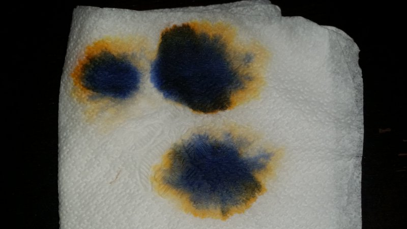
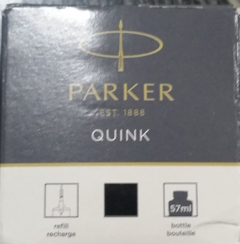
So then next I tried dipping it in the Dip Quick methanol, same as Giemsa, & then leaving the slide flat & letting it dry for 2-3 min naturally. When I saw it was dry, then I added the Parker's Quink ink & stained for 2 min & then rinsed w/distilled water. That is how I made the slide & pics above, with the yellow rbc's & blue wbc's & platelets. You can see when I put in 1 drop of ink & 1 drop of water on a tissue the yellow & blue dispersion of the ink. That is how the rbc's stain yellow & others stain blue.
Good to know about your end only really needing excitation filters. I may play with it again eventually.
Procedure for staining with Parker's Quink ink: 1) Make peripheral blood smear & let dry for 15 min. 2) Fix with methanol & let dry. 3) Add NaCl (0.9%) to wet the surface evenly on the slide (helps better absorb into cells). 4) Add a few drops of ink on top w/wet NaCl & let sit for 1-2 min. 5) Rinse the slide with water & let dry.
Lymedin2010
Frequent Contributor (1K+ posts)
Member # 34322
posted
So these are tick culture spiros in 0.2% AO a few hours into it & one can see many blebs/spores/granular forms & you can also see many dumbbells. The dumbbells are the juvenile spirochetes that undergo splitting. Again, the fuller strength AO in tick juice causes EVERY SINGLE ONE of them that is a juvenile or adult to either undergo SOP or 2 daughter splitting (dumbbell formation). In my blood AO staining I also notice that they undergo SOP & 2 daughter splitting & that is when they stain. So they might be more of the persister types & only in time does the AO force them to undergo change & that is when they take up the stain. Mind you at full 0.2% strength AO the blood spiros do not appear & must either form cysts or just lyse and die out quickly, but when I lower the concentration of the AO then they take up the stain when undergoing SOP & dumbbell creation.
Lymedin2010
Frequent Contributor (1K+ posts)
Member # 34322
posted
Here is my 2nd Co-infections video & on there I put a link to Malaria Fish Fluoro probes & you can see my tetrads look exactly like the Malaria tetrads.
TNT
Frequent Contributor (1K+ posts)
Member # 42349
posted
quote:Originally posted by Lymedin2010: Here is my 2nd Co-infections video & on there I put a link to Malaria Fish Fluoro probes & you can see my tetrads look exactly like the Malaria tetrads.
Great work again, Lymedin! Those DNA & RNA organisms definitely look like piroplasms.
The only thing that keeps me slightly less interested in AO wet mounts for co-infections is that there seems to be so much lighting up besides what you point out with the arrows. I too noticed the same thing with my wet mounts. What you are seeing inside the cells has to be pathogenic, but since there is so much of it, I wonder how much is Borrelia and how much is co-infections. What you pointed out with the arrows absolutely looks like piroplasms, but what about the stuff you didn't point out but can be seen?
Are you seeing anything with the distinct typical morphology such as shown in the ID Fish video at 3:43 that you linked to? It's possible that the chromatin dot and ringform morphology is only seen in a dry smear, but I thought I'd ask.
As a side note, you must be sealing with immersion oil again because I see a number of oil droplets in that video.
Great job again! I definitely think we are learning and building upon what we have found and upon what has already been proven.
Posts: 1308 | From Eastern USA | Registered: Oct 2013
| IP: Logged |
I haven't been here for a few months. I started BVT 4 months ago, 4-6 bees every other day. Today, I took out my microscope, my blood is still loaded with spirochete, a few of them in every field view at 100x. TNT, didn't you start BVT too, and you said that the load went down? Anyone here saw the load going down consistently with BVT or any other method?
Despite the heavy load, I am functioning at 70-80%, even went on an international business trip for 20 days with a demanding schedule.
In live blood, I see that roulough has gotten better. White blood cells are abundant and some are lively, and some are nearly dead (dark, and not moving).
Has anyone here clearly see improvement in live blood and/stain? and How?
Thank you for an update!
Posts: 41 | From NJ | Registered: Apr 2015
| IP: Logged |
I haven't been here for a few months. I started BVT 4 months ago, 4-6 bees every other day except for weekends. Today, I took out my microscope, my blood is still loaded with spirochete, a few of them in every field view at 100x. TNT, didn't you start BVT too, and you said that the load went down if I recall correctly? Anyone here saw the load going down consistently with BVT or any other method?
Despite the heavy load, I am functioning at 70-80%, even went on an international business trip for 20 days with a demanding schedule.
In live blood, I see that roulough has gotten better. White blood cells are abundant and some are lively, and some are nearly dead (dark, and not moving).
Has anyone here clearly see improvement in live blood and/stain? and How did you do it?
Thank you for an update!
Posts: 41 | From NJ | Registered: Apr 2015
| IP: Logged |
posted
Btw, does anyone live in the bay area who knows how to stain? I'd like to get my blood stained.
I haven't gotten around to staining, maybe I have a mental block about doing it myself. Just can't bring myself up to do it.
Posts: 41 | From NJ | Registered: Apr 2015
| IP: Logged |
TNT
Frequent Contributor (1K+ posts)
Member # 42349
posted
quote:Originally posted by rainboworiver: Hello Everyone,
I haven't been here for a few months. I started BVT 4 months ago, 4-6 bees every other day. Today, I took out my microscope, my blood is still loaded with spirochete, a few of them in every field view at 100x. TNT, didn't you start BVT too, and you said that the load went down? Anyone here saw the load going down consistently with BVT or any other method?
Despite the heavy load, I am functioning at 70-80%, even went on an international business trip for 20 days with a demanding schedule.
In live blood, I see that roulough has gotten better. White blood cells are abundant and some are lively, and some are nearly dead (dark, and not moving).
Has anyone here clearly see improvement in live blood and/stain? and How?
Thank you for an update!
I'm glad to hear you are able to do as much as you are!
Yes, I've been doing BVT for 18 months now and it has really helped. I didn't notice as much improvement as when I went up to 10 stings per session, but I did have improvements from the very beginning. These have been clinical improvements, but I have noticed improvements under the scope as well. Earlier on I did notice decreased loads of spirochetes, but I haven't checked my live blood for a while now. I am functioning better overall compared to then. I am also on meds and herbs/supplements, too.
One of the biggest improvements have been the increased numbers of CD57 white cells (natural killer lymphocytes). I saw almost none when I first started staining (before starting BVT), but more plentiful as I continued stinging and doing conventional meds.
BVT was a game-changer for me in spite of doing it with antibiotics, because before starting BVT the antibiotics were not helping. I think doing them together has been synergistic--contrary to what some believe. That's been my experience anyways.
One thing I did notice which was pretty profound was NO SPIROCHETES in a sample I did late last year when I was on Omnicef and Tetracycline together. I was doing BVT at the same time of course, and that timeframe was about a month after getting to 10 stings per session. So, a pretty powerful combination that was. I wasn't able to do that very long (three weeks maybe) until the Babesia moved to first place and I had to switch protocol with the meds only.
I have found staining to be quite easy....at least with Giemsa. Acridine orange has been problematic for me still. Giemsa staining can be quite helpful for finding some of the co-infections. But you have to be careful not to confuse artifacts and platelets for pathologies.
I get a one-step Giemsa solution that works beautifully for me. I get free samples from the company. If you are interested I can private message you with the info on how to get some.
Posts: 1308 | From Eastern USA | Registered: Oct 2013
| IP: Logged |
do you guys see spirochetes coming out of the white blood cell? What else do you see that I haven't noticed?
Posts: 41 | From NJ | Registered: Apr 2015
| IP: Logged |
do you guys see spirochetes coming out of the white blood cell? What else do you see that I haven't noticed?
I cannot view the videos...it says they are unavailable. I think your videos are not set to public viewing. You'll have to change your settings before we can see them.
I'll send you a link privately about the Giemsa as soon as I have a little more time.
Posts: 1308 | From Eastern USA | Registered: Oct 2013
| IP: Logged |
Btw, I am so happy to hear you have improved! Keep up with the good work!
Posts: 41 | From NJ | Registered: Apr 2015
| IP: Logged |
Lymedin2010
Frequent Contributor (1K+ posts)
Member # 34322
posted
I only read the first 2 posts for now & coming back to read the rest later.
They do look like babesia morphologies & many times appear in groups of 2 or 4, but more can appear within the red blood cells. I can't believe it has taken me this long, but I still have not checked AO in a normal persons blood & this is vital for our knowledge. The many other things that are floating in the plasma are wbc's that lyse & scatter the granules, so I usually dismiss anything in the plasma unless it is a SOP or 2 daughter splitting spiro.
The only thing that has taken the spiros away was Cowden Protocol & I felt better & had more energy. But it wore off after 4-5 months into it & I could not tolerate it anymore. I could only see very, very small what might be juvenile spirochetes & no adult ones at that time. A few weeks/months after the Cowden then I saw once again my blood was loaded & I felt a whole lot worst. That is the only thing that has ever changed the blood presence of these spiros & I have taken so many different abx & combos of abx/herbs. Also, this is considering if what we are seeing is really true & they are all true spirochetes. All the evidence to me suggests that they are, aside from the false spiros that I pointed out. And of course I am always looking for more clues & to either prove or disprove as the evidence comes in.
TNT, do you see the same type of babesia forms in your blood? Have you done any normal blood with AO & do you find anything within the rbc's?
I will come back to read the rest & check the new video posts.
Posts: 2087 | From NY | Registered: Oct 2011
| IP: Logged |
Both of these samples are fresh: one week I look in my blood and find only spirochetes, no cysts, the next week I look and find only cysts, no spirochetes. I'm not sure why, it's very peculiar.
Next step for me is to get some Acridine Orange and see if I can time-lapse the conversion!
Posts: 71 | From Canada | Registered: May 2016
| IP: Logged |
Lymedin2010
Frequent Contributor (1K+ posts)
Member # 34322
posted
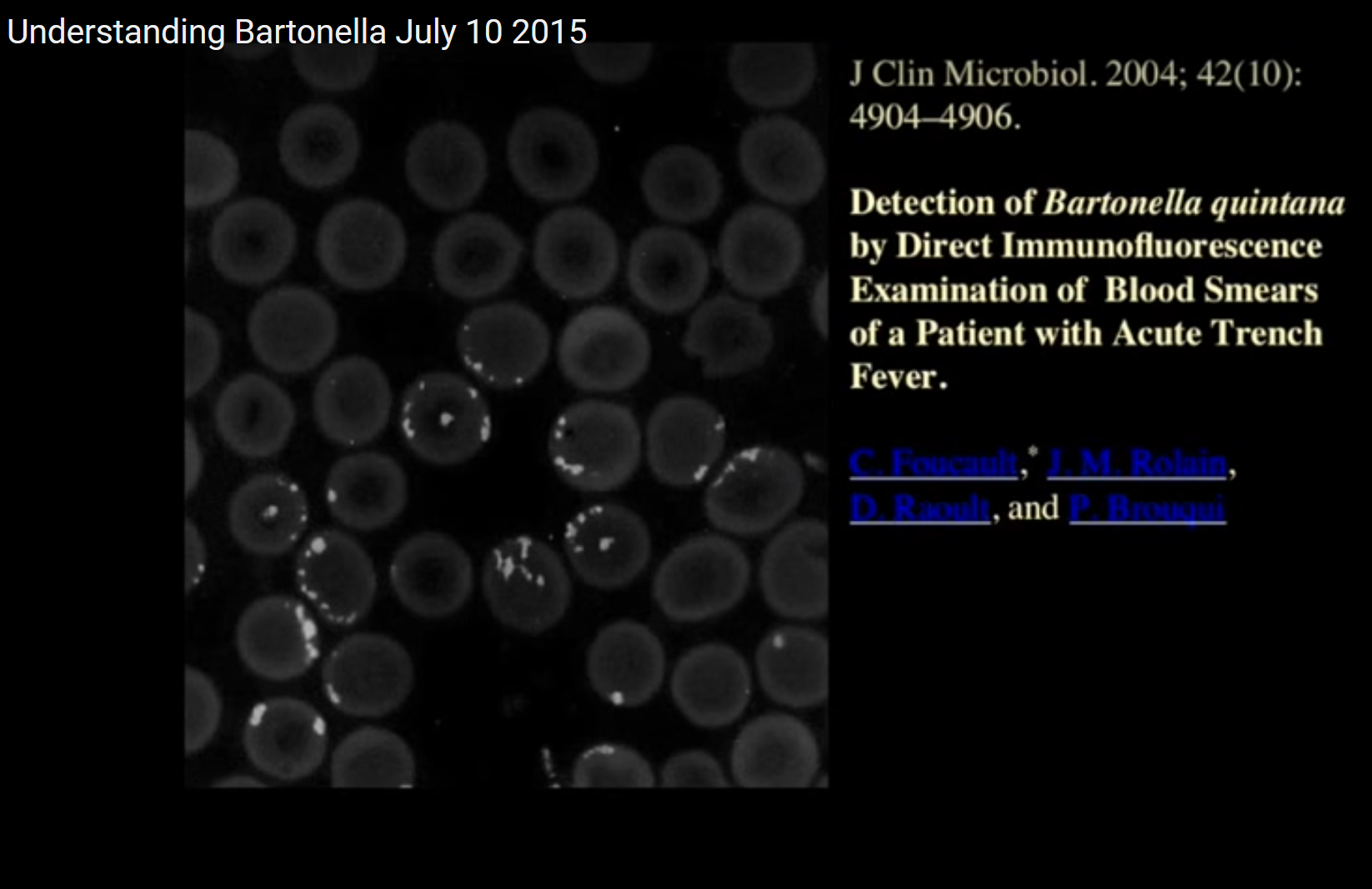
At time 50:05 detection of Bartonella quintana by Direct Immunofluorescence examination of blood smear.
Lymedin2010
Frequent Contributor (1K+ posts)
Member # 34322
posted
Hey Mustard, those look like what we have been calling spiros for sure, but of course testing is king. Cysts I could not be too sure most times & they could be other particles too. The discoid gemma shape in most cases seem more convincing to me, but yours does not look like it though. Hard to know for sure with cysts.
Posts: 2087 | From NY | Registered: Oct 2011
| IP: Logged |
TNT
Frequent Contributor (1K+ posts)
Member # 42349
posted
quote:Originally posted by mustardseed2:
Potentially 2 cysts, one of which looks like it's mid-conversion:
Both of these samples are fresh: one week I look in my blood and find only spirochetes, no cysts, the next week I look and find only cysts, no spirochetes. I'm not sure why, it's very peculiar.
Next step for me is to get some Acridine Orange and see if I can time-lapse the conversion!
The little round "balls" in that second video are almost certainly granules from a Granulocyte such as a Basophil or Eosinophil. I've seen them before, usually near a disintegrated Granulocyte.
That's interesting about seeing spirochetes one sample, and cysts the next. Like Lymedin said, only the Gemma cysts should be considered true cysts. I have seen what I have earlier referred to as granules, and these look a little like tiny salt crystals or like miniature "Everlasting Gobstoppers" (from the movie Charlie and the Chocolate Factory). I have only seen these in mine and other's blood when NOT on antibiotics.
The acridine orange will be very helpful in seeing the conversion. With AO, I noticed a lot of conversion to typical l-form structures, but some conversions to Gemma cysts and various-sized little round forms.
Good job, Mustardseed! I hope to get my scope out more in the next several weeks.
Posts: 1308 | From Eastern USA | Registered: Oct 2013
| IP: Logged |
posted
Interesting, I hadn't considered WBC granules. I don't know if I've ever seen a granule that big before.
I wish I had better magnification... the reason I suspected cyst was because the inside of the "granule" looked hollow and didn't light up. It reminded me of the gemma cysts in this video:
You might be right though, I'll know for sure if I can catch some mid-conversion, which I hope to do soon!
Posts: 71 | From Canada | Registered: May 2016
| IP: Logged |
TNT
Frequent Contributor (1K+ posts)
Member # 42349
posted
quote:Originally posted by Lymedin2010:
Great find, Lymedin! This pic also resembles Mycoplasma. These resemblances can make discerning microbes via microscopy difficult without the aid of DNA fluorescent probes. Even with acridine orange (non-specific fluorescent stains) it can be hard to discern between bugs unless there are specific morphologies.
Posts: 1308 | From Eastern USA | Registered: Oct 2013
| IP: Logged |
posted
Have any of you guys ever put your Giemsa stains and/or Dip Quick stains under darkfield?
I tried it for the first time today just to see what it would look like, and I had several small granules stuck to the perimeters on RBCs light up... almost looked like those flouro stains that were posted before. It was really cool
Anyone tried this?
Posts: 71 | From Canada | Registered: May 2016
| IP: Logged |
TNT
Frequent Contributor (1K+ posts)
Member # 42349
posted
quote:Originally posted by mustardseed2: Have any of you guys ever put your Giemsa stains and/or Dip Quick stains under darkfield?
I tried it for the first time today just to see what it would look like, and I had several small granules stuck to the perimeters on RBCs light up... almost looked like those flouro stains that were posted before. It was really cool
Anyone tried this?
No, I never have. Someone on healingwell mentioned this to me a while back, but I haven't done it.
So, you say darkfield gives it a fluorescent-stained look? What do you think the granules were? Just some loose WBC granules?
Were they only noticeable in darkfield or were they just easier to see under darkfield?
Posts: 1308 | From Eastern USA | Registered: Oct 2013
| IP: Logged |
posted
I'll try and redo it today and take some pictures and/or video.
Ya, it looked like a normal sample, except there were little dots spread out that lit up like bulbs on a Christmas tree.
I don't know if they were WBC granules, but it seemed like if that's what there were, there should have been more.
Posts: 71 | From Canada | Registered: May 2016
| IP: Logged |
posted
I know there aren't a ton of people left looking at this thread, but I'm wondering if there was any meaningful discussion of Lymedin2010's "false spirochete" video:
Specifically look at 1:23. If I saw that in anyone's blood I wouldn't hesitate to call it a spirochete. But keep watching the video and it becomes clear that it's likely the leftovers of a RBC. In short, this video (which I saw for the first time today) really has me questioning if what we're seeing (outside of flouro staining, or cyst conversions) are actually Borrelia.
Is there any way to tell spirochetes apart from these "false-spiros"? Are a lot of the videos were seeing of Borrelia actually not? Has anyone seen false spiros in healthy people?
I feel like everything I've taken for granted in the last year has been thrown into question...
Posts: 71 | From Canada | Registered: May 2016
| IP: Logged |
posted
Hello people, checking in with you guys after a long break.
I have had a break from microscopy since the initial letdown when I did not find anything from my blood.
Still, I have had this in my back burner for a while, and now I also have possibility to have the microscope out of the closet all the time.
As my time is limited, my aim has been to automate as much of the process as possible.
To automate it, as you all know, I would need time lapse or video for several days. The major problem with this seems to be keeping the image in focus.
One part of the focus problem seems to come from the heat from the lamp.
Some people may recall that I have Olympus BX-4. Since I wrote here, I have retrofitted my scope with LED like was recommended here. It is Retrodiode 20 watt LED system.
I also have a solution for controlling the lamp on/off by computer.
However, in any case, the image will eventually go out of focus unless there is a way to autofocus it.
So my next step is trying to build autofocus for my microscope.
This will probably consist of Arduino controlling a motor for the focus, and then using the photos taken on the computer and simple autofocus algorithm to do it.
Anybody here who has done the same?
Posts: 31 | From Finland | Registered: Sep 2016
| IP: Logged |
posted
Also, in a little bit longer term, I am interested in researching whether it would be possible to automate locating certain things, such as the spirochetes, from the images/videos using computer vision/deep learning. I am not seeking for 100% accuracy, but rather ways of lessening the manual work of observation. Anybody else interested in this or working on it already?
Posts: 31 | From Finland | Registered: Sep 2016
| IP: Logged |
Lymedin2010
Frequent Contributor (1K+ posts)
Member # 34322
posted
There are systems used for automation in microscopy. There are some expensive full blown microscopes with built-in systems that can reference points of interests. There are also systems that systematically take pictures & move the slide along & then you can digitally zoom in & out and do your searching. Most beneficial in stained & fixed prepped samples.
_________________________________ So can the Lyme bacteria really be found in our blood?????
New LM-PCR from whole blood, serum & urine in 2017..... "Greater sensitivity is particularly important for detection of BB, because the bacteria are typically found in very low concentrations in blood and urine samples from Lyme disease patients."
I would argue in some patients after long-term illness they can be found in more substantial quantities as well.
quote:There are systems used for automation in microscopy. There are some expensive full blown microscopes with built-in systems that can reference points of interests. There are also systems that systematically take pictures & move the slide along & then you can digitally zoom in & out and do your searching. Most beneficial in stained & fixed prepped samples.
Yeah, I asked around, but the prices for just retrofitting my scope with x,y,z axis started from over 5000 USD.
So I am taking the DIY route.
Posts: 31 | From Finland | Registered: Sep 2016
| IP: Logged |
TNT
Frequent Contributor (1K+ posts)
Member # 42349
posted
quote:Originally posted by BorreJaakko: I got the autofocus to kind of to work. The software is preliminary, but if anybody is interested how I did it, here is a link to the code.
I also installed a darkfield condenser in my scope.
Here is some images from the darkfield from from fresh blood.
Do you people usually see something with the darkfield right away, or does one need to wait the five days or so like with brightfield?
That's great you are equipped with darkfield now! It's so much easier to see spirochetes with DF than with brightfield.
To answer your question about how soon we see ketes in darkfield, it depends on your load of infection (but it's the same as with brightfield). If you are very debilitated and chronically ill, then you should see them almost immediately if you have Lyme disease. If you are still pretty functional, you may not see them until 48 hours post draw, or you may need to traumatize the slip cover. That crushes the red blood cells and makes the intracellular ketes visible.
Thanks for sharing your autofocus program.
May I make a suggestion concerning your darkfield image? Do you notice how your red blood cells appear to smear toward the bottom of the picture? It appears like your darkfield condenser needs centered on it's mount, or your light source needs centered. The screws that hold the condenser unit on the fork are how you center the condenser. As for the light, you should be able to center the collimator lens below the condenser (where the light comes up out of the base). Making sure both of these are centered (especially your condenser) will make your darkfield images crisper and more even.
Keep up the good work with the programming and improvising!
Posts: 1308 | From Eastern USA | Registered: Oct 2013
| IP: Logged |
TNT
Frequent Contributor (1K+ posts)
Member # 42349
posted
quote:Originally posted by TNT: Do you notice how your red blood cells appear to smear toward the bottom of the picture?
I didn't make myself very clear. What I mean is that your red cells appear to be melting. If you center your condenser and/or your lamp collimator that should take care of it for you. The outlines of your cells will then be crisp and distinct.
Posts: 1308 | From Eastern USA | Registered: Oct 2013
| IP: Logged |
Lymedin2010
Frequent Contributor (1K+ posts)
Member # 34322
posted
Awesome that you can DIY your own & looking forward to some discoveries. If I had extra energy I would probably try as well, but I am running on a limited budget.
Guys I finally did AO stain of some normal blood & not a single spirochete or SOP was found. Not even any organisms within the rbc's, such as what looks like Babs tetrads from my AO stains. I need to do a few more samples & a few people to get a larger sample size. Not finding it in another persons blood & finding it in mine hits home.
I also found spirochetes & SOP's in my wife's blood & she has been hit harder with body/muscle/joint pains the last few months. Seems it has gradually increased over time in her. She finally got 2 bands a few months ago on her standard & crappy Lyme tests and she just said yesterday she is not feeling well and she wants another test. I do not find any babesia or bart subjects within her red blood cells like I do in mine and so the unusual tetrads & other subjects hit home harder that they are actually some pathogens in my red blood cells. I have the night sweats & shortness of breath, but she does not. I think she just has Borrelia.
Posts: 2087 | From NY | Registered: Oct 2011
| IP: Logged |
quote: To answer your question about how soon we see ketes in darkfield, it depends on your load of infection (but it's the same as with brightfield). If you are very debilitated and chronically ill, then you should see them almost immediately if you have Lyme disease. If you are still pretty functional, you may not see them until 48 hours post draw, or you may need to traumatize the slip cover. That crushes the red blood cells and makes the intracellular ketes visible.
Yes, what I actually was trying to ask was that does one see them sooner with darkfield than brightfield, for example is one able to see them inside the RBCs.
quote: May I make a suggestion concerning your darkfield image? Do you notice how your red blood cells appear to smear toward the bottom of the picture? It appears like your darkfield condenser needs centered on it's mount, or your light source needs centered. The screws that hold the condenser unit on the fork are how you center the condenser. As for the light, you should be able to center the collimator lens below the condenser (where the light comes up out of the base). Making sure both of these are centered (especially your condenser) will make your darkfield images crisper and more even.
Thanks for the comment. This kind of comments are exactly the reason I posted the picture, so if you have more comments, they are more than welcome. I don't yet know what darkfield should look like, although I have watched most of the pictures and videos from here.
It seems easier to set the condenser screws properly for the brightfield. It is kind of intuitive. For darkfield I seem not to know when it is centered. But I'll try playing with it based on your comment.
Posts: 31 | From Finland | Registered: Sep 2016
| IP: Logged |
posted
I have been reading the Mysterud and Laane's paper on using Acridine-Orange solution, and 450 nm excitation and 570 nm barrier filters, as well some of the comments in this thread.
It seems it is even easier to see the sketes using this method, but I am intersted to know, what do I really need for it?
The dark field condenser was quite expensive for my scope (although I found an used one), so I am trying to avoid bying unnecessary stuff.
The paper mentions phase-contrast condensor, apochromatic phase optics, and even hints about fluorescence microscopes. These are quite expensive, so I was thinking what would be the minimum setup to try it out.
I have very strong LED lamp (20watts) with 5500K color. I read a long article about fluorescence microscopy. It seems, like it is suggested int his thread, that it is possible to do fluorescence microscopy just one or two filters, without needing other necessary equipment. But it depends on the liht source whether it is possible.
So the first question is, does the 5500K LED light contain the necessary wavelenghts for 450 nm excitation and 570 nm barrier filters to work?
My second question is, would I be needing phase contrast condenser, or would I be able to try this out with a regular brightfield condenser?
My scope seems to have a place for filters just above the objectives. I guess this is the place for the barrier filter.
There is also place to put a filter with diameter of 45 mm on top of the light source. I guess this would be the excitation filter.
And lastly, what kind of filter materials have you tried out? I guess glass would be cheapest. Do you have any ideas where I could buy those filters?
quote: To answer your question about how soon we see ketes in darkfield, it depends on your load of infection (but it's the same as with brightfield). If you are very debilitated and chronically ill, then you should see them almost immediately if you have Lyme disease. If you are still pretty functional, you may not see them until 48 hours post draw, or you may need to traumatize the slip cover. That crushes the red blood cells and makes the intracellular ketes visible.
By the way, thanks for this reply.
My problem I still do not know whether I had Lyme or if I did, whether it is cured. My condition is in general nowadays pretty good. I have taken a 9 months Cowden protocol. But my condition was never so bad that I would have had to stop working, for instance.
By traumatizing, do you need like really pushing them hard? Currenly, I am just gently spreading the blood drop.
Posts: 31 | From Finland | Registered: Sep 2016
| IP: Logged |
TNT
Frequent Contributor (1K+ posts)
Member # 42349
posted
quote:Originally posted by BorreJaakko: By traumatizing, do you need like really pushing them hard? Currently, I am just gently spreading the blood drop.
Some people spread the drop and then top with a cover-slip, but I simply put a drop of blood on the slide (actually a drop about a fourth of the way in from either end...so two drops to maximize my chances and to make the most of each slide) and then gently lay a cover-slip down on top of it. The blood spreads out on it's own.
After the blood has equalized under the cover-slip, simply take an ink pen and push down on the top of the cover-slip. That will crush the cells and release the intracellular bacteria.
If you want to see it as naturally as possible, then let the blood dry around the borders of the coverslip (it takes hours) to form a light seal. Then place a drop of immersion oil along the edge of the cover-slip and allow to travel the whole way around to make an airtight seal. The dried blood will make enough of a seal to prevent the oil from contaminating the sample, and the immersion oil will prevent the sample from drying out. You can view the blood for at least a week this way. Of course the spirochetes will be long gone (turned into cysts and granules), but this method gives you the ability to view the sample over a long period of time to view as much spirochetal activity as possible.
Posts: 1308 | From Eastern USA | Registered: Oct 2013
| IP: Logged |
quote:Originally posted by TNT: In some of my videos I have shown possible merozoites. The organisms could very possibly be bartonella (or BLO). Their shape suggest apicomplexans, but their size would suggest BLO.
What I am about to post resemble released apicomplexans from apparent ruptured red blood cells. In these pictures, the shape AND size both resemble merozoites, not to mention the proximity and position of the ruptured RBCs. The only anomaly is that these objects did not take up the Giemsa stain.
It is impossible to determine what these objects are. The only clues I have are my positive lab tests and my correlating symptoms.
That all said, these objects could be platelets beside ruptured or maybe even lopsided RBCs. But the scenario and placement (and shape/size of the objects) would suggest my earlier-stated possibility as well.
So, basically two possibilities.
I'll let you be the judge.
These pictures were taken from my Giemsa slide. I simply toggled the neutral density filter below my condenser to get more contrast on the specimen.
TNT
Frequent Contributor (1K+ posts)
Member # 42349
posted
Hi norri, welcome to the microscopy thread. Would you like to comment, or maybe you have a question?
Posts: 1308 | From Eastern USA | Registered: Oct 2013
| IP: Logged |
TNT
Frequent Contributor (1K+ posts)
Member # 42349
posted
Hey guys, I just came across these videos by Alan MacDonald and somehow didn't see them before! They are awesome. His 3-part series on the Biology of Lyme Disease was great, and it looks like many people viewed those. But these two videos have relatively very few views and are EXCELLENT! If you listen closely you'll hear that he even validates our amateur microscopy! I think those comments are in Part 2.
quote: Some people spread the drop and then top with a cover-slip, but I simply put a drop of blood on the slide (actually a drop about a fourth of the way in from either end...so two drops to maximize my chances and to make the most of each slide) and then gently lay a cover-slip down on top of it. The blood spreads out on it's own.
After the blood has equalized under the cover-slip, simply take an ink pen and push down on the top of the cover-slip. That will crush the cells and release the intracellular bacteria.
If you want to see it as naturally as possible, then let the blood dry around the borders of the coverslip (it takes hours) to form a light seal. Then place a drop of immersion oil along the edge of the cover-slip and allow to travel the whole way around to make an airtight seal. The dried blood will make enough of a seal to prevent the oil from contaminating the sample, and the immersion oil will prevent the sample from drying out. You can view the blood for at least a week this way. Of course the spirochetes will be long gone (turned into cysts and granules), but this method gives you the ability to view the sample over a long period of time to view as much spirochetal activity as possible.
Thanks, lots of things to try out.
Posts: 31 | From Finland | Registered: Sep 2016
| IP: Logged |
posted
Hello guys/girls, I haven't been here for a while. I thought I would post an update on my end. I was on 6 months of on and off bee venom therapy. I also did 6 sessions of HBOT at 2.4 ATA for 60 minutes. For the first time ever, the spirochete long forms have disappeared! However, the malaria/babesia (or babesia like oganisms) growth have taken off. My symptoms match those of malaria/babesia: swelling of my brain, pressure, headache, stiff neck, memory loss, insomnia, clamy feet, chill I don't seem to have any lyme symptoms.
I also had metagenomic testing done on my stool and blood. The stool DNA testing identifies Plasmodium falciparum (which is malaria). The blood DNA testing identifies Babesia. Either way, I have these protozoas. what is the most effective herbal treatment for this?
In the following pictures, you will see that a good percentage of my RBCs are infected with a pathogen in the middle.
Hi everyone, I spoke too soon. After letting the slide sitting for 3 days, the long forms of spirochetes came out of hiding, probably from white and red blood cells.
Still the load is much less than before.
I am currently starting treatment for babesia using the CSA complex from woodland essence.
Posts: 41 | From NJ | Registered: Apr 2015
| IP: Logged |
TNT
Frequent Contributor (1K+ posts)
Member # 42349
posted
quote:Originally posted by rainboworiver: TNT, can't see your posts.
Hi everyone, I spoke too soon. After letting the slide sitting for 3 days, the long forms of spirochetes came out of hiding, probably from white and red blood cells.
Still the load is much less than before.
I am currently starting treatment for babesia using the CSA complex from woodland essence.
Interesting. When I reposted them the pics were visible. Your links work just as good so I think I will delete my post. The pictures reposted were very large anyhow and made the page larger and harder to read. To post a pic to the page, simply copy the image url and use the Instant UBB code "IMG" before and "/IMG" after the url of the image.
Yeah, it sometimes takes a little while for the spirochetes to come out if a person is not heavily infected. So I guess you can take that as a positive.
I honestly don't think what you see in your pictures is Babesia. I think it is just the light refraction from the concave of the red blood cells. Some cells show more refraction than others. I'm not doubting your Babesiosis diagnosis, just saying it's usually more distinct than that. But it's hard to say with absolute certainty without a stain.
Good luck with the Babesia treatment.
Posts: 1308 | From Eastern USA | Registered: Oct 2013
| IP: Logged |
TNT
Frequent Contributor (1K+ posts)
Member # 42349
posted
Just published online ahead of print what we all know to be true.....
Persistent Borrelia Infection in Patients with Ongoing Symptoms of Lyme Disease
Version 1 : Received: 7 March 2018 / Approved: 8 March 2018 / Online: 8 March 2018 (07:08:02 CET)
Introduction:
Lyme disease is a tickborne illness that generates controversy among medical providers and researchers. One of the key topics of debate is the existence of persistent infection with the Lyme spirochete, Borrelia burgdorferi, in patients who have been treated with recommended doses of antibiotics yet remain symptomatic. Persistent spirochetal infection despite antibiotic therapy has recently been demonstrated in non-human primates. We present evidence of persistent Borrelia infection despite antibiotic therapy in patients with ongoing Lyme disease symptoms.
Materials & Methods:
In this pilot study, culture of body fluids and tissues was performed in a randomly selected group of 12 patients with persistent Lyme disease symptoms who had been treated or who were being treated with antibiotics. Cultures were also performed on a group of 10 control subjects without Lyme disease. The cultures were subjected to corroborative microscopic, histopathological and molecular testing for Borrelia organisms in four independent laboratories in a blinded manner.
Results:
Motile spirochetes identified histopathologically as Borrelia were detected in culture specimens, and these spirochetes were genetically identified as Borrelia burgdorferi by three distinct polymerase chain reaction (PCR) methods. Spirochetes identified as Borrelia burgdorferi were cultured from the blood of seven subjects, from the genital secretions of ten subjects, and from a skin lesion of one subject. Cultures from control subjects without Lyme disease were negative for Borrelia using these methods.
Conclusions:
Using multiple corroborative detection methods, we showed that patients with persistent Lyme disease symptoms may have ongoing spirochetal infection despite antibiotic treatment, similar to findings in non-human primates. The optimal treatment for persistent Borrelia infection remains to be determined.
Middelveen, M.J.; Sapi, E.; Burke, J.; Filush, K.R.; Franco, A.; Fesler, M.C.; Stricker, R.B. Persistent Borrelia Infection in Patients with Ongoing Symptoms of Lyme Disease. Preprints 2018, 2018030062 (doi: 10.20944/preprints201803.0062.v1).
Hopefully this puts the dagger through the heart so to speak of the Denialists' propaganda!
Posts: 1308 | From Eastern USA | Registered: Oct 2013
| IP: Logged |
Lymedin2010
Frequent Contributor (1K+ posts)
Member # 34322
posted
Awesome find TNT! I will come back to read some of the other posts on the next round....for now this....
Here is another VERY valuable video. Lab grown Borrelia has been added to blood & then Giemsa wright stained. This shows us that they can be stained & that the "lab grown" ones have nice spirals.
BUT I wonder about 2 things; how many of the Borrelia were lost in the staining process & whether the other atypical forms might come across as artifacts to us in human LD blood stains.
The Lyme Disease Network is a non-profit organization funded by individual donations. If you would like to support the Network and the LymeNet system of Web services, please send your donations to:
The
Lyme Disease Network of New Jersey 907 Pebble Creek Court,
Pennington,
NJ08534USA http://www.lymenet.org/


 UBBFriend: Email this page to someone!


![[Frown]](frown.gif)


 Printer-friendly view of this topic