First off, I would like to just stress the importance of doing microscopy. It can buy clues as to what is ACTUALLY going on in your body.
So many of us have difficulty knowing what is going on. Are we experiencing herxes, disease progression or something else?
I cannot imagine being a Doctor & not having the passion or desire to look at what is going on from within. I would not be able to sleep at night!!!
I have looked at my blood for quite some time & have gained considerable knowlege. Something that I can teach someone else in a day & save them some time. This way we can continue to progress forward and make new discoveries.
I bought my Microscope on ebay for $100 & other equipment for well under $100 (Glass slides, slip covers, oil immersion, diabetes kit for finger pricks).
We cannot waste more time. Had we done this years ago, we might be in a different place today. It is important for those of us who are trying various methods to see what the results are in front of our eyes & share them with one another.
To possibly help those professionals who are trying to help us.
Posted by Lymedin2010 (Member # 34322) on :
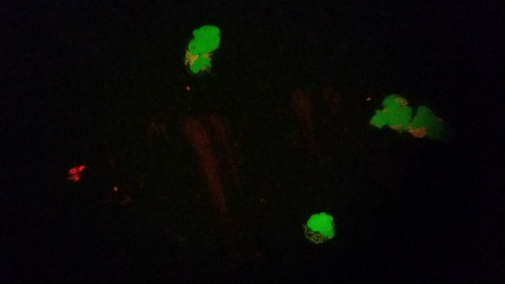
I believe the organism we are looking at to be spirochaeta gallinarum. A very hardy and clever specimen that avoids ABX by encysting, budding spores, invading your RBC�s & and your WBC, as well as just about any tissue in the human body.
These are just a few videos that I had just made after getting my new camera 2 days ago. They in no way reflect the best of what I have seen.
I have taken notice that they come in various sizes, shapes, and morphologies. Some are short and stubby dumbbell shaped & are very active and move in a Brownian motion.
Others are medium dumbbell, with a longer middle line (thread). As if the older and matured version of the short and stubby dumbbell. Basically a line with two bulbous tips on either end.
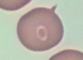
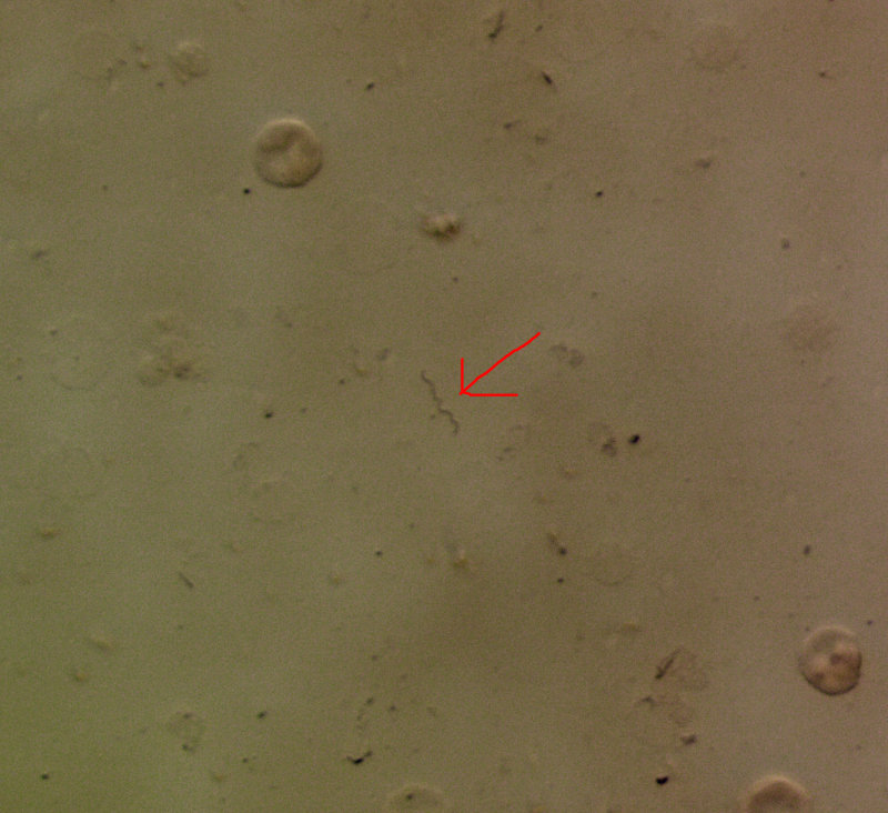
At times they appear as a long dancing string & can be seen exiting RBC's. One end is exposed and is bulbous, while the other end is still within the RBC. Initially not much can be seen, but if the live smear is left for an hour or more, they start to appear in the plasma & some can be caught in the act of exiting RBC's.
If the smear is left overnight, more can be seen the following day. The trick to see them the next day is to spread the oil immersion around the peripheral of the slip cover & let it spill over to the slide. In essence creating a vacuum seal for the smear & decreasing plasma evaporation. With this method the RBC's can be kept whole, but shrunk for days & those organisms can be seen for quite some time.
Others have a dotted body, besides the two dots on either ends, as if they are giving rise to new spores (dots).
The progression might be DOT--> DOT WITH TAIL-->STUBBY DUMBBELL-->MEDIUM DUMBBELL-->LONG DUMBBELL (STRING).
The LONG DUMBBELL can be even longer & the MEDIUM DUMBBELL can be dotted.
Some of these dots (spores) can be seen dancing around freely in the plasma & yet some have a very small tail. Basically a tear drop, or cigar shape with a tail.
Others are extremely long and wrap around. They appear to have grown longer with time. When I first started getting symptoms they were smaller & incidentally I felt the twitches within me smaller. Months into the progression I was able to observe how long they can actually get & this coincides with the feeling that something bigger and something that has grown within me. They have also grown in abundance and prevalence. This produces various burning pains, twitches and movement...Fibromyalgia type symptoms.
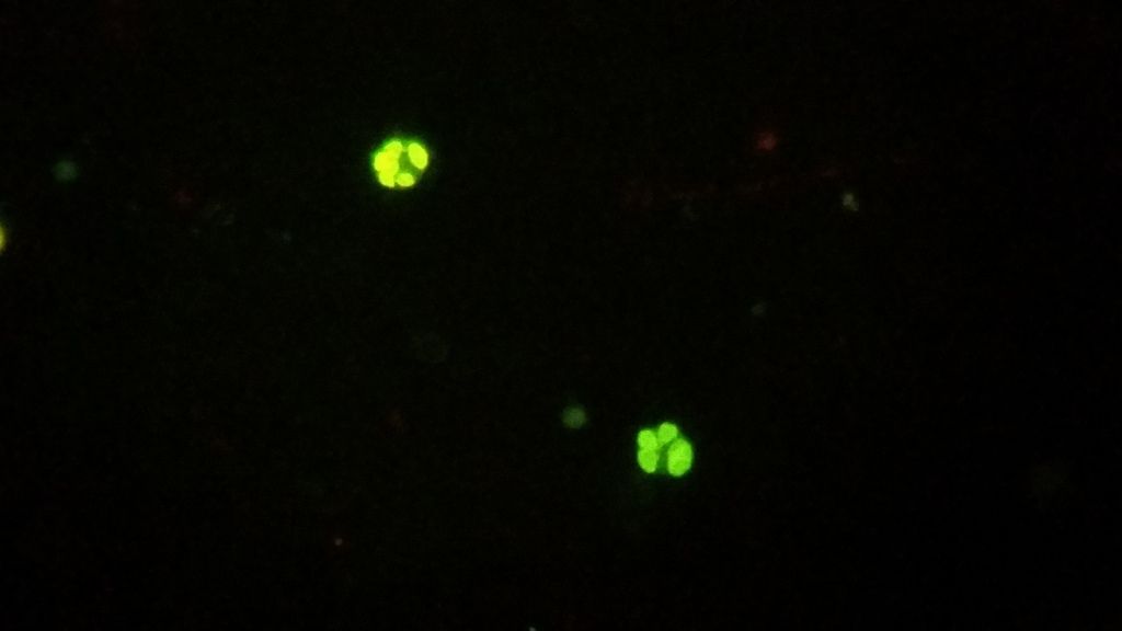
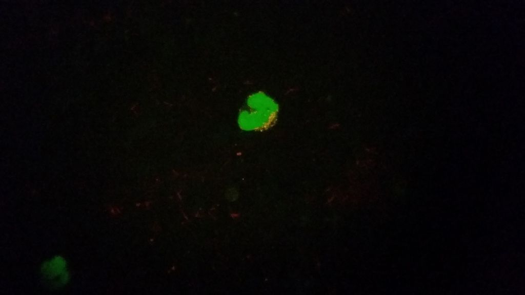
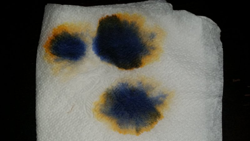
Here is an interesting find. I had been watching my blood for many hours & months. I had seen so many of those things come out of RBC's, but never once from a WBC. I had started to do Hot Baths & on day two I checked my blood. I noticed that my blood was more watered down and fluid. When I pricked myself for the smear, my finger did not want to stop bleeding. When I got my Bacillin LA shot, that too gushed blood (which is out of the ordinary).
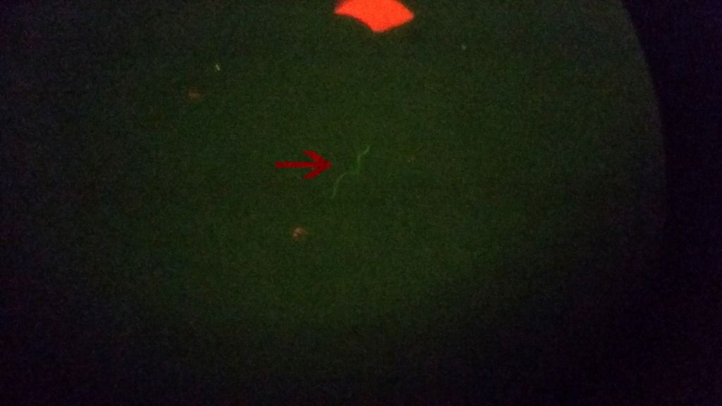
Here is a poor video from my hand held camera of this, but it is hard to tell. You may need to expand the video fully. There are so many coming out of the WBC for the first time ever & this is with multiple WBC's.
I am very, very interested in this!!! I have not even looked at the links yet.
This is my next step. I am currently in a Biol lab at school simply to learn how to use a microscope. The students are half my age and do not have borrelia in their brains, but I am determined to do my own research. I occasionally have a synapse in my brain fire, I plan to get them working again.
I will send you a PM and also give you my phone number. Maybe you can walk me through which scope to buy etc..
Also - maybe we could start a facebook page for people that are doing this. They could post their results there.
Posted by lax mom (Member # 38743) on :
Amazing!
Posted by Lymedin2010 (Member # 34322) on :
Haley, yes we can talk.
Also note that my blood smears are using a standard Phase Contrast microscope, without the phase use & can be seen with a standard compound microscope. They are live & slightly more wet (so I can see things freely swimming) & NO STAIN!
Yes, the BB is hard to see inside RBC's without a stain, but once they come out they are easy to spot. If you look closely you can see some of the RBC's dancing around, indicating there is something inside. One can also see the cysts within WBC's, that look like specks.
Here is Dr. Andy Wright (from UK) making similar observations
http://lymerick.net/video/AndyWright2004-640.wmv (52 Mb, 4 minutes) video of spirochetes in more lenghts, granules, a moving granulated cellular structure, thus illustrating all the phases complex spirochetal lifecycle drawn on page 475 in this article by Hindle 1912 (PDF) printed in Parasitology (1912), iv, pp 463-477. So far (June 2005) 98/98 of Andy�s ME/CFS patients with such structures in their blood tested positive on direct fluorescent antibody test for Borrelia burgdorferi ANTIGEN!
[ 10-03-2012, 03:22 PM: Message edited by: Lymedin2010 ]
Posted by Catgirl (Member # 31149) on :
Very cool!
Posted by Lymedin2010 (Member # 34322) on :
-Things to look for: -Get a Binocular instead of a single one. It makes things easier on your eyes when looking long term. If you can get a Trinocular, even better. It will allow you to place a camera without hindrance to the Binocular portion. I have a binocular.
-Make sure you get one with 100x (Oil Immersion) objective lens (the long tubes you swivel). Usually if there are 4x lenses they may be (20x, 40x, 80x, 100x). You need a min of 100x & coupled with the eye piece (usually standard 10x), you will be able to see 100x * 10x or 1000x.
-It would be nice to get anything over a 100x if you can. I don�t even have one yet, but eventually will get one. Maybe a 200x, so I can see 200x 10x = 2000x.
2) Glass Slides: $10 (with ship) Do a search on ebay for �microscope glass slides� You get 75 slides & 100 slip covers with these. I bought more & a bulkier batch & have plenty left over. �72 Blank Microscope Slides and 100 Square Cover Glass - Karter Scientific
4)A Lancing device, such as this Delica Onetouch system (under $20). You can draw blood anyway, but this makes it easy & pain free and can be bought at RiteAid or Walmart. It is a good idea to swab your finger with alcohol before drawing blood.
Here is a youtube video on how to make a blood smear. Follow the video & then add the slip cover on top. Then you add the oil immersion on top of the slip cover (1-2 drops). Personally, I use the slip cover to do the smear & it works out fine & is fresher.
I will make a video of a smear eventually & post.
Posted by Lymedin2010 (Member # 34322) on :
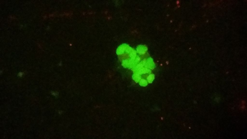
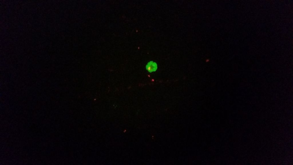
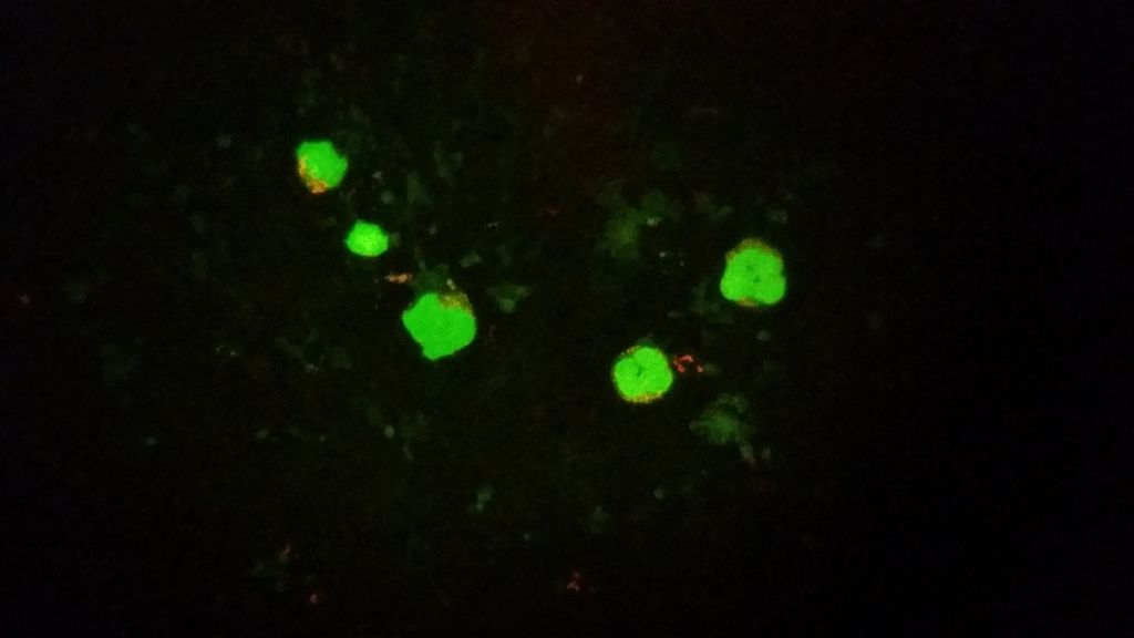
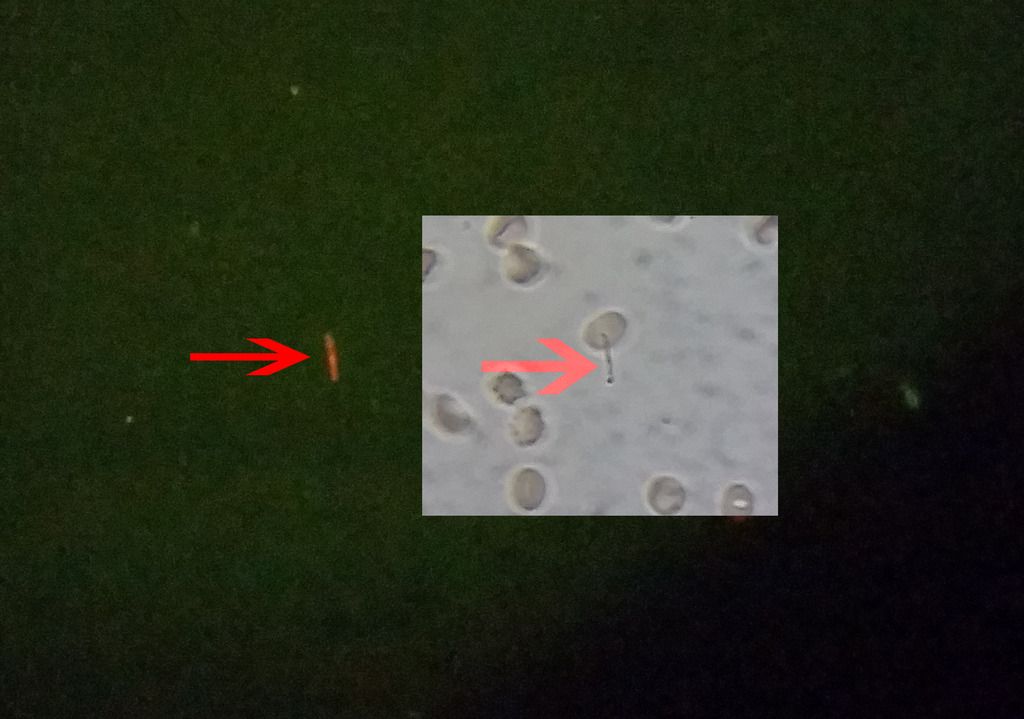
Here is another video of my Phagocytes in action.
Notice one of the Phagocytes has larger speckles within the body. This is most likely the Borrelia Cyst or Speck. The other phagocyte has no such specks.
do you just use a drop of whole blood and view under the microscope or do you place a drop of blood on the slide, traumatize the blood cells by pricking with a needle then put the cover slip back on and view under the scope.Traumatizing the cells makes the bugs jump out of the cells into the extracellular space. If you are not doing this and u see the bugs in the extracellular space using a drop of whole blood then you are toxic, your extracellular space needs to be cleaned up before more detox.
Posted by Lymedin2010 (Member # 34322) on :
I prick & manage to get about a drop out of me. Place it on the slide and smear it using the cover slip. I don't induce any trauma.
The blood smear looks clean and nice while fresh. Give it an hour & things start to appear. Give it a day & one would be suprised and perhaps SHOCKED.
I have seen the blood of others & have seen things come out of many people's blood smear. At one point I was similar to others, but recently things appear to have gotten worst.
Posted by seibertneurolyme (Member # 6416) on :
Have not had time to read the whole thread or view the videos.
But one quick comment. Was on the phone tonight with hubby's LLMD. The doc tried something I had read and took some pictures of their own blood with an iphone.
Do a finger stick and then do an ear stick and compare the results. The ear stick may show much more. At least that is supposed to be best when looking for babesia in dogs.
Bea Seibert
Posted by lymenotlite (Member # 33166) on :
Is this a darkfield microscope? I'm wondering why the link you gave is a preferable microscope.
Is it possible to identify most of what you see? Can it help you diagnose exactly what is infecting you and so enable you to create a better protocol for yourself?
Wondering whether you really need a microbiology class to be effective.
Posted by Lymedin2010 (Member # 34322) on :
Never heard of an ear test, would be nice if it held water.
Darkfield would be nice too. �I have seen some Lyme blood online under Darkfield. �You can actually buy a dark field condenser and turn a compound into a dark field.�
I think Darkfield is better for identifying Babs and Bart. One can more clearly see through RBC. I had my blood viewed at the peak of my sickness with a Darkfield, before a good set of ABX that actually worked for me. Nothing was spotted. Maybe now might be a better time to check.
It could help make a better protocol. For instance, I had shortness of breath and the one thing that held it back was Buaxin. I was on it for over a year.
I have switching LLMD and have now switched meds. Because of interactions I had to stop Biaxin. I had never seen Borrelia in my WBC, only after 2 hot baths did I see them AND after one week of stopping Biaxin.
Biaxin is a good intracellular ABX. Perhaps I released more Borrelia from skin surface and they were picked up by macrophages or mybe I removed Biaxin, what prevented my WBC's from backing up?
If we had an individual who we knew only had Babesia or Bart, it might be easier to distinguish based upon shared video. Right now it is hard for me and for doctors. I am trying to make it easier.
The Borrelia is clear cut to me, nothing else mimics a string with a bulbous tip. But for instance, can the specks inside WBC be Bart? Yea, it could be and only until we do a DNA test can anyone know with certainty.
My next step is to transfer some of them over to new media and observe them for a longer term. This should buy further clues.
Posted by seibertneurolyme (Member # 6416) on :
Another suggestion from hubby's LLMD -- do not use alcohol pads to disinfect your finger before the blood draw. Doc suggested using a red cleaner instead -- could not remember what it is called -- not betadine but something else that just washes off. Said results would be different if you use the alcohol pad.
Bea Seibert
Posted by sparkle7 (Member # 10397) on :
That's really cool (pardons that you may be ill from it) but how do you know it's spirochaeta gallinarum as opposed to something else?
Posted by a mom (Member # 23920) on :
Bea, were you able to get the video clips to the parasitologist in NY?
Posted by a mom (Member # 23920) on :
Bea,
Oh arrrggghhhhh....I can't figure out which button to click on to send you a private message. Would you call me when you have time and I can give you Dolores phone number?
HUGS,
Janet
Posted by seibertneurolyme (Member # 6416) on :
A mom -- Working on it. My computer is old as the hills and it would have taken over 2 hours to download dropbox. Am at Kinko's -- sending some of the video clips by yousendit.com -- still is taking over 5 minutes for each video clip (there were 27 in total). For now just sending 5 and will try to send more later today. If it works I will post the link in this thread -- think I can do that but not 100% sure.
Bea Seibert
Posted by Lymedin2010 (Member # 34322) on :
I am always willing to try. What is the red substance called?
There is no doubt in my mind what this is.
Did you guys watch this video from a DOCTOR who has seen and has described the same thing one sees in my blood smears.
He also makes references to this species by other individuals in the early 1900's.
Posted by a mom (Member # 23920) on :
HELP!!
Bea called and asked me to post a New Thread. I don't know how to do that. Can anyone start a New Thread for Bea's info:
Steve is dying from ARDS or lung failure from unknown causes. Docs think his heart will give out but can't say when. For example something as simple as turning him over in bed can cause his oxygen levels to fall -- even on a ventilator at maximum (100 % oxygen).. Other times his heart rate slows to dangerously low levels instead of the oxygen falling.
The docs are pushing for me to change Steve's code status (now at full code) or even to pull the plug on the ventilator. Undecided what to do about code status right now -- but pulling the plug is NOT an option at this time.
I am trying to decide what to do next to help Steve.
Once I get the Clongen blood smear results I need to come up with a new plan of action.
Addendum:
Babesia meds are still on, WBC down to 11,000. She did not say if he had fever.
Bea is trying to reach a doctor Dolores recommended: Dr. Thomas W. Nash (Columbia Presbyterian). Pulmonary and ID.
Bea called, but office was closed. I sent an email for HELP. Is anyone friends with him and could reach him for Bea?
Posted by a mom (Member # 23920) on :
More Prayers Please. When i called Bea, the Pastor was in the room with them....
Posted by map1131 (Member # 2022) on :
a mom look towards bottom of this page see post new topic. Click that.
Pam
Posted by a mom (Member # 23920) on :
map1131: Thanks!
Can everyone see the 27 clips listed in this playlist?
Part 2: Traversing the field of view down the length of the organism. It did not appear to move, perhaps because it was sandwiched between the glass slide & the slip cover.
This could be the something that I feel in me that has grown in size & or colonies. Something moving inside of me throughout my body.
Posted by derk diggler (Member # 31903) on :
that is crazy crazy, how you gonna kill it, or them inside of you, just prves that parasites play a bigger role in this than we think, would you teach someone how to do what you do on a scope
Posted by dal123 (Member # 6313) on :
Are you sure this is not an artifact, or food fiber? to tell if it's a parasite, staining would be more helpful, ie to differentiate between this better, can see the scolex & segments, etc. that's the best ID aspect of parasites in the bloodstream, tell if it's microfilaria.
by the way are you taking any enzymes? I noted lots of rouleaux, possible leaky gut issues. digestive enzymes with each meal, ie TPP Digest and then on a very empty stomach, Interfase plus or serrapeptase.
Posted by Lymedin2010 (Member # 34322) on :
Yes, I am willing to teach, just let me know.
I don't think a fiber would normally be:
-transparent, just like some of our own cellular material.
-have folds and ripples like it does.
-such finely tapered ends.
-looks to almost have a central tubule running across the one side. Almost as if a digestive or perhaps a reproductive tract.
I have never seen one of these before, but have seen bits and pieces of material, which I always associated with dead skin tissue and debris.
I just started serrapeptase a few days ago on docs orders. Here is my total regimen:
Doxy, Biaxin, Plaquinil, Malarone, Nystatin, Cryptolepis, A-Bio, & Colloidal Silver.
Posted by glm1111 (Member # 16556) on :
Someone here who had a microscope posted a stained blood smear a year or two ago and identified them as microfilarial worms.
Ivermectin and doxy were taken by people in a study who thought they had these microfilaria and said they had success with that protocol.
Remember that Willy burdorfer found Filarial Worms in the ticks he dissected, and also Dr Eva Sapi is finding them in over 40% of the ticks she is dissecting.
My own experience with this disease has shown me that I was LOADED with these parasites and then some. It amazes me that even the ILADS community has overlooked this and is soley focused on BB.
According to Dr. K. once the parasites/worms are eradicated, bb and the other co-infections are easier to kill. Antiparasitics have to be taken if anyone wants to get rid of this disease. Abx alone will keep you on a treadmill and never eradicate this infection known as Lyme disease.
Gael
Posted by debilyn (Member # 35753) on :
Wow LymedIn, that is amazing. Looks like a parasite to me. How do we kill blood parasites?
If this is a filarial worm, how do we kill them in our blood? Babs medicines?
Posted by Haley (Member # 22008) on :
I'm bringing this back up. Does anyone have pictures of parasites in their blood?
What do you think of this microscope and do you think I can do video with it also?
I personally don't trust AmScope. Their units are sometimes unpredictable, but at that price range I would expect it to be ok. I believe it is the lower end ones that are riskier.
I believe LymeNurse or LymeTwister on here also bought an Amscope. It was unusable and he had to return and upgrade it. He got an awkward one with a built in video screen, but it did the trick. He posted his vids on youtube & you can check them out. Here is one of them: https://www.youtube.com/watch?v=BXRNH0lkiNg&list=UUR9kWYE08hN1pbvzIzwL3Dg&index=7
The clarity is not that great, but it could be due to his video recording.
I caved in and bought a Amscope 9MP microscope camera. I figured they can't go wrong with that, can they? The build quality is great, but the software and auto refocus is pure crap. At the end the worst piece of hardware I have bought in a very long time. My point & shoot camera took clearer images and video & was better with frame rate.
The only problem with my PNS cam is that I had to hold it by hand & it was shaky.
Posted by Lymedin2010 (Member # 34322) on :
Haley, your PM box is full. Try & delete your older messages to make some room.
I don't see any Zeiss Trinocular microscopes on Ebay as of now, but here is a good & cheap alternative.
American Optics is a good brand used by many professionals. I would be bidding on this right now, if I needed a scope.
Lymedin, my LLND says if we have blood parasites, we also have them in tissues and organs.
Posted by Lymedin2010 (Member # 34322) on :
I believe it. I have many disturbances and movement all over my body. In particular under my rib cage is a hot spot, that seems to have grown in size and disturbance.
This is right where the spleen & Liver is located. Once the spleen goes, I would imagine the immune sys gets knocked down hard & I feel as if mine is. Most of my blood work shows fine, except for slightly elevated Liver enzymes & they did notice immature granulocytes.
I think a source for parasites for me might have been the mosquito bites that ensued after starting treatment. I didn't know we had to be wrapped in a bubble while treating. If ABX knock down ones immune system, then you can really pickup anything and everything.
I feel like an AIDS patient, but with much more pain and inhumane disturbances.
I highly recommend watching the TV series "Monsters Inside Me." This will give you an idea of what others have been exposed to & how easily they got sick without any Lyme. With Lyme there are so much more dangers out there & they cannot necessarily all be attacked by the ABX we consume.
Posted by Haley (Member # 22008) on :
Thank you Lymedin2010. I will bid on that scope. Can a camera be attached to that one?
I'm so spaced out that I have no idea how I will do any on this, but I am very interested in looking at tissues in addition to blood.
I will read through this entire post later.
My mailbox should be open now.
Posted by Lymedin2010 (Member # 34322) on :
Larger trinocular openings, such as that on this scope usually are meant for CCV cameras. I believe they are called C mount & size to be 30mm.
They also make adapters that will allow you to attach cameras and video cameras the size of the eye piece (23mm for example) to that opening.
Once you get it the best thing to do is find the dimensions of the opening & get the dimensions of the camera you are using. You can give a microscope store or B&H photo a call & provide them with your dimensions & they can help you find an appropriate adapter.
My Trinocular is a narrower openeing, same size as my eye piece, 23mm & the Amscope camera fits right into the trinocular adapter tube.
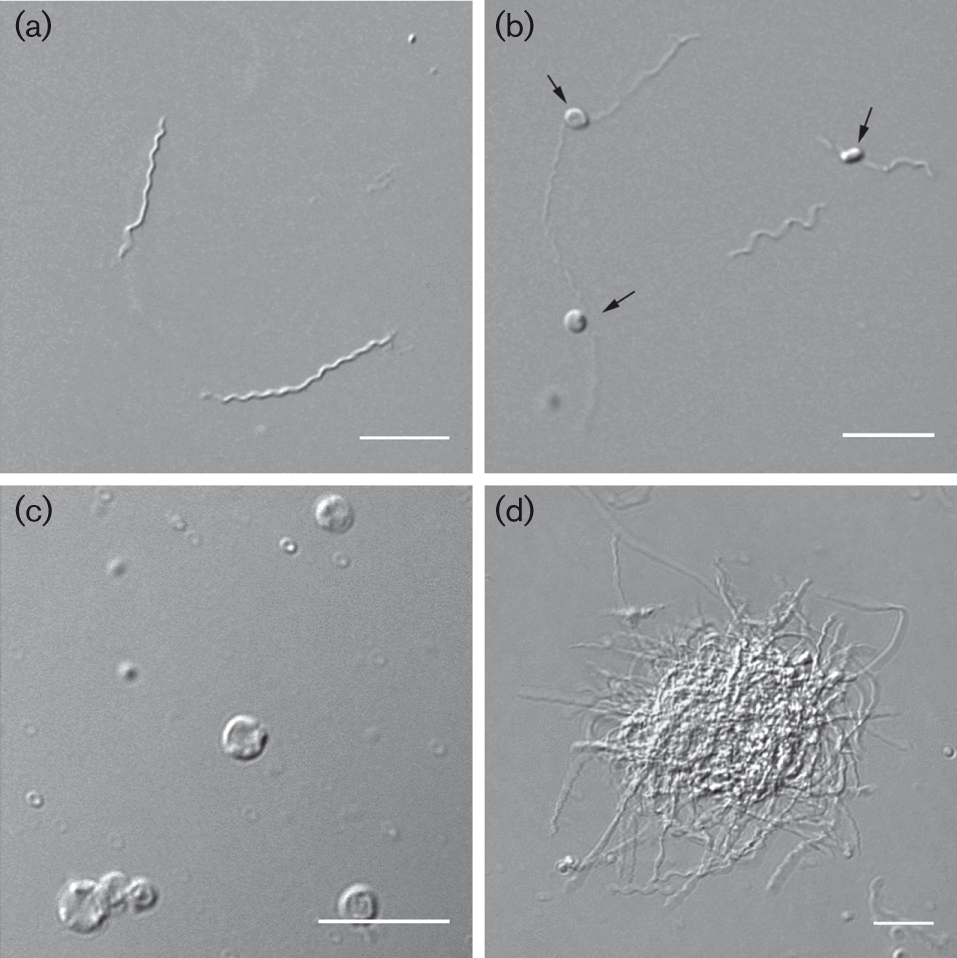
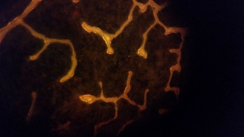
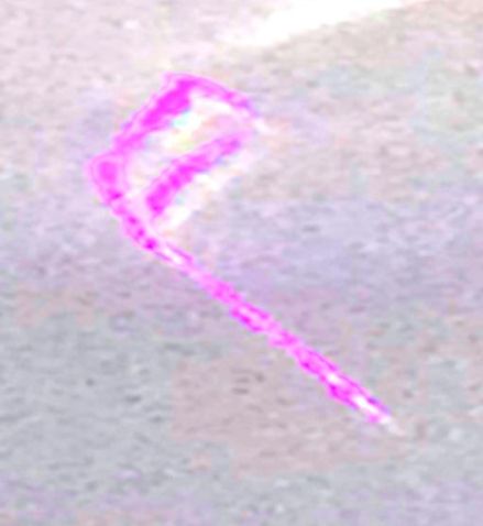
Here is a WONDERFUL video showing you spirochetes coming out of RBC's. It shows you the cyst forms, which have various shapes all depending on how they curl up.
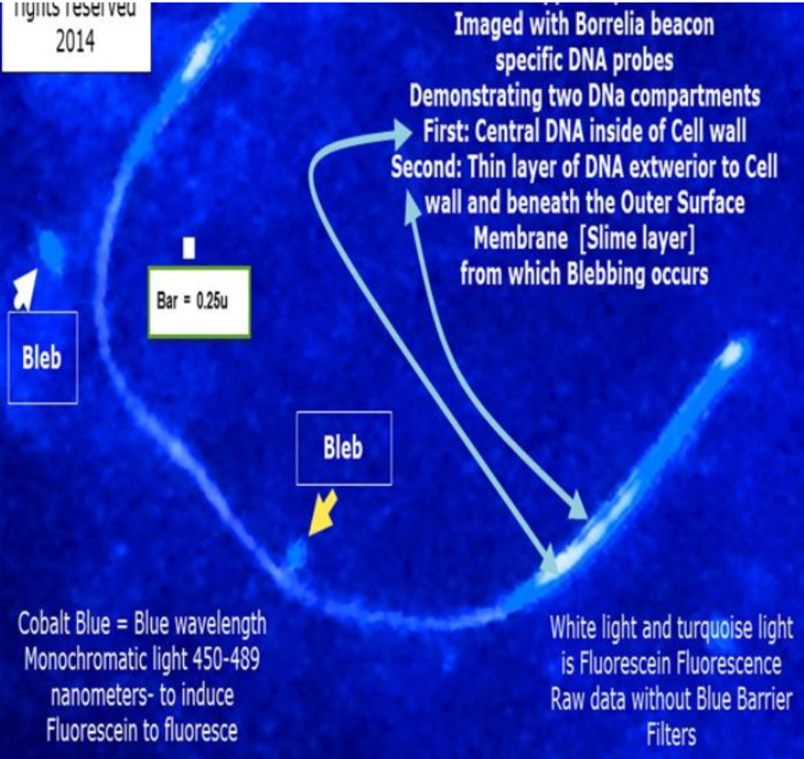
It even shows the bleb or granualar forms, which is the smallest unit of the spirochete. In this form it basically consists of DNA packaged in a cell wall. This form is reminiscent of a virus, but much larger in size.
I have seen all these forms in my blood. I have seen them in individuals who have been bitten & function overall, despite some minor symptoms.
I also see them in individuals who claim perfect health.
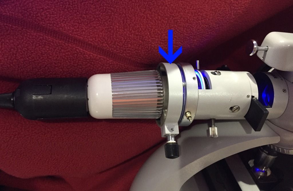
Here is one of the best ways to capture video & images from your microscope. It is a device that attaches directly to the eyepiece or trinocular port of the microscope. My trinocular is 23mm, so it is perfect.
The Samsung Galaxy S2 & S3 are amazing for picture & video quality. The iPhones & iTouches as well, but you can use many smartphones that fall within their size range. Keep in mind that some phones have better quality cameras. I give high praise for the Galaxy series of phones...amazing quality for a phone, at times better than your point & shoot cameras.
I did a manual shot using my Samsung hoovering over my eyepiece & despite my shaky hands I was impressed with the image clarity.
I have ordered mine & can't wait to get it & post many vids soon. Price of the unit is $75 + 5 shipping. Compare that to the $250 piece of garbage 9MP Amscope video camera I bought.
It looks like the world is catching up & are now realizing that Borrelia IS IN OUR RED BLOOD CELLS. For me Borrelia exists & continues to grow in numbers DESPITE ABX treatment & it is clearly & elegantly visible in blood smears.
Also a warning to those taking Doxy. It is the only ABX that keeps me from being in extreme pain, but over time it only does produce cyst forms & leads to more chronic lyme disease. I now stand by Dr. Sapi's test on the ABX.
A QUOTE & LINK:
"Filming the bacteria
The two scientists are pretty much alone in what they are doing: investigating samples of living blood under the microscope - over time. They have looked at the blood of a large number of people who suspect that they are chronically ill following a tick bite.
"We study the behaviour of the bacteria directly through the microscope. We have taken thousands of pictures, and we film their movement pattern in real time or with a time lapse. This allows us to see how the bacteria enter and leave the blood cells and swim around in the blood plasma. These are powerful methods, but nobody uses them any longer", says Laane. He himself is regarded as one of the foremost microscopy experts, and he has been studying bacteria for over 50 years.
I apologize for not understanding this but what are the implications of it being in the RBCs?
Are the abx we take not sufficient to kill it?
[ 02-06-2014, 11:40 AM: Message edited by: terv ]
Posted by oxygenbabe (Member # 5831) on :
This seems unprofesional to me (just viewed the latest video).
Posted by Lymedin2010 (Member # 34322) on :
Here are some of the implications:
____________________ 1) You may not need expensive test or to be bounced around in the medical field. A half borrelia brain dead like myself can look under a microscope to find these.
It would be great to find someone who gives a hoot & have them do PCR on these organisms. It would be nice to know what particular species they are. As we all now know there are many species of spirochetes.
It would be nice & easy to simply place a blood sample in BSK spirochete growing medium & culture them. One can clearly see borreliosis in this manner.
____________________ 2) Despite aggressive & varied ABX, with herbs, spirochetes in the blood still exist. We can break it down to the basics. If one cannot eliminate the infection in the blood, then there is no hope of eradicating it from the body. Just because you don't have symptoms does not mean you may not have borrelia in the blood. It is a good early warning sign.
It is easier and important to first discover how to kill it in the blood. The blood acts as the highway to infections throughout the body. What hope do you have getting rid of this, without clearing out the blood first?
Before I started treatment I only had a few symptoms. While treating I have over 2 pages now & what I have observed is an increase of organisms in my blood.
Blood can be kept alive for weeks outside of our bodies & experiments can be conducted on the blood. These would be cheap & easy experiments with blood drawn from Lyme subjects & would result in no harm.
____________________ 3)Gives us the possiblity that we can treat the blood externally. We may be able to treat & get rapid improvement with things such as frequencies, ultrasound, & UV/LED spectrum lighting. Attempting to disrupt the various stages of borrelia in the blood first, so that your body can once again fight the rest of the infection in your body.
Without the proper use of your blood & immune system, the rest of your body is defenseless.
____________________ 4) Indications of transmission. If borrelia is in your blood, that gives you clues as to where else it may be & how it can be transmitted. If it can be easily seen in the blood, then it is in every square inch of you, since blood & capillaries are spread throughout your body.
It is in your saliva, on the surface of external soft tissue (vagina & penis), eye fluid, mucous, and most likely is being shed as blebs on your skin.
Posted by SLML (Member # 42986) on :
Lymedin2010 - what antibiotics have you taken for Lyme to date?
Posted by Lymedin2010 (Member # 34322) on :
Here is what I can remember & is a pretty solid list of what I took. I may come back here to edit in the future.
2011 (May part & June) Month 1: IV Rocephin 2g/day (at the end of 3-4 weeks I was 90-95% better with only head pain & I wished I had stopped there) (I could not find ANYTHING in my blood smears after HOURS & HOURS of looking)
2011 (July) Month 2: IV Rocephin 2g/day, Zithro & Mepron
2011 (Aug) Month 3: IV Rocephin 2g/day, Biaxin & Mepron
2011 (Sept) Month 4: Ceftin, Biaxin & Mepron
2011 (Oct-Nov) Month 5-6: Doxy, Biaxin (More & more Lyme symptoms with each passing week prior to adding Doxy. 2 pages worth of symptoms) (I started seeing things in my blood smears & accidentally discovered that if I cover the slides in oil, I preserve the smear longer & then I can see the spirochetes existing the red blood cells when conditions are properly met). In Nov I took my first major slide back after running out of Biaxin for 1 week.
2012 (Dec-Feb) Month 7-9: Doxy, Biaxin, Tindamax, I added Rulide the last month here (After 3-4 weeks of Doxy improved from 45% to 80-85%, still many minor symptoms but those I could handle. I was very gradually going back & some new symptoms)
2012 (March-April) Month 10--: Amoxicillin w/Probenecid, Biaxin, Flagyl, during the last week I added Rulide to this since I was just feeling worst & worst with new returning symptoms. This was a big decline on this combo for me & that I could never really recover from some of the new symptoms.
2012 (Oct-March 2013) Month 15-21: Doxy, Plaquinil, Nystatin, Malarone, Bacillin LA shots, Cryptolepis 1 teastpoon 3x day, Colloidial Silver(stopped in Feb), Detox 2, NT Detox
Posted by SLML (Member # 42986) on :
Wow, you are awesome! What a detailed list! :-) The one thing that I am seeing though is that your doctor only had you on a cyst buster for a couple months so based on my understanding of how lyme is most effectively treated (always being on a cyst buster) maybe that is why the spirochetes are still showing up in your blood??? My doc has me on a cyst buster for the whole duration of treatment. I know some doctors just prescribe cyst busters intermittently but to be honest, I would be scared to treat that way because of the potential for baby spirochetes to be formed during the months a cyst buster wasn't used. I understand cyst busters are really hard to tolerate. I was really scared to take them too because of their reputation. I did feel worse for a few months and have progressively gotten SO much better. Anyways, I would love to see my blood under a microscope. I wish I knew of someone here that could do it with the level or expertise yours was done with!
Posted by Lymedin2010 (Member # 34322) on :
Here is another person who has also found spirochetes in their blood sample:
When I took tindimax I felt nothing. I took Flagyl for 4 months the first time, just can't remember how the time frame overlapped.
Then again they gave me another 4 months in 2013. I must say that by the 4th month I did have slightly more energy, but by then they took it away.
I don't know why LLMD's are so reserved to give this right off the bat & I don't know why they don't keep us on it for longer? Maybe they know something we don't, perhaps there is great risk of neuropathy & cancer for them to withhold is as they do.
So I am taking Doryx, Penecillin VK, Nystatin, Plaquinil, Cryptolepsis plus, A-Babs, & detox. I decided to try Amoxi on top of all this. I remembered that I herxed on Amoxi the first time & even months after, but the herx did not last long.
I took 1g & within 45 felt different. I then took another 2g & then came the herx. So here we can be on many different ABX & it will not get to all the forms & get to all the tissues of the body. It is so important to rotate more often & aggressively.
I wish they would just give us flexibility to try this & that on our own.
[ 02-20-2014, 03:40 PM: Message edited by: Lymedin2010 ]
Posted by Lymedin2010 (Member # 34322) on :
Good deal on a solid microscope for anyone who is looking.
I am going to have darkfield done out of state on 3-19 Wed.
I have seen videos of the Bb forms. Does anyone have the links to videos or pictures of other pathogens like Bart or babs or filarial worms that I could look at before I go?
Thanks.
This darkfield is in KY and if it is very helpful I will share the location if someone is interested. I found this on a site that lists darkfield locations/docs.
Posted by Lymedin2010 (Member # 34322) on :
Babesia is harder to spot, but sometimes these crosses can be seen. I have never seen definitive babesia or crosses in my blood.
It is much easier to spot in dead blood stained samples.
I don't see many babesia videos & this one is rare.
"My name is Dr. Allen Anderson. Almost four years ago, at age 65, a stroke rudely interrupted my busy ear, nose, throat and allergy practice. None of the physicians in the multi-specialty clinic where I worked could pinpoint a cause for the stroke.
I was not over weight, did not have high blood pressure, exercised regularly and did not smoke. My cholesterol was mildly elevated at 240. My diet was standard American with plenty of steak, french fries and diet soda.
After 3 months of rehabilitation I returned to practice. My problem was that I could barely drag myself through a work day because of chronic fatigue and severe pain in my shoulder and neck. These symptoms did not improve with time. I also noted that I was depressed along with problems sleeping at night.
With no help from my colleagues other than pills, in desperation I consulted a naturopathic physician who looked at a smear of my blood under the microscope. He pointed out many spiral like organisms called spirochetes along with yeast parasites. This was my introduction to the epidemic-like malady that is affecting thousands of Americans yearly.
I had Lyme disease, an infection caused by the bite of a deer tick infected with the spirochete organism Borrelia burgdorferi. "
Posted by Lymedin2010 (Member # 34322) on :
"Sickest patients have most abnormal findings
This abnormal chemistry of the blood (the low amount of oxygen present, coupled with high levels of oxidative stress), then allows proliferation of what Professor Ali calls ‘Primordial Life Forms’ such as yeast, which clump with the microclots and worsen the problems outlined above.
In my experience, there is a correlation between the amounts of these anaerobic bugs seen and the severity of the illness.
Certainly my sickest patients have the most abnormal blood appearances on the video microscope. However these appearances are not only seen in CFS (ME) and Fibromyalgia (FM), but in many chronic illnesses. This is because at a cellular level the oxygen problems also exist in these illnesses. What I feel is important though is that the clots and yeast are a very important co-factor. "
SLML, GSE reputedly pops Bb cysts. I use the nutramedix brand and I have no bad reaction.
On the microscope screening, there is a place in Idaho if that is close enough to you in Wash.
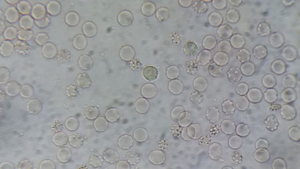
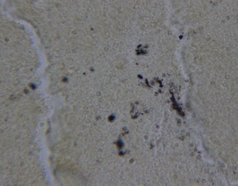
I had dark field Wed and was amazed. I saw one long lyme spirochete, 2 short stick bacteria with knobs on each end (maybe newer Bb), some yeast blobs, some type of immune cells loaded with waste & proteins on surface, mycoplasm (did not know I had this also).
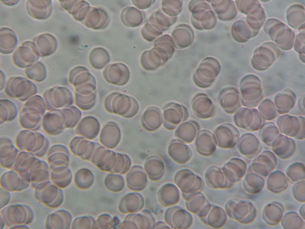
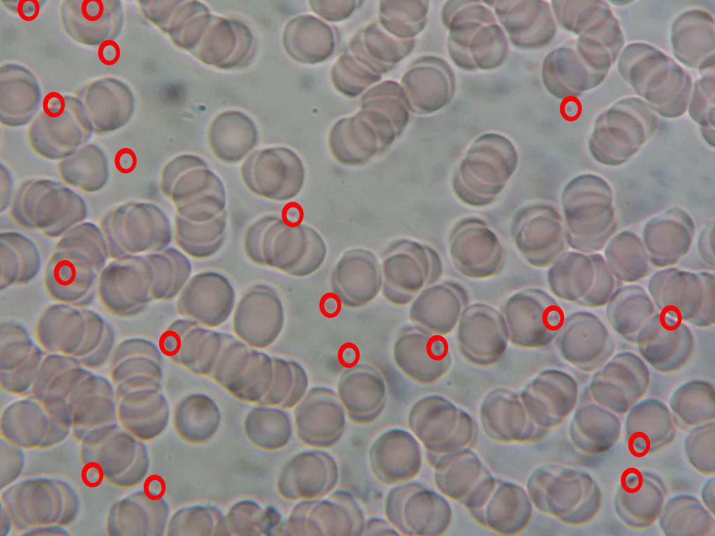
Also, I had a number of RBC's with "target" centers, which is from having thin-membrane outer cell walls. And a number of RBC's that were shaped like tear drops and some were paired with their points touching.
The tear-drop RBC pattern was explained to me as caused by incomplete protein digestion, and food enzymes must be taken with meals to correct this.
Dark Field microscopy is amazing to watch. You can really see the health/non-health of your red & white blood cells, and pathogens.
Posted by Lymedin2010 (Member # 34322) on :
Be careful with GSE. If you are on Doxy or Zithro it can reduce the effectiveness of those ABX.
I am Doxy dependent & I know it definitely looses effectiveness when I try GSE.
It is always good to have visual verification & it is so easy to find. Look at this video from "Under Our Skin" & it comes on after Alan MacDonald speaks.
Doxycycline (Doryx®, Periostat®, Vibramycin®) Especially with doxycycline hyclate (Doryx®). Drink a FULL glass of water to avoid stomach irritation.9 Tetracycline (Tetracyclin®, Sumycin®)
DO NOT take the above medicines with some drinks containing calcium or iron. These can make your medicine less effective.
Herbal teas Popular flavored water Smoothies Orange juice Grapefruit juice Milk Milk of magnesia
In another article on grapejuice & ABX. This applies to clarithromycin (Biaxin), erythromycin derivatives (Azithromycin / Zithromax).
Lymedin2010 - You said in an early post that you thought you had spirochete gallinarum. Is this a lyme spirochete?
Do you still think you have this type?
I have some pics of my blood now, and could forward some of them to you by email, if you think you could identify a couple of things.
I am computer-illiterate and do not know how to post on Utube or anything like that.
Not sure if the pics sent show this, but there were two short-stick (thin stick) with rounded ends like dumbbells, but not too much fatter than the stick. Not stubby in width of stick. Were yours stubby in width of stick?
Posted by Lymedin2010 (Member # 34322) on :
I would like to answer this better, but I am actually on a new protocol & herxing and feeling worst..yippeeee.
Here is the short answer.
The best & really the only definitive way to know is via PCR (Polymerase Chain Reaction). Basically, we use a small portion of the organisms & it's DNA and we amplify & and make millions of copies of it's DNA. Then it is compared to known DNA sequences & the exact type of bacteria can be identified.
Being that I cannot perform PCR, I look for best fit descriptions. I also look for something visual in the form of pictures, illustratiosn or videos for comparison. It basically acts like a mug shot when there are no other resources
I have pondered how to approach this on many nights & concluded that we must approach things like our forefathers where we give best guess hypothesis on observations. Then we continue to find evidence & research on what others have to fill in the gaps & come to a more clearer truth. Ultimately this is how truth is reached in many fields of discipline. In my case it has to be more so with limited resources.
When I was hunting for answers on Borrelia I could not get a complete picture of what I was seeing. It does not mean it is not out there, just that I had not come across anything that completely satisfied what I was seeing. I saw the blebs, cysts, various size spirochetes and the string of pearls. I also hypothesised that we could be multiple spirochete co-infected (different types of spirochetes in the blood) & always looked at multiple angles.
There was nothing that I came across that mentioned string of pearls or gave me a clear & cut PICTURE of blebs or how they formed. I knew before I even read anything on blebs that these things HAD TO come from spirochetes. Over the course of months I saw more & more of spirochetes in my blood & these dancing dots (blebs).
More sickness-->more Lyme symptoms-->more spirochetes-->TONS more blebs in my blood. I knew with all my being there was a correlation & especially when I saw what FULL BLOWN LYME patients blood looked like (from multiple youtube videos).
I came across this one website with information from a DOCTOR. This DR. was angry at other docs & even mentioned spirochetes coming out of White Blood Cells. I fell in love with this guy, since this is exactly what I saw in my blood. This even showed blebs (they called them coccoid bodies). The link may be in my old laptop now, but I did download one of his links to a pdf document & paper written by another individual.
A quick search for this document name & here is the link. Check the diagrams of the spirochete formation. http://lymerick.net/1912-01.pdf
At the time this doc & paper best fit what I was looking at. I thought to myself even if this was not exactly the spirochete, at least it gives us clues as to how other spirochetes function. Just like Alan MacDonald did between Borrelia & Syphilis.
The only trouble I had were these two. The cysts were not really mentioned & I assumed that he just did not focus on these & we all know spirochetes go into cyst mode. The other issue is that the spirochete drawing did not have bulbous tips, like what I have seen. I attributed this to artist drawing or the scientist trying to fit the spirochete to look more closely to what spirochetes look like.
All the older common notions of spirochetes are these aggressive & burrowing/drilling machines. What I see is more passive & go with the blood flow organisms. Perhaps that is what makes them so much harder to kill?
A few weeks ago I came across Alan MacDonalds videos & I nearly jumped off my chair. String of pearls, blebs , videos of passive spirochete with bulbous tips and with the title on the video that said "BURRELIA BURGDORFERI." This thing moves & looks EXACTLY like what I see in my blood. Now I can take what I learned from Gallinarum & apply it to know VISUAL proof from another Dr. or scientist & consider this as my most recent explanation of what I see.
I also think that one of the next important things we will learn is that the baceria is EVERYWHERE in the body & this will give more credence to contact transmission. Just because there is contact does not mean you will get Lyme. Look at it as if someone had just gotten bit & we know how differently everyone reacts. There are many other factors that lead to full blown Lyme than just getting the bacteria in your system. A simple enzyme or normal presentation of vitamins can keep it at bay.
Most people may have spirochetes in their blood, but they may not have all of the co-infections, metals, mold, or detox issues combined to cause complete disease.
Reportedly there are 3 types of Borrelia that can cause lyme disease: Borrelia burgdorferi, Borrelia garinii , & Borrelia afzelii. I would not bet my money that these are the only ones. This is the new uncharted frontier or realization for humanity.
I would be more than happy to look at your pictures, videos & even look at your blood if you send it to me. I have a lot more to say, but I will leave it at this for now.
Posted by Nancy L (Member # 42733) on :
Lymedin2010 -
The spirochete shown in my dark field microscopy was also relaxed, long (not straight), some slight curves that changed, but definitely not spiral. And they ends (small, so might have missed some tail), looked somewhat bulbous.
I understand that the Bb go into spiral form mode when they are trying to move quickly. The spiral shape is for motility, to move faster.
Latest research (can't give link right now) shows that blebs somehow turn into small cysts and then form their dna into a spirochete that then leaves this form as a new spirochete in about 9 days.
There may be other cysts that are protective forms for the spirochetes too, when threatened. I'm not sure if that is an "also" or just older info.
Thanks so much for the link.
About contact transmission. If you have an active cut, perhaps that would be a source of contact Bb, but since Bb is mildly anaerobic, it would stay away from the surface of the skin, I believe.
I saw a picture of a test tube with different forms of bacteria in solution (still active and alive).
The aerobic bacteria were clustered at the top of the solution in the test tube, the completely anaerobic were at the bottom of the tube, and the Bb bacteria were about 1/3-1/4 of the way down the test tube solution, in a cluster band. So they stay away from direct contact with air.
Thanks for all the good info Posted by Lymedin2010 (Member # 34322) on :
There is a BEAUTIFUL video of Borrelia releasing a BLEB on one of it's end. So it looks like the bulbous ends might have two functions.
1) Burrowing into cells 2) Bleb formation
Don't forget blebs can also be formed throughout the length of the spirochete in the "string of pearls" formation.
Look at 6:40 on this video, the link below will take you right into the scene:
At 4:12 you can see the one of many, many, many forms of the cyst. So many & depends on how big the spirochete is when it folds in on itself & how it folds in on itself. The longer it is in cyst form I believe the longer it develops an outer protective layer & fortifies itself. Looking more bean or slipper like.
I made a video on how to create your own blood smears for anyone who is interested in seeing borrelia spirochetes. I made dozens of blood smears & it did not come out the best in this video, but there were some waiting on me to make one for a while now so I just hit record & did a quick one.
I was going to wait for upgraded equipment, including a tripod, but enough waiting. I will make a better one in the future & more borrelia videos in the future.
My Lyme brain won't let me get out everything I wanted to say & I am too tired to do another take.
Bigstan, I would buy this one right away (first link below) & then search for alternate means to make videos. I have a Zeiss microscope & got mine for $100, but I waited for 2-3 months before buying, as I scouted Ebay on the perfect bid. Then down the line I replaced the binocular head with a trinocular & then bought a camera.
I use an Amscope MU900 9MP camera, but it leaves much to be desired. I think my next camera will be a Sony Nex6, which is a standard point & shoot camera with HD video capabilities. This will be placed with an adapter on the trinocular & the built in time lapse capture will be used.
I have finally done some time lapse work & found it to be more revealing. Things opened up that I just could not spot before & it is amazing how much is missed otherwise. I think I finally found free swimming babesia in the blood.
Now answer this question. Do you know why the tick was able to pickup spirochetes from the mammal when human observation of tissues & tests could not pick it up?
Because the spirochetes are READILY IN THE BLOOD!!!! Think about it they must be readily available for the tick to be able to pick it up & spread the disease.
Also, how many spirochetes must one have for a small tick to take a small random bite, such a miniscule amount of blood, & from anywhere in your body & pickup lyme? BILLLIONS & BILLIONS of them!!!!!
Posted by lymenotlite (Member # 33166) on :
When I first got into microscopy because of Lyme I had looked at Amscope microscopes as well. Just simply debating whether I should go with this one vs an older but true & tested model (like the Zeiss or Nikon). I have always been impressed with Zeiss lenses on camera equipment & so that it might be the best choice on a microscope.
There is another poster here (LymeTwister) who checked his blood with an Amscope. I don't see him posting anymore. I did tell him how to do it briefly, only he did not have the benefit of my slide video. I have an older private message from him & he says "Nothing would be moving on a smear after 24 hrs. Impossible ! "
I would imagine either he was not completely infected yet or by his comment that his blood smears dried up & as a result he could not see the spirochetes dancing around when this happens. This individual was a nurse & he made that comment, yet I am able to see my blood cells live after days & progressive exiting of spirochetes. I experienced the same thing though, in the beginning I could not see anything because my blood would dry up & by the next day it looked like a debris blood cell war zone.
Also keep in mind if the outside/indoor temps are cooler, then it will be preserved longer. I always have central AC running when it is 80 or above.
It was a learning process for me, but so easy for anyone to learn quickly once you tell them the details & what might have taken them a long time otherwise.
This lymenet poster bought an Amscope & he reported it was no good. He then called Amscope & reported this to them and they sent him another model that worked out for him.
My opinion of Amscope is that they are cheaper scopes & all depending which model you buy, it is hit or miss. I have their Amscope MU900 9MP video camera & it is the best option I have now, but it is a horrific camera overall. My old point & shoot cameras & cell phones take better quality video.
If there is any Amscope microscope that I would buy, it would probably be that one. Call them up & tell them what you want to do & tell them that you want to see the blood cells clearly. Also buy directly from them & if there is a problem make sure that you can exchange/return it for another one.
Microscope I use & I will make a video sometime this week.
This guy uses the AmScope T490B. His images are at 10x & 20x (so 100x-200x what the eye can see). Video & images look better the bigger the object viewed is & lower the magnification. He does not provide us with samples of 100x oil (1000x), but based upon his lower mag slides, you should be ok with this microscope.
I like the adjuster on the trinocular, I wish I had that & you can see things smaller than I can. I can only go to 1000x, whilst you can hit 1500x & 2000x. Easy for me to get a 15x or 20x eyepiece & they are rather cheap. This higher mag is not always beneficial because of the VERY VERY narrow depth of field, but always nice to check & see.
Your biggest issue with this camera will be recording video, nothing to do with microscope, but the adapters & recording camera equipment choice.
Now look at video #1 & you can see how clear & beautiful the macro fauna is with his GoPro video camera. He has a DSLR video adapter on top of his trinocular port & just sits the camera on top...temporary setup for him.
Now look at video #2 & his first video as he shoots indirectly the video on his computer from the software. Horrific with the MU1000 (10MP Amscope) camera. Remember I have the 9MP version & I also have the same issues with my camera. The refresh rate & the AUTO exposure are HORRIFIC, it is like going back to when digital cameras were first invented.
You will end up using a cell phone & adapter or camera/video camera & adapter to get better quality video. If pictures tell a thousand words, then video can tell millions. I would stick with video recording if imaging & sharing is your intention.
To recap, I would take the risk on this EXACT model microscope, but stay far away from their cameras and be weary of some of their other model microscopes. Also, don't forget that your equipment will retain a certain value & you could resell for $150-$200 after Ebay fees.
So I have been experimenting with time-lapse photography & it looks like since things move a lot slower that more info can be extrapolated/observed via this method.
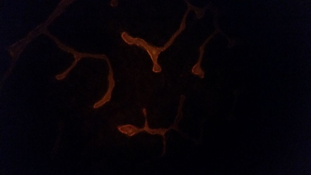
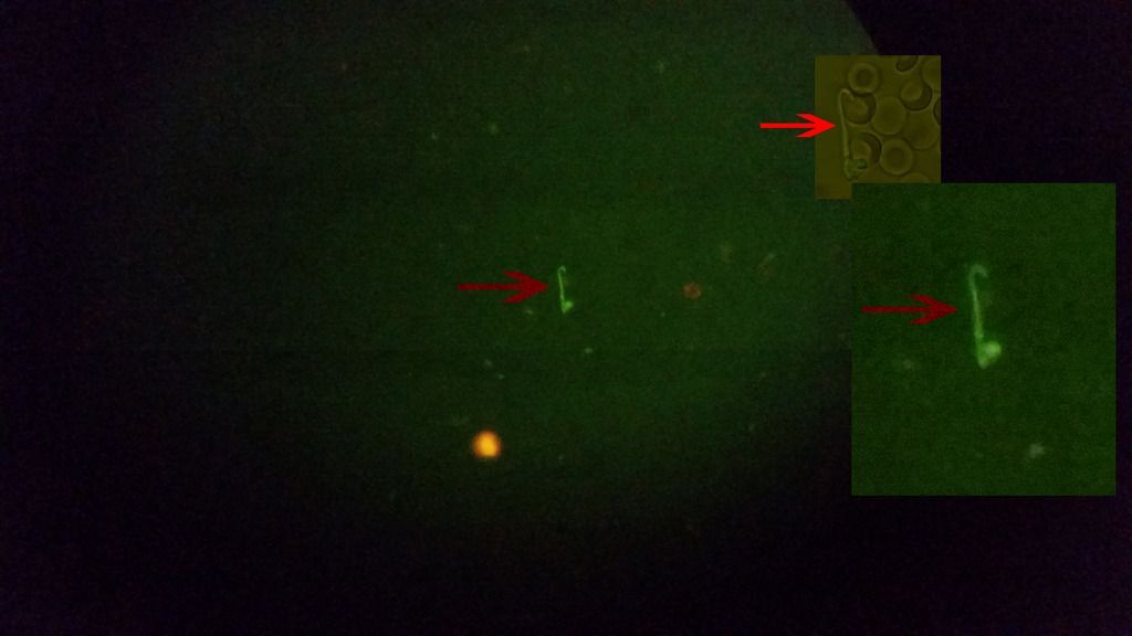
Expand the video to the fullest, look at the RED arrow & then the spirochete burrowing into the WBC.
123 images taken 15s apart (123 * 15s) = A little over 30 min of pictures, so it took it that long to burrow into the WBC.
I actually took a break from antibiotics 2 days a go. The blood was drawn today. I allowed the blood to sit for 6 hours on the slide before the video was made. The spirochetes seem to come out of hiding after a few hours. I can only imagine they hide in the red or white blood cells like Lymedin2010 has also shown.
Posted by Lymedin2010 (Member # 34322) on :
Beautiful video & it is very clear cut what that is.
It is so funny that HUNDREDS of years worth of doctor experience can't tell us much & such a simple technique can be so revealing.
These things simply won't die. I honestly believe that certain cells do not allow for (or allow much) contaminants (ABX) within the cell, doing so would disturb the integrity of what it was designed to do....carry O2 to the rest of the body.
I am doxy dependent & if I go off of Doxy, then I slide backwards rather quick. What I have noticed is the spirochetes are more motile, they move around a bit more rapidly, when I am off Doxy.
I have done this off & on a few times to be able to say it with confidence. So Doxy does appear to inhibit protein synthesis, making it more sluggish, but does not kill it. If they are sluggish within your cells it may be a lost cause, your WBC never get to it to kill it.
The biggest impact to the spirochetes that I have seen is when on Doxy AND Penicillin VK. They appear to be more sluggish & just all bent out of shape and look very deformed. There are more quantities of smaller spirochetes & they too look deformed.
Some reports indicate an antagonistic affect between the two, but experienced LLMD's know from REAL WORLD lymie tests that they work synergistically.
Posted by S13 (Member # 42830) on :
I agree, the damn things just wont die with conventional abx.
Btw, i used an old cheap Olympus binocular E-series microscope. Dark field up to 400x (and bright field up to 1000x immersion) which is good enough for clear recognition of spirochetes.
Posted by Lymedin2010 (Member # 34322) on :
WOOOOOOOHOOOOOOOOO!!!!!!!!!!!!!!
I have the recognition & attention of a well known Lyme researcher. This person sees the same thing in my videos & one of their students is trying to capture the same thing with the WBC's & spirochetes.
[ 09-08-2014, 10:57 AM: Message edited by: Lymedin2010 ]
Posted by Lymedin2010 (Member # 34322) on :
One of your posts you mention treating from outside body with rife or led or lasers type things. Have your tried anything like that and looked at blood as a response. Curious
Posted by Lymedin2010 (Member # 34322) on :
I actually exchanged some information with someone on here that was not too far away from me & who had a rife. I could not wait to start testing on that actually & then at one point we lost contact. I don't think this person ever responded to my last PM.
The above testing I mention would be too tedious to do on the blood initially & directly. It takes time to prep, hunt, record, edit all the material. It is so much easier to do things for your own eyes, but at the end we don't exchange info & others don't learn anything.
I have seen so many more things, but I have no proof of it because I did not have a camera at that time to record it.
What I would first do & this requires more money & resources, is to buy BSK medium & culture the bacteria from my blood. Which means I also need a temp controlled refrigerator.
Then we can just focus on the bacteria & see how they respond to external stimuli (LED wavelengths, ultrasound, rife, & UV light....and my other one heat or maybe a combo of these). Even if one or combo methods work, it does not mean it will cure you. It will AT LEAST clear your RBC's & WBC's & let your body exchange O2 more easily, giving you a fighting chance.
Then the next set of experiments would be strictly on infected human blood externally. We treat with what we observed to work with just the bacteria & see how the human cells fair & the bacteria within cells. This part takes longer & that is why we do it in 2 phases. You may have to do this gradually too, all depending on how much of a herxer you are.
If successful we then we can focus on the rest of the body & other methods.
My other thought & what I could do right away is HEAT. This idea comes to me from the fact that many of the sickest people who recovered from Lyme also incorporated sauna, so then why not just heat the blood up. I heard 108 for 20 min should do the trick
Basically I could work with blood directly with this method. I would need something safe & sterile to contain the blood. I will have to make a video describing all this.
Posted by springshowers (Member # 19863) on :
I have though heat would work well but how to safely get body up to temp is the hard part.
I have been using cold laser for same reason to affect the cells and give them accelerated oxygen and also cleanse and detox and so the body can do the job.
I have a rife as well and led applications and Infared done for heat. The cold laser is most recent and has been getting to what others haven't or in ways they couldn't. It is rough due to herxes but as I treat I can see the herxes slightly less per area I treat each time. I know hope to change the terrain but who knows if it will put my over the line where the body can kick back in. They are smart and strong and resistant.
Your doing cool stuff. I wanted to get some equipment at one point and I just had too much going on.
I admire your hard work in this and posting it and reporting too. Would be cool if we could starting a bank of patients blood to post and just grow it so big nobody could ignore it.
Why doesn't one of these docs studying the blood just ask for us to send in blood. To do something at that level. We all would do it. Money I assume. But.....
Posted by Lymedin2010 (Member # 34322) on :
I am trying to get LLMD to do it. I spoke to someone & at one point they had that aha great idea moment. What happens is that LLMD's are sooooo busy & inundated with patient load & work that it is not feasible for them to do it. Not to mention some of them go to conferences, rally's & interviews.
I also mentioned that they did not necessarily have to do it themselves. The microscopy methods become rather robotic once you get the initial know how & they can easily pay someone minimum wage. Then it is just a matter of the one behind the scope hunting & hitting a record button. The doctor can then more quickly review a recap from the recordings & not have to spend a whole lot of time physically sitting down & hunting on their own.
This is another video I was planning on making to inspire & spark a fire for them to do so. This is also why I have chosen to add references from other docs or scientists to my newer videos to support what I see & give credence.
There is no doubt that what I see microscopy wise is continuation of infection & it is clear cut. Call it whatever organisms you want at the end, it simply continues to grow in number & pleomorphic forms. I can't believe to this day I am still discovering new forms. It is easy to show you one organisms or a few, but much harder to show you what I can barely find at the beginning of chronic illness vs continued infection and what shows up in the blood.
This goes back to what I said at the beginning of this thread. I CANNOT STRESS ENOUGH THE IMPORTANCE OF DOING MICROSCOPY!!! It provides us insight into the guessing game & would provide immeasurable insights to those directly in contact with numerous Lyme patients.
Posted by Lymedin2010 (Member # 34322) on :
I have now captured borrelia coming out of a RBC & releasing a thin filament. There can also be seen other borrelia moving in the blood plasma.
Good work Lymedin!
Posted by Lymedin2010 (Member # 34322) on :
Thanks.
Patient #2 is my wife & lately she has been feeling very tired. Always tired & tired and some sleep disturbance on & off. When she rests she feels better, but she quickly tires out & is nowhere near what she was. Gradually & over the years (as gradual as my progression was) she is becoming more like me.
I have also noticed her joints started to make some light noise & crackle here & there, where she never had that before.
I did not see this many spirochetes in her two years ago & I only saw a very few & mostly they were small ones.
She is like..."I don't have Lyme!." She does not like the outdoors like I like the outdoors & rarely goes. Me on the other hand I jumped into anything & everything. Not anymore though. Then she goes on to say "if I do, then you GAVE IT TO ME!!!!"
You don't have to believe me on contact transmission, but listen to this NOBEL PRIZE winner on contact transmission. Just because you acquire the bacteria does not mean you develop Lyme right away though.
Lida H Mattman, PhD, has spent seven decades studying the different forms that bacteria can take. Her contributions to medical science can be summarized best by noting that in 1998 she was nominated for the highest honor attainable in her profession: The Nobel Prize in Medicine. Professor Mattman graduated with a M.S. in Virology from Univ. of Kansas and a Ph.D. in Immunology from Yale. She has taught Immunology, Microbiology, Bacteriology, Virology, Pathology, and for 35 years worked in these fields at various schools and institutions including Harvard Univ., Howard Hughes Institute, Oakland Univ. and Wayne State Univ. where she is Professor Emeritus. She is currently working for the Nelson Medical Research Institute studying the relationship between spirochetes involved in MS, Lyme disease, and ALS.
Posted by lymenotlite (Member # 33166) on :
Is it possible to examine your spit instead of blood. The idea of being punctured frequently doesn't appeal to me a lot.
I might be taking a microbiology class this summer. Perhaps they would allow me to culture my blood if that is available. Do you know what constant temp would be required?
Posted by Lymedin2010 (Member # 34322) on :
I tried viewing saliva once or twice, but deemed it too difficult to find anything, due to the mixture of debris, bubbles & the likes. It may also be that the saliva enzymes force the spirochete into cyst mode.
A high school or college microscope would be perfect for many, if they had access. You can obtain the slides, cover slip & oil on your own. Make your slide & go in with slide already prepared for viewing.
I leave my blood smears at room temp, which is typically 70-75 in my house.
Posted by lymenotlite (Member # 33166) on :
I went to a local community college today and spoke with the microbiology teacher about microscopes so I thought I'd do a rundown.
The school has a new science building with some new microscopes. There are bigger microscopes but the smaller ones are Olympus CX 31. Apparently this model has a blue filter but I don't know what that means. Anyhow, they were very pleased with the optics and with the microscope. Magnification is 1000x and a halogen light is used. They did not like the LED lights.
The teacher showed me how to do an oil inversion on a slide that I looked beforehand. She used a gram stain and put a drop of oil on it and the oil really made the bacteria stand out, kind of like 3D.
A couple of places where one might get a microscope for less money are Carolina Biologicals and Edmund Scientific. Another suggestion was that I might try to get ahold of a demo scope. Then they gave me the number of a company rep.
The teacher will try to get ahold of a retired professor who knows a lot about microscopes and see if he will speak with me. The guy has a lot of his own microscopes and he is in love with them and with microscopy. Sounds like just what I need.
Posted by Lymedin2010 (Member # 34322) on :
That is great!
That looks like a good microscope to be able to check for borrelia, just use 100x oil.
Stains are great for babs, bart & many other organisms. However, you do not get a high hit ratio most times. The more infected you are the better, but nothing compares to lack of stain.
So live blood on a slide, no stain, & oil. Some stains may prevent the bacteria from coming out of cyst mode or force them into cyst mode. What you want is to see them swimming in your blood, then you can clearly say AHA that is a spirochete and there is no confusion with fibrin or other string like substances.
I would prepare a slide before you go in, this way you give time for the spirochetes to come out. When you get there you can make another fresh slide, so that you can see both the few hour old smear & the new fresh smear.
[ 05-23-2014, 11:20 PM: Message edited by: Lymedin2010 ]
Posted by TNT (Member # 42349) on :
Has anyone seen Lymedin2010? Hope you are doing well! Hope things with you or your wife have not gotten so bad you aren't posting.
Good thoughts and concern heading your way!
Posted by Lymedin2010 (Member # 34322) on :
Hi TNT, thanks for the concern. I have not been on here for days. I have been out of my house after a mold outbreak that caused my lungs, throat, & nose to burn & I became toxic & ill.
I was looking up some new mold nose spray info today & by sheer coincidence found your post & IM here.
It had slowly developed & I was not aware why I was feeling worst & then I found the leak.
I planned on doing some great microscopy work & collecting new video proof, but instead my microscope is collecting dust & mold spores back at the house.
Posted by TNT (Member # 42349) on :
Sorry to hear that. What a nightmare! I hope you can get it completely taken care of. I am much more sensitive to mold or anything toxic like that. Car exhaust is terrible; there are days I can't go in my laundry room even.
Thanks for taking the time to respond to my PM in spite of your circumstances.
Keep posting new videos once you get things taken care of and are settled back in. I am subscribed to your channel!
I had live microscopy done in Feb. by an amish man who supposedly has a lot of experience. He didn't see any spirochetes, but I don't think he did it with oil like you do, and didn't look at it very long.
He said I had low levels of candida, and there were crystal like things that looked like salt I thought. I did wonder if those were from him not swabbing my finger before sticking me. I didn't like him sticking me like that, but that day I was quite out of it.
Keep up the good work!
Posted by Lymedin2010 (Member # 34322) on :
Yea, me too. After this mold exposure my chemical sensitivity went through the roof & everything bothers me now & makes me feel more toxic & sickly.
Thanks for the positive feedback.
On another note.....WOOOOOOOOHOOOOOOOOOO (look at the link below)!!!!!!!
This is exactly what I have been preaching all along & I cannot stress enough the importance of checking your blood via microscopy for Lyme patients. I have diagnosed my wife & simply by looking at her blood & observing the many few & subtle, inconspicuous and intermittent symptoms.
Doctors & LLMD's in particular can see your blood & know for a fact that the ABX that they are giving you are not doing squat if you still have many symptoms & are still questioning herx vs disease progression.
I also think that the future of Lyme will be in blood treatment. So one can use an IV line & let the blood be stored in an external container, heated & treated (with things such as ozone), dialysis type filtration & then transfused back into the blood stream in a continuous loop.
One can start out with small volumes of blood & depending on your responses, increase the volume to one pint, then one one quart at a time. This may not be a cure for all, but at least it will clear your Red Blood Cells & White Blood Cells & afford your body the opportunity to fight back. Otherwise the infection has hijacked your blood & if you cannot clear the blood, there is no way in hell you can clear it from your body.
About the paragraph on Lida Mattman: that must have been written a while ago. She died a number of years ago, sadly.
The process that you describe doing with the blood being treated outside the body and returned via a circulatory treatment has been done quite successfully.
But it is not done in the US, and requires 2 medical personel and very specific equipment. It's called Reciruculatory Hemotherapy.
Anyone trying to do it in the US would be prosecuted, sadly, unless it is completely below the radar. And I do mean completely!! Otherwise, you're looking at jail----arghh!
Posted by Andromeda13 (Member # 8314) on :
This is the best microscopy I've seen so far - and it's very important because it shows the borrelia are intracellular, inside the red blood cells. The string of pearls morphology is amazing to see, especially on the videos.
Nothing Steven Spielberg could conceive of could outdo these films and photos for HORROR - all the more horrible because it's real.
Elena ps It seems that shortly after the British microscopist Peter Kemp published this web-page, which also contains a link to the excellent work of Norewegian researcher Prof Laane, further sabotage was done to the page in the medical journal which has Prof Laane's article.
The Professor had described in great detail a method for seeing Borrelia in live blood and related it to chronic Lyme borreliosis.
I think we need some kind of response to this defacing of his site and will write about that in a separate thread.
quote:Originally posted by Andromeda13: This is the best microscopy I've seen so far - and it's very important because it shows the borrelia are intracellular, inside the red blood cells. The string of pearls morphology is amazing to see, especially on the videos.
Thank you to those who mentioned my page on intracellular spirochetes. There was a lot of luck in getting those results, but in general, observing spirochetes is not difficult with a bit of practice - as Lymedin and others have helped to show. I am writing a page on how to do blood microscopy with pictures and (hopefully) simple instructions. Will let you know when it's ready. Best Wishes, Peter
Posted by Lymedin2010 (Member # 34322) on :
I will have to look into Reciruculatory Hemotherapy & yes Lida Mattman has been dead for a while, but she knew a great deal & at such an early stage in the Lyme game & she was able to cure herself of Lyme as well.
Interesting that one can get prosecuted for this, yet no one has been sentenced for scientific & medical acts of crime against humanity and the misinformation on Lyme and stealth diseases in general.
This is a video clip from Under Our Skin 2: Emergence. A biologist from Oslo University (Oslo, Norway), Morten Laane, discusses his findings & observations.
Those ARE great videos, BUT we will have trouble getting the non-believers to accept:
1) That spirochetes can appear less spiral & more docile in movement. The solution that they are in affects their ability to spiral or drill.
2) Accept the "String of Pearl" very existence. I have seen many morphologies myself, but have only seen the string of pearls about 3 times or so in the blood plasma (outside of the RBC's) & perhaps because this phase exists more readily within the RBC. My method of preparation & observation does not lend itself to ample strings in the plasma.
These great videos show mainly the string of pearl morphology & I wish they would show more of the standard spirochetes as well. It would be too easy for the non-believers to discredit these very important findings.
3) Accept that they are within the RBC & can be found so readily, especially in chronic infection. They would particularly find it shocking to discover the spirochetes in the RBC's after extensive ABX therapy.
Peter, I look forward to your microscopy instructions. I find the bulbous tips on the spirochetes rather interesting. It is almost as if they are similar to the pearls & are made of phospholipids that can more readily penetrate a cell wall. I have not witnessed the spirochetes burrowing out of RBC's any other way other than bulbous tip first & this may hold a key to controlling the spirochetes. Inhibit the bulbous tip entry/exit into cells & we can hope to control it more efficiently.
The resulting breakup of the string of pearls is the blebs. At times when the blood is left long enough, I see thousands of these blebs, which must have burrowed out of the RBC's. They must have similar properties & composition to the bulbous tips. Perhaps the bulbous tips are indeed blebs or modified blebs that remain?
Posted by PeterKemp (Member # 35585) on :
Thanks Lymedin. I haven't time at present to address all your interesting remarks, but there is one that I think is really important. In thousands of observations I've seen only a few 'corkscrew' spirochetes in blood. There are actually a few on this page from an experiment I did earlier this year: http://counsellingme.com/microscopy/ExperimentLgfont.html
The known morphology of spirochetes goes back over 100 years. I would be surprised if anyone who knows spirochetes denied that the videos show them, but it might actually help if they did; especially if they got their own microscope out of the attic!
Posted by Lymedin2010 (Member # 34322) on :
Thanks for your feedback. Actually I have been trying to contact the youtube user with those spirochete videos. If they are yours, then it appears I have been unknowingly trying to contact you.
Those are collectively the BEST photos & videos I have seen to date!!!
Prof. Laane's blood smear concoction lends credence that spirochetes behave more or less spirally in motion depending on the solution they are in. You & I both see the spirochetes readily in blood, but Prof Lanne's solution allows him to see them in their full spiral mobility.
He too readily finds spirochetes in Lyme patients blood, but when the general scientific community looks at this evidence, they are more inclined to believe when they see the spirals. The typical spiral behavior in blood is a notion we have to dispel & allow the floodgates that accept that chronic Lyme patients blood is inundated with less active (less spirally) spirochetes.
I would imagine when the solution becomes less viscous & more watered down, such as with Prof Laane's solution or BSK media, then the spirochetes assume their "typical" & widely accepted, but archaic movement.
Lanne has been met with resistance & understandably so, from a community designed to mask the truth.
Have you encountered resistance as well? I would imagine that the medical & scientific champions of Lyme see your work as God sent, while the opposition continues to follow suite at any cost?
Posted by PeterKemp (Member # 35585) on :
Thank you! I do get some supportive feedback from Lyme doctors/researchers, though I have yet to hear from any who think they are artefacts.
I think there are a number of reasons why the establishment Lyme people are against microscopy, and none of them are good.
Microscopy has the potential to show that the 2 tier testing system is completely useless. That would hit the manufacturers of test kits; who currently make claims for sensitivity and specificity based on how they compare with other kits that work in an identical way - which is completely crazy.
It would also harm the reputations of those who have claimed that the 2 tier testing is reliable. I suspect that these include people who consider their egos are more important than the lives of patients.
Microscopy might be able to show whether the new vaccine actually works. Vaccinated people might have antibodies, but they might also have spirochetes as well, due to borrelia's ability to evade the immune system.
These and other possibilities have patients asking why microscopy is not used more. Even if it were not used for testing, it could be used for testing other tests; monitoring the effects of treatment and learning about the behaviour of borrelia inside the body.
Conversely, there are no good explanations for not using microscopy. They may frown upon alternative therapists who use microscopy, and in that I am somewhat in agreement; but it is no reason to discard such a valuable resource entirely when the world is dealing with an uncontrolled epidemic.
Best Wishes, Peter
Posted by S13 (Member # 42830) on :
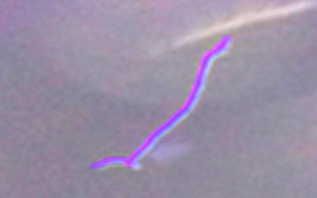
Ive seen the same shapes in my own blood, the thing that resembles the string of pearls:
There is actually a smaller motile borrelia visible (the lower arrow). The upper arrow shows the string of pearls. The left and right arrow show larger "pearls". Some of these strings i see are not made up of pearls, but look like longer worms actually (a single object). One thing that strikes me is that these objects grow quite fast. Basically within 6 hours it goes from seeing nothing (the time the blood is drawn), to these large strings. Im not sure if borrelia is capable of growing this fast? Could there perhaps be other parasites or fungal forms involved with these string like objects?
Posted by Lymedin2010 (Member # 34322) on :
I am glad that you share with us the importance of microscopy in chronic infection. Imagine where we would be if LLMD's monitored their patients blood, especially after administering ABX. They can know with some relative degree of certainty whether the ABX are having a positive or negative effect.
I have also noticed in some of the videos that the spirochetes are fairly long. I have seen some jaw dropping long spirochetes & I am amazed that they can grow to that length.
Here is one such spirochete that I captured, but in no way represents some of the longer ones that I have seen. At times I see them 40-60+ microns long. Do you notice that at times some of these spirochetes are longer than what you thought possible? At one point the spiro in the video loops on itself & then two ends spiral into each other. In the video you can see the spirochete line double, thicken & darken where they meet & spiral into each other.
Also, how readily do you find them in WBC's? Early on in my investigation, I would see WBC's with rather large specks. I was always suspicious of their origins & wondered if they could be cycsts.
One fine day I happened to check my blood right after a hot bath soaking. I managed to get my body temp to 102+ that day & after some recouping from the after effects, I made my blood smear & noticed that many of the WBC's had a large quantity of specks. More so than what I have ever seen before this hot bath.
I had left the smear out for a few hours & then checked again & I could not believe it. For the first time ever I so many of the WBC's with spirochetes burrowing out & again some of these were VERY long.
I tried to capture one of them with a hand held camera, the only way I had at that time, but they are difficult to discern in this video.
This might give evidence to the belief that many spiros & cysts lodge themselves on skin surfaces & perhaps heat forces them back into the blood stream, where they can be more readily picked up by WBC's.
Posted by Lymedin2010 (Member # 34322) on :
S13, I have seen your work before & I think they are spirochetes as well. You have a beautiful image there showing the spirochetes emerging from the RBC's. Looks like some blebbing from the tips & blebbing from the string of pearls. Even that beautiful more spiral like smaller one.
Whenever possible, I would always do video first. It is so much easier for the naysayers to dismiss these as artifacts without them desiring to investigate this further.
As far as I am concerned, if one sees the bulbous tips, the undulating motion, and sheer abundance of such objects, then that is a dead giveaway. I think it is hard to logically dismiss that as random artifacts & should force the general scientific/medical community to AT LEAST be interested in further investigation.
Also, do you see that many of your RBC's are studded & bumpy? In the very beginning I thought they may have been cysts trying to burrow out, but after finding no evidence of them coming right out of the cells succinctly, I then concluded that it must by the hypertonic nature of the blood being out of the body.
The blood is at 96-98.6 & some h2o evaporation occurs. Over time the blood plasma becomes hypertonic & water travels from a higher concentration to a lower one. So in this respect water rushes out of the internal RBC & finds its way to the blood plasma. As a result the RBC's shrink a bit & appear studded.
Posted by S13 (Member # 42830) on :
I also made a timelapse from what i think is a spirochete burrowing out of a RBC:
The timelapse is done manually, with intervals of 30mins to an hour. Its interesting to see that the RBC deforms where the potential (bleb-form?) spirochetes burrow from the RBC, and in the final image the RBC takes its normal round shape again.
Also some morphology in the next picture.
Red arrow: shows a long solid worm-like entity initially, which then breaks up in segments (string of pearls?) over time. Green arrow: shows a segmented string of pearls which converts to a more solid spirochete over time. The endpoint remains a bleb or cystform?
There is about an hour between each image.
These spirochetes are weird and "intelligent". Looking at it alive under the microscope is like seeing a very bad horror movie. Especially when you consider this is taking place inside your body!
Posted by S13 (Member # 42830) on :
quote:Originally posted by Lymedin2010: Also, do you see that many of your RBC's are studded & bumpy? In the very beginning I thought they may have been cysts trying to burrow out, but after finding no evidence of them coming right out of the cells succinctly, I then concluded that it must by the hypertonic nature of the blood being out of the body.
The blood is at 96-98.6 & some h2o evaporation occurs. Over time the blood plasma becomes hypertonic & water travels from a higher concentration to a lower one. So in this respect water rushes out of the internal RBC & finds its way to the blood plasma. As a result the RBC's shrink a bit & appear studded.
Yeah, a lot of RBCs become dehydrated over time and will show bumps and crenation, but that is different from the cyst burrowing out like in the timelapse above. If all RBC's that show these bumps would have cysts inside them, then my blood is extremely infected. Virtually all my rbc's show the bumps over time.
Posted by TNT (Member # 42349) on :
These are just great! I'm fascinated, but totally freaked out. Definitely the worst horror shows I've seen in some of those youtube videos.
Have you ever seen any biofilm?
Posted by Lymedin2010 (Member # 34322) on :
Crenated RBC's is what I see in normal folks blood & without any spirochetes, as well as those with Lyme. Here is a "normal" cell after it has been outside the body for some time.
Your beads of string time lapse is precious. On the first picture from the set, I wouldn't have spent much time without seeing clearly defined bulbous tips. Yet the specimen identifies itself as a spirochete via blebbing.
The stringy fibers are just that, fibrin.
Note that the bulbous tips can bleb themselves & that is why I say they must be underdeveloped or modified blebs. Both the blebs & spirochetes (via bulbous tips) can readily penetrate the RBC's & perhaps many other tissues (as they have been found in many varieties of tissues).
He may have some biofilm at the top right clusters of cells. I have seen patches of goo & what appears to be biofilm in my blood. At times they accompany spirochetes & other times they appear to be absent.
Posted by S13 (Member # 42830) on :
quote:Originally posted by TNT: Have you ever seen any biofilm?
Not that im aware of. The biofilms would be difficult to diagnose from a simple live bloodsample. I think it requires a blood culture with fluorescent markers to identify clear biofilm colonies of spirochetes and blebs / granules. Like what Macdonald has done before.
quote:Originally posted by Lymedin2010: Your beads of string time lapse is precious. On the first picture from the set, I wouldn't have spent much time without seeing clearly defined bulbous tips. Yet the specimen identifies itself as a spirochete via blebbing.
Thank you! Yes, it was not a specific RBC i was tracking with the timelapse, but rather a large area at 400x mag. Within this area there was a lot of growth of these blebs and string of pearls. Literally in the hundreds. Strangely it is only this one area on the slide where it happens. Other areas stay clean without any growth. Somehow the infected RBC's seem to clump up together on the slide? Ive seen these infected areas on more slides before, but it puzzles me what the mechanism is. Perhaps while making the slide, the diseased RBC's stick to a certain area and the healty RBC's flow away to other areas?
Posted by Lymedin2010 (Member # 34322) on :
In reference to the biofilm, that is exactly why I use the word "may" & "appears." At the end anything that we simply observe without testing can be discredited & obvious testing is required. We as simple microscopists continue to make observations that hopefully allow us to gather evidence that increases the likelihood of what we believe is there to actually be there.
What I have noticed is when I start a fresh smear, that there are relatively few biofilmed looking areas. The ones that don't have spirochetes and/or blebs I tend to dismiss. The ones that have them, I am more likely to accept & be further suspicious of.
If that same fresh smear is left over time & I make observations a few hours later, then I notice that there is a significant # of WBC's that have lysed & release their lysosomes and internal contents.
Over more time the WBC's lysosomes degrade adjacent RBC's, initially causing them to be sticky at their contact area to the resulting debris & at times the end result causes ghosting of the RBC.
Ghosted RBC's loose their internal content & it can appear rather dramatic at times. At times the end result of ghosted cells looks like candida all mixed in a field that can be mistaken as biofilm.
I found this more evident after doing some time lapse on WBC's & making comparisons from initial cell death to hours later. For this reason what I thought might be candida from my initial findings a few years ago, I now have a reference point of what might be their true origin.
Posted by Lymedin2010 (Member # 34322) on :
On another note, I am surprised that we can't simply add a sample of our blood to BSK media & watch the spirochetes flourish? Why do we need methods such as xenodiagnoses, of a sterile tick biting a human to pickup the infection & then testing indirectly. There must be some mechanisms at work, or more likely something missing in the BSK solution?
What if we take a combination of BSK media & rabbits or sheeps blood filtered through the smallest filter possible? Or the heck with it, just take the very infected persons blood & filter it & mix that with BSK media.
Perhaps then we can culture them from our very own blood & use that as a basis for proof.
Posted by PeterKemp (Member # 35585) on :
Blood added to BSK will allow spirochetes to grow, but so will other things that are much cheaper. BSK is really useful when you want to do long-term culture, perhaps aiming to grow corkscrew spirochetes which I found can take months.
For short-term culture on a microscope slide, normal saline can be bought ready made or prepared at home with distilled water and salt. Sodium Citrate (as used by Mysterud and Laane) is cheap to buy on Ebay and this can also do the job.
Using a duluent separates the cells which makes for easier observation, but as others have remarked, it can also affect the shape of the cells.
Just putting blood onto a microscope slide subjects the cells to numerous stresses which can break them or make them misshapen. Other than doing thin-film smears which dry instantly (videos on how to do this on Youtube); it is questionable how much can reliably be told from blood cells in a wetdrop - though they are still fascinating to observe.
Posted by S13 (Member # 42830) on :
What i would be interested in, is where to get the BSK medium? Also fluorescent markers like the ones macdonald uses would be very interesting to experiment with!
Since string of pearls is an interesting topic, and i seem to find a lot of in my blood, ive decided to share some videos. The first video is actually the same string of pearls as the one in the first picture from me, a couple of posts back:
Also a rather large colony which ive found, really horrific to see this in your blood. Again, all videos and pictures are taken after several hours of culturing, so you wont see this in freshly drawn blood!:
What you can see is; crenated RBC's (from the dehydration), many string of pearls, some medusa heads, probably lots of cystic forms (the smaller roundish forms? approx 1/2 the size of the RBC?), and perhaps it is clumped together by biofilm, but that is just speculative. Fluorescent microscopy would be very helpfull in this case!
So im starting to think now my blood is literally filled with borrelia cystic forms. Blarghhh... Good thing is that there are hardly any motile spirochete forms.
Posted by PeterKemp (Member # 35585) on :
Hi S13. I found your photos and videos very interesting. The time-lapse morphology is amazing.
Sigma-Aldrich stock BSK but will not sell to the public as it is shipped frozen with dry-ice. I found a laboratory supplier that ordered it, thawed it out and then sent it on to me.
To prevent drying of the slide, the edges of the coverslip can be sealed with immersion oil (a pipette is handy for this) or I sometimes use Bostik glue which can be removed. These should keep a slide usable for at least a week. To avoid getting immersion oil underneath the coverslip a sample is needed that takes up the whole area. Sealing the coverslip might prevent it from settling-down and giving a nice shallow sample that is easy to focus. Blotting the slide with paper-towel before sealing the edges can help.
Posted by S13 (Member # 42830) on :
Thank you PeterKemp. Im looking in to more advanced camera system for my microscope, so that i can do automated long term timelapses. Now i have to make each photo by myself each hour or so, which is very time consuming, inaccurate and i get only a very limited amount of photos.
Thanks for the tip on immersion oil. I think it could prove helpful. But is it possible the immersion oil is pulled into the bloodsample?
Posted by PeterKemp (Member # 35585) on :
Timelapse can be done with a computer program if a camera is connected to the computer via a TV or Video Capture card (PCI card). The program is free and called VirtualDub.
In capture mode it displays the AV output from the camera and can record frames at the required rate. It can also do editing of the recorded videos or videos uploaded from the camera; though it does produce very large AVI files and needs plenty of hard-drive space.
I have made timelapse videos by manually recording a few seconds of video each 30 minutes or so. It is a nuisance to do, but the results have the benefit of showing any motion present which can be quite informative.
Lymedin and myself have both found problems with the scope losing focus when doing timelapse, so it might be that regular checking is necessary. I find this is due to the light being left on. The lamp I use heats up the scope which expands, losing the focus. It probably does not do the microbes much good either!
I have not had contamination problems using immersion oil to seal a slide. If there is enough volume in a sample to cover the whole area beneath the coverslip, the edges will dry out quite quickly and form a barrier. If you try it, make sure to blot the slide before hand to make a shallow sample for observing as drying will be slowed down.
Posted by Lymedin2010 (Member # 34322) on :
I have the Amscope MU900 camera & at first it was horrific. When they finally updated the software it was much better at auto detect, but the lag & video refresh was intolerable for this type of work, where we are constantly hunting & changing views on the slide. It would simply take too long for the video to refresh on the monitor.
You would love the time lapse on the Amscope software though, it is very convenient.
Another researcher has told me that clear nail polish can be used to better affix the slip cover, but I have not tried that as of yet.
Here is a link to the BSK from another researcher, but I have never tried to order it.
ATCC Medium: 1914 Revised BSK Medium ATCC currently uses Sigma # B - 8291 BSK - H Complete medium. This medium is purchased in 500ml volumes and stored frozen at 80ºC. The medium is thawed and dispensed as required. If prepared from components: HEPES (Sigma H - 3375)...................................5.64 g Neopeptone..................................................4.7 g Sodium citrate................................................0.7g Glucose........................................................5.64 g NaHCO3......................................................2.0 g
TCYeastolate (BD 255772)..............................2.0 g Sodium pyruvate............................................0.75 g Nacetylglucosamine.......................................0.37 g Bovine Serum Albumin, Fraction V.....................47.0 g CMRL 1066, 10X (w/o Glutamine or NaHCO3)......100.0 ml Rabbit Serum (heat inactivated).........................60.0 ml DI Water........................................................840 ml Dissolve ingredients up to and including bovine serum albumin one at a time in distilled water. Adjust to pH 7.5 with NaOH and filter sterilize. Aseptically add CMRL 1066 and rabbit serum. Mix well and aseptically dispenses into appropriate vessel. Final pH of complete medium should be 7.5-7.6.
Posted by PeterKemp (Member # 35585) on :
I think it worth remembering that although the BSK formula looks very complicated, borrelia can grow successfully just on a drop of blood and whatever else it can find in a tick's gut.
Ticks regurgitate some of the liquid part of their meal, which I guess probably gets rid of some of any antibodies present and concentrates the cells. Borrelia can convert chitin (which the tick's exoskeleton is made of) into glucosamine; which suggests that it likes/needs it. Prof Brorson found that it also likes extra zinc.
Posted by Lymedin2010 (Member # 34322) on :
Do they use a standard media, such as BSK, as a reference point, which allows for all things being equal other than the variables in question? In theory a great concept, albeit it is a pitfall with the complications of Lyme & in particular when making observations of virile drilling spirochetes vs more docile ones and the efficacy of growing spiros directly from human blood. The long growth rate of BB only adds to the complication.
Personally, I was going to obtain rabbit blood from a local butchery & make do with that using micro filtration. If that failed then I would have sought out BSK.
It is interesting about the chitin conversion & perhaps that is why LLMD's suggest glucosamine for joint support. What supplements do you think BB is more likely to deplete in the body & would you supplement at the risk of feeding & propagating infection?
Also, I never quite understood the notion that O2 would kill the spirochetes, especially when they are lodged into the very cells that act as O2 transports???? Isn't this contradictory, unless they enter cyst mode or bleb lengthing (forming blebs along the length of the spiro) upon making contact with in the blood?
Posted by Lymedin2010 (Member # 34322) on :
S13, given that you have many spirochetes in your blood, what are your symptoms like?
I have checked the blood of various people who have been bitten & some only have a very few symptoms. For instance one person has only headaches & stomach pain that come & go. They have pains from time to time, but they come & go and it is difficult for them to consider ongoing infection.
Yet when I check their blood, I am left to wonder why they don't have consistent, more, & more pronounced symptoms. It is during these times that I often wonder how important detox pathways are to the manifestation of the disease or perhaps some other factor such as inflammation & immune response?
Posted by S13 (Member # 42830) on :
Thats the thing, i dont have many spirochetes in my blood. With fresh blood i can hardly see any spirochetes at all! I think they are all cysts and blebs and they emerge after a short while of culturing.
My symptoms are very diverse. Mainly neurological, derealization, brain fog, cognitive difficulties, emotional/rage attacks, severe fatigue, nausea, twitching muscles, weight loss, jaundice, palpitations, SOB, night sweats and a few others. When im doing antibiotics or mhbot i dont have any pain at all. Besides borrelia, bartonella and babesia also play a big role in my disease. The borrelia, im thinking, is just keeping the other infections ongoing without doing too much damage by itself. But knowing what i know now, i will be focusing more on borrelia as well. First the cysts (never had a cyst buster before) and then the cell wall.
With the people you are helping, do you find loads of motile spirochetes in freshly drawn blood?
I think borrelia mostly causes symptoms in already weakened parts of your body. Parts that me be inflamed from previously existing infections or inflammation, or coinfections. If the detox pathways function properly and you eat well enough i think you can compensate a lot of the damage that borrelia does by itself. Like collagen replacement.
Posted by Lymedin2010 (Member # 34322) on :
MOST do not have motile spirochetes in FRESH blood, but they come out eventually. Even with my own blood most of the time I find a few motile spirochetes, but every now & then I find a suprising amount from a fresh culture. I have realized that I have not paid much attention to when the blood has been drawn (before/after eating, caffeine consumption, abx usage...etc) & my next goal is to be more mindful, since it could buy clues.
This one person was on 2x ABX & with only 2 major symptoms, headache & stomach pains. When I drew their blood, I was shocked to find so many free swimming spirochetes from a fresh smear. I only drew this persons blood once & it was early on during my microscopy ventures & it just left me bewildered as to why not more symptoms & why so many free swimming? This person does indulge in extensive amounts of carbs & sugars and could be one possible explanation.
Maybe one explantion is that their immune sys is not compromised YET. The carbs & sugars bring out the chetes to the plasma & the WBC's are able to pick them up & rid of them because the immune system is still functioning normally.
I have all of yours except derealization, which I did experience for one day & it was such a crazy & inhumane experience, so I feel for you.
I also don't have emotional/rage attacks, BUT I do have exacerbation of anger. So for instance if I get angry or stressed, then the chemicals that calls for anger or stress get released & since the body does not detox or process them quickly enough, they linger at greater concentrations for longer periods of time.
The end result is that anger in me breeds anger 10x to the point where I can rage and I find that I can get explosive. Before chronic Lyme I was a very subdued individual & people knew me as a laid back guy & knew that nothing bothered me.
This I can also associate with chemical & food sensitivity. So if I eat or breath something & it enters your blood stream, it takes my body much longer to detox & remove the foreign particulates, so I go on feeling sick & toxic for hours after exposure or food intolerance.
Also with sleep. I used to not be able to sleep & it was difficult for me to sleep for years. Now if I lay down & sorta force myself, I can feel the melatonin kick in & I then feel as if I just took a sleeping pill & it knocks me out to the point where I have to sleep.
All these things that I mention above are more pronounced after the mold exposure that I just experienced. Just a few months ago, before the mold exposure, sleep was hard to get to, but falling asleep was seamless & now it feels like I have been drugged to sleep. Again because the concentrations build up too quick & are not handled properly by their respective pathways.
Posted by Lymedin2010 (Member # 34322) on :
Do you have any of the following that I have?:
-food allergies -chemical sensitivities & mold sensitivity -internal vibrations -sudden onset of stroke like symptoms -sudden onset of muscle tightness, for me it is the neck, which then leads to brainfog, but it then releases. -jaw tighness -ear ringing -migrating joint pain (it took me years to develop joint pain & only after 1-2 yrs of ABX usage did it become pronounced).
Do you have any one of these too?:
-You engage in physical activity & if you perform it too quick, then you get shortness of breath & at times accompanied heart palps? To me that is a sign that the blood is loaded & parts of the body get devoid of oxygen & call for the heart to compensate.
-At times do you talk to fast & all of a sudden you can get shortness of breath?
-If you don't sleep enough then you get aggravated symptoms & in particular vibrations through the roof?
Posted by PeterKemp (Member # 35585) on :
Although borrelia prefers oxygen at low levels, inside the red blood cells the oxygen is bound to the heme so it probably does not have much effect on the bacteria. Babesia (which also infects erythrocytes) has very similar oxygen preferences to borrelia, i.e. very low levels.
I have found the same phenomenon of spiros sometimes being present, but mostly needing some culture time before they appear on a slide. One of my pet theories is that the drop in temperature, pressure, movement etc on a slide 'tells' the spiros that they have been ingested by a tick - so it is safe for them to grow.
Posted by Lymedin2010 (Member # 34322) on :
So it does not easily "accept" the O2 from the hemoglobin...hmmmm.
So HOB therapy binds more O2 to heme or makes more free floating o2? My oximeter used to read 99% before the load of bacteria grew & now 98% mostly & at times drops to 97%, but that is not such a big change? I know others who run around in their 60%'s due to other medical issues.
Posted by Lymedin2010 (Member # 34322) on :
Peter, we have seen the new research on sexual transmission. Knowing all that you know & all that you have seen. Knowing that borrelia can be found, all depending on what stage of the infection one is in, in the RBC's readily & that blood flows through the entire body. What do you feel is taking place?
Is sexual or contact transmission likely? I have seen the studies that describe contact transmission in animals & our microscopy observations lend itself to this likelihood of transmission?
Is Lyme perhaps by itself not as potent, but has a greater impact with co-infections? A person who has been bit, can go on for months, years, & even a lifetime without many symptoms, even if multiply co-infected. So is it not possible if contact transmission occurs that the partner may not necessarily acquire all the co-infections & therefore may take longer or never be symptomatic?
Posted by PeterKemp (Member # 35585) on :
Hi Lymedin. I have no medical or science training so any ideas I have should be taken with a big pinch of salt.
I'd say that the areas you mention need a lot of urgent research. Borrelia is in the blood. That is something that the CDC/IDSA, PHE and other authorities seem to be avoiding. So either they know that it is in the blood but have reasons for suppressing this information; or they simply haven't bothered trying to locate borrelia in the blood - which seems incredible.
Incredible.
I think that every point you make is credible or even likely - so where is the large-scale research needed to protect the population; to understand P2P transmission, other vectors, infection that starts with virus-sized propagules rather than motile spirochetes?
There are no good answers to these questions. The pittance spent on researching LB suggests to me that the CDC and others have cobbled together their theories and are sticking to them. They either already know the answers, or don't want to know them. We are left trying to figure them out for ourselves and for the sake of others including our loved ones.
Posted by Lymedin2010 (Member # 34322) on :
Yea, it truly makes one wonder how the CDC & company do not bother to take a closer look at the blood of those chronically ill, especially in a BLOOD BORNE disease. It is just pure abhorrent idiocy on their part & I don't think they are that stupid and for that reason I think they MUST BE withholding what they really know?
I think the reality might be that they have identified this organisms as being:
-widely spread.
-involving and influential to billions of medical dollars.
-very hard/impossible to kill. Those who are in remission might be pushing time back to a phase where they were bit but asymptomatic & this can go on for months, years, & even lifetimes without any major complications. I have even heard of a story just this year of an older (70's) year lady coming into the hospital with only slight symptoms from a lifetime of Syphilis, but these types of stories are know near & far.
-Can create resistant specimens from ABX treatment readily.
One only need look & there is consistency across the border for what we see in the blood. The blood of those who are not treated look a lot worst & it makes one wonder...how is it that they are alive?
So coming from a psychotherapy background, what prompted you to take that first look? For me it was 2 months of entering chronic stage, realizing what my daughter was developing, the symptoms of many family members, & the people around me. It seemed only logical that I look.
Posted by glm1111 (Member # 16556) on :
Excellent thread and info Lymedin! Just thought I would mention what Bryon Rosner has to say about parasites protecting the spirochetes and other bacteria. I will bring up the thread that gigimac posted for those interested in reading what he has to say about the antiparasitics being effective.
Gael
Posted by Lymedin2010 (Member # 34322) on :
Thanks Gael!
I have heard of the theories of parasites protecting borrelia. I can find borrelia in the RBC's despite aggressive & varied ABX treatment & it looks like borrelia does not need any parasitic help, although it is quite possible given the aggressive & tactical nature of the spirochete.
Very cunning & surprisingly adaptable in a host of creatures. So I wouldn't be surprised if they are found to be protected in masses within the parasites.
Peter, have you done any more MMS experiments? So in essence, MMS had an affect on the spirochetes & many could be found with their motility impacted? Where the blebs affected at all?
[ 09-17-2014, 01:22 AM: Message edited by: Lymedin2010 ]
Posted by PeterKemp (Member # 35585) on :
Hi Lymedin and All. The MMS experiment used the chemical (chlorine) as a highly toxic antimicrobial. The aim was just to demonstrate that the spirochetes were living organisms and not 'artefacts' which Lyme denialists claim. To test its effect on blebs would be much more complicated and require reculturing the sample; though chlorine is so powerful that it is probably reasonable to speculate that with the levels used they would no longer be viable but neither would human cells.
Propagules are very tough. I attempted some DNA extraction (for PCR) on a sample full of these tiny round-bodies. The bacterial lysis kit I used only worked when I accidentally used 20x the specified amount of enzyme to dissolve the cell wall.
I also tried a microwave protocol which called for 6 minutes at 600 watts under pressure - to break endospores (very tough organisms). I could only simulate pressure by using a tightly sealed tube but gave it 10 minutes at 900 watts. Whilst many RBs shattered, around 25% of the propagules looked perfectly intact under the microscope (though they might actually have been dead).
Some nematodes have extraordinary immune suppression abilities. If memory serves, there were some doctors considering giving people with arthritis a single strongyloides worm which could reduce the inflammation in the joints of a person with arthritis. Just one worm could suppress part of the immune system!
Posted by S13 (Member # 42830) on :
So, ive got my new automated timelapse microscopy setup running. Like you said Peter, the image will go out of focus when the lamp stays on for a long time. So i made a system that turns the lamp off in between the frames. That also should preserve the sample.
I was fiddling around a bit, and decided to try your method of sealing the blood sample on the slide with some immersion oil. That works perfectly! I do notice the borrelia bacteria is not forced out of the RBC as fast as before. I think the dehydration in my previous samples triggers the spirochete to mobilize and seek another living area. So for a fast culture i will use the dehydration technique (so no immersion oil seal on the slide) and for long term culture i will use the sealed technique.
I found a bizarre phenomenon today when looking at the long term culture: http://youtu.be/rlAYKAR9X2Q I have never seen this before, and it could very well be contamination from my finger or something. But it looks like fungal growth of the blood. Perhaps a lost candida cell? Each frame of the movie = 5minutes. Total of 7,5hours of growth is shown.
Posted by S13 (Member # 42830) on :
Im wondering if this picture i have taken a while a go actually shows a budding yeast cell? If so, perhaps that eventually causes the fungal growth from my previous video.
Posted by PeterKemp (Member # 35585) on :
Hi S13, I use 50mm coverslips and often just seal the long sides which keeps the sample viable for up to a week, but still allows it to 'breathe'. Mysterud and Laane attribute their successful culture on a slide to the fact that certain regions of the sample develop the correct oxygen levels. This is what Dr Lida Mattman said years ago, that at a certain depth in a culture the oxygen level would be ideal for borrelia growth.
The video is very impressive and looks like it could be candida albicans which might have come from the air or could have been present in the sample. I used to find a lot of fungal growths on blood slides, but strangely, after taking long course of abx I rarely see them now.
Your timelapse set-up gives wonderful results. It would be fantastic to see a spirochete actually emerge from a blood cell and morph into a string of pearls. That would really stand the accepted 'wisdom' about borrelia on its head. I have tried to do this, but no luck yet.
Best Wishes, Peter
Posted by S13 (Member # 42830) on :
I have smaller coverslides, 18x18mm, so i will try leaving open 1 side for breathing.
I think the fungal forms are probably a result of a suppressed immune system. So by dealing with the bacterial infections, the immune system should become more active, and fungal forms will be eliminated more quickly from the blood. Im still not sure if my fungal hypha is the result of contamination, or if it was present in the sample.
I will do some more experiments with the time lapse setup, so i hope to provide more results soon.
Posted by Lymedin2010 (Member # 34322) on :
Here are 2 good candida vids & yours may as well be just that.
_______________________________________ The video reminds me of this Streptomyces coelicolor video. This is a gram-positive bacteria that is typically found in the soil.
Do you guys see smaller versions of the spirochete, as represented by a dumbbell. It basically looks like a rod with bulbous tips on either end, sorta like this but connected together •-•
I see all sorts of these at various sizes & when we consider them all collectively, they can represent the various growth stages.
Also, do you guys see blebs with tails, basically a footed type of bleb? I think these are the blebs forming into the smallest spirochete, a dumbbell, & I am trying to get time lapse proof of this. There are various thickness & thinness of dumbbells too.
It is so difficult to do, since they constantly move in the Z field via Brownian motion and escape consistent focus. With time they also escape the entire field of view & force me to re-position & re-adjust the slide constantly.
Posted by Lymedin2010 (Member # 34322) on :
S13, thought of your candida video when I found this article.
"First direct blood test for yeast, T2Candida, gets FDA nod"
Great video lymedin! More people should know about this. You have almost the same microscope as i have. But i dont have the trinocular option, so i have to use one of my binocular tubes for the camera. Great tip about the led light too! My bulb is old and gives a yellowish color. I think the white led produces a fuller white spectrum right?
I have also seen the dumbbell shapes you speak of. Could be a spirochete i suppose?
In the my series of timelapses, i have captured a string of pearls emerging from a cyst: http://youtu.be/ObT6IHXHVFw Watch in HD to see the details. Im not sure, but could the process be triggered by the touch of the RBC???
Posted by Lymedin2010 (Member # 34322) on :
Thank you sir!
You can easily replace with a trinocular head & it would make life much easier for you. Buy used & then when you resell, you will get back most of your money.
Before that I used a soft white CFL, only because that is what I had in the house, but it resulted in yellowish light. I expected to get a white/daylight CFL & fortunately bumped into the Cree LED's. It is cheap enough for you to give them a try in darkfield & it should suite you well.
Oh man, I LOVE, LOVE, LOVE that video! That is exactly what I had wanted to do. So this is all manual time lapse? Do you find yourself having to refocus every few minutes, like I do? I won't have the stamina for 13 hr sessions & hence my lack of microscopy video production.
I wonder if it is really coming from a cyst or a RBC? It certainly is the right size & shape to be a cyst & I think more likely so. Also notice, that as the string of pearl emerges from the cyst, it absorbs the cyst, so that the cyst gets smaller & smaller over time.
Precious work, please keep it up!!!!
Posted by Lymedin2010 (Member # 34322) on :
I KNEW they had to bleb out, but could never provide microscopy proof. I knew when I first looked at fresh blood I can only see a few spiros if any & then as time passed more & more spiros emerge. A few hours later I see a boatload & then MYSTERIOUSLY a few hours after that all of a sudden they disappeared from the plasma & there are far fewer left. So instead of many spiros, they become fewer, but there is a ton of dancing debris in the plasma. As if the blood conditions change further & don't allow for the prospering of larger spiros, only the formation of cysts & blebs.
For me it is hard to prove that the dancing dots are cysts or blebs, but with a video like yours people are more willing to listen. They would be more inclined to take a second look, if not anything else. Try to take it even further next time. Let the blebs break up & scatter in the blood plasma. I bet when you do, you will get something like this video I took a few months ago. Your video will show the missing link between the spiros blebbing & them scattering with TONS of blebs/cysts in the plasma over time.
As far as the dumbbells, just think about it logically. They break up into cysts & an adult spirochete has to come from somewhere. Even when you look at the smallest spirochete, it had to develop from something smaller.
When I string together the quantities of all that I see collectively it only makes sense that the sequence is bleb-> tailed bleb -> dumbbell spirochete -> spirochete w/slight undulation -> longer spirochete with more undulation -> very long spirochete & a break up into blebs. Of course there are so many variations in between any of stages as well.
Posted by S13 (Member # 42830) on :
I have not seen the strings break up and scatter yet. I will try to do a longer timelapse to see what happens with the strings over time.
I dont do manual timelapses any more, no that is too time consuming. I have automated the process. Ive made a simple program on my laptop that forces the image-capturing software to take a picture every few minutes. It also turns the light of the microscope on a few seconds before the picture is taken, and turns it off a few seconds afterwards. This way the bulb of the microscope doesnt burn out so fast, and the blood sample is not heated up by the light. And it prevents the image from going out of focus. I think it would be even better with a LED light, since they dont mind being switched on/off all the time.
Posted by Lymedin2010 (Member # 34322) on :
So Peter was correct then, the heat DOES force the scope to go out of focus? My discussions with other microscopists lead me to believe that it was the top weight that affected out of focus issues & that the camera + mounting contributed to this effect?
So you see a difference when turning off the light? Prior to keeping the light off when not needed, you had focus issues? And now that you are doing just that, you have managed to resolve focus issues? Also, how old is the blood smear from the first minute you begin the time lapse video recording?
When I press down slightly from the very top of my scope (from the camera), it gets out of focus.
Posted by S13 (Member # 42830) on :
Yes Peter was right! Its not the static weight of the camera, its the heat that slowly builds up in the frame of the microscope causing it to warp.
And yes i had the same focus issues when i kept the light on. It has completely resolved with switching the bulb off in between pictures.
My microscope also goes out of focus when i press down on the top, but it needs s fair amount of weight to do it. Like 1 lbs of force doesnt make it go out of focus yet, but 4-5 lbs does.
Im not sure, but i think my blood smear was about 10 hours old before i started the time lapse.
Posted by Lymedin2010 (Member # 34322) on :
S13, your last video is one of the most impressive videos that I have seen to date. I have distributed your video in various sites & shared it with key people.
I would also like to use your video in my future video with commentary & I will give you & your youtube channel credit.
I will do a lamp/focus test today & hopefully that will inspire me to do something about it. The out of focus issue is such a huge issue when doing time lapse & it is super awesome to know the real source of the issue.
Also, check out these DIC (Differential interference contrast microscopy) blood videos. Really expensive equipment, but I wonder if anyone has done any DIC Lyme microscopy? It really gives it that 3D edge in viewing/recording.
quote:Originally posted by Lymedin2010: S13, your last video is one of the most impressive videos that I have seen to date. I have distributed your video in various sites & shared it with key people.
I would also like to use your video in my future video with commentary & I will give you & your youtube channel credit.
Thank you! Please share it, i think more people need to be aware of this.
Those DIC images look very nice. You really get a sense of the 3D geometry. Expensive equipment probably Posted by Lymedin2010 (Member # 34322) on :
"...i think more people need to be aware of this."
Haha, that is exactly why I created this thread. I went from having just a few symptoms & checking my blood for days & hours. I could not find anything & I thought they were right, that the infection was imbedded deep in the tissue.
I checked days & weeks after & slowly started to see things. As I got sicker, I saw more & more of these things in the blood. At that early time, I knew whatever it was, it was responsible for making me & keeping me sick.
It is coming soon, check your blood at 1000X & you can see borrelia in your blood & this will make it available to EVERYONE & no need for scopes. Tie in time lapse apps on the gadgets & bingo!!!
They are using this to check for Anthrax too.
3D Printed Microscope for Mobile Devices that Costs Pennies
The forming of the cyst happened at the edge of the screen which is a bit out of focus (problem of my microscope).
Posted by Lymedin2010 (Member # 34322) on :
Another beautiful capture. At the end you can lineup all your captures & a full story emerges.
After a few days of the smear being out, I find surprisingly less spirochetes coming out & more blebs/cysts. And it works out this way a majority of the time.
Are you finding the same thing? Also, are you waiting 20 min between each shot?
For anyone looking, there is a cheap microscope for sale on Ebay, the power supply is not included. The lamp/LED bulb technique I use in my video will more likely be a better substitute for nice clear & bright white light than the darn halogen on the scope.
It has a trinocular port & you can always add the attachment & cam in the future.
[ 10-05-2014, 02:06 AM: Message edited by: Lymedin2010 ]
Posted by S13 (Member # 42830) on :
Yeah im finding the same thing. I think its because the environment in the smear is deteriorating and thus most spiros will have converted to cyst. I think the string of pearls is also some kind of passive cystic form, because they seem to persist for long periods of time.
Im waiting 2 mins between each shot. So my previous movie was created from almost 800 separate images.
Having a darkfield condenser on your microscope makes it easier to detect the spirochetes. So anyone who is looking to buy a microscope, try to get one with a darkfield condenser. Most of these microscopes will only do 400x mag with darkfield, but that is enough, and it is what i use in my videos. Somehow 1000x oil darkfield requires a different (probably more expensive) condenser.
Posted by Haley (Member # 22008) on :
I have not read all of the posts, but have checked out a few of the photos and one time-lapse video. Very impressive. I have considered getting a microscope, but still have not made a purchase. I'm hoping to get a bit more strength, both mentally and physically before I mess with the microscope.
Would it be possible to use a time-lapse ap. from an iPhone ? I have not even seen what it does, just curious if the time-lapse feature could be used on the iPhone.
I am using an oxygen chamber. I thought it would be interesting to take before and after photos. The oxygen does seem to be helping quite a bit, fingers crossed.
Posted by Lymedin2010 (Member # 34322) on :
Yes, you can, but any slight movement will easily disrupt the video. So if you have patience, then you can do it...I found out that I don't for long term hunting.
There are pre-made phone adapters, like this SkyLight, but I bought one & did not have any patience with the sensitivity from any movement & the plastic holder warped over time, so that it does not hold the phone properly anymore. Easy fix with some clamps, but for an expensive piece it should not lead to this & that is why I say just build your own. https://www.youtube.com/watch?v=H6WBeB0kvUM
So it is good if you just want to see it in your blood & make a quick capture. If you are serious & want to do long term hunting & proof, then just do a USB cam or video cam connected with an adapter.
Posted by Lymedin2010 (Member # 34322) on :
I've had a new & very exciting video for some time. It is taking me forever to finish up the commentary & I just got a new mic which should help. New video will show:
-Medusa spirochetes
-Many change form into the String Of Pearls (SoP)
-Break up of spirochetes into multiple infinite forms
-Formation of 2 cysts
ALL IN ONE VIDEO!
Posted by Lymedin2010 (Member # 34322) on :
-Break up of spirochetes into multiple infinite forms
-Formation of 2 cysts
ALL IN ONE VIDEO!
Posted by TNT (Member # 42349) on :
Hey Lymedin2010,
I just watched the new video.
I saw the prominent kete, but didn't see it convert. I did notice it changing a little at the very end, but I will have to look at it again.
I did see the funny looking red blood cells. I think you have referred to them before as "crenated" red blood cells, but in the HD format, they sure looked (to me) like bartonella bacteria on the cells.
Posted by Lymedin2010 (Member # 34322) on :
Hi TNT.
There is a lot going on in that video & the damn spiros don't stay still, bouncing around all the time.
Notice one of them break off & I show you with an arrow & then use the caption "A SoP spirochete broke off from the medusa."
I slowed down the video & showed you frame by frame & you can see it shrink at first for cysting & then break into 2 pieces that remain in the smaller cyst attempted configuration.
When I watch other spiro videos, I often find myself going back & forth to see things a few times in order to more clearly satisfy myself.
The most important take home lesson is this. Look at the plasma surrounding the medusa before & after the time lapse. Do you see all the dancing pieces of broken up spirochetes?
This is what we see when we see the wretched debri infested blood smears of those who are chronically sick with Lyme.
Posted by TNT (Member # 42349) on :
Just realized I didn't see the video you just mentioned up above at 7:20 pm. I saw a different one that I was notified through Youtube that you had recently posted.
So, I will go back and look at these two again sometime soon.
You're doing incredibly awesome work! Keep it up. I am inspired to get myself a good trinocular once I am more able to.
Right now my family either laughs at me or gets grossed out about the things I look at under my 30x lighted magnifying glass.
I was eating organic spinach from Walmart one day and kept noticing this funny taste and then noticed these tiny creepy crawlies.
So, I got out my magnifier and looked at some of these things and had my wife and children look at these "neat" little bugs I had been eating.
No sooner had my wife stuck her eye into the lens that she shrieked, "YOU ARE EATING LITTLE STINKBUGS!!!" All my children were equally grossed out, and now remind me of the time this summer that daddy ate baby stink bugs.
I think they were some kind of aphid. But I'm no insect biologist.
So, I would love to get a good trinocular to look at things and take videos of things with. Particularly of blood. Hopefully in time.
Keep making strides and giving out inspiration!
Posted by Lymedin2010 (Member # 34322) on :
Thanks TNT.
That is exactly why I steam or lightly boil my veggies. If you need help choosing a microscope, I can send you links on ones I would buy from time to time. I always buy used, which can be a gamble, but you get most of your money back when you resell it. So it is an investment & place holder for your money until you sell.
Below is my response to phone cameras on microscopy in another thread, in case anyone needs this in the future:
The short answer, Yes & you will get better images/video than I have.
The longer answer is dependent on you. The "magnification" will be dependent on the objective lens & eyepiece of the microscope & not the phone really. The phone will act & see the same thing your eye does when it looks into the eyepiece of the microscope. So in this respect it will be dependent on your microscope & any good lab micro will be good for this.
I know the Galaxy S4 & S5 & the iPhone 5c were reported to have a narrow Field Of View (FOV) & focus to infinity & hence better at maximizing the video capture. How much better or worst they are compared to each other is dependent on a phone by phone basis, but most phones should be able to capture video.
*****TRICKY part #1:***** This will depend on you. All dependent on the attachment method (Skylight vs DIY setup) the phone may be susceptible to movement, shakiness & displacement, which may force you to constantly readjust.
For instance I tried the Skylight & each time I go to hit the record on the phone the whole setup gets displaced by the slightest movement, forcing me to readjust. The DIY setup should hold it in place better, but it is still up to you to create a solid DIY. I always wanted to make one, but have not as of yet.
*****TRICKY part #2:***** When you hold the phone up to the eyepiece manually, you don't just hold the phone up to the eyepiece. You have to hold it up & move it up & down until you get the optimal spot. Too high & you get video of the black tube in the peripheral & you lose sight of subject. Too low & you lose more video of your blood cells, so for example you capture 100 cells instead of the normal 200 cells with optimal distance.
The DIY tube will rely on your precision at optimal distance & that is DEPENDENT ON YOU & how well you can do this. The Skylight too, which has clamps for up & down (Z direction) & sliders that EASILY move in X & Y direction (don't forget it can easily get bumped & moved).
If you make a DIY setup, you have to find a PVC or plastic tube that is the right size to fit the eyepiece & just the right size/distance from the eyepiece (precise length cut).
If you build the DIY perfectly to your specs, as based on your test from manually holding the phone, then it will be a more solid choice...less movement.
*****TRICKY part #3:***** Which is LIGHTING & is true for just viewing with a microscope, as well as with any type of camera attached. You can do a DIY LED light setup or use the bulb setup I showed you in one of my videos. BE WARNED that LED lights are powerful & can burn your retina, especially the small DIY ones. The Cree bulb I show in my video & use has frosting on the bulb & it does not bother my eye, although still use caution.
The same is true for any camera you have (Point & shoot cameras (PNS) or DSLR's), in that you should be able to hold it up with your hands to the eyepiece & record video. There are devices that you can purchase to hold it in place though.
Now compare all this to the other method, which is what I use a Amscope USB cam that just slides in the eyepiece or the trinocular port. Advantage is that it is cut to specifications & all I have to do is just slide it in. Disadvantage is that I cannot capture as beautiful images with crappier resolution & price for better resolving ones.
So all this depends on you!
Posted by PeterKemp (Member # 35585) on :
Dear All,
My latest sets of pictures (and some oldies) are now on flickr. I've been using an FITC antibody stain for b.b. which has worked on some extracellular agents and on white blood cells; the latter being of particular interest to me. There is some text with the 'Albums' explaining what the photos are about: https://www.flickr.com/photos/76898309@N08/sets/72157648320849440/ If anyone has experience/skill at identifying WBCs, I have included a brightfield/geimsa or Diff Quik stained photo with some sets. I would love to know exactly what some of these cells are. Best Wishes, Peter
Posted by Lymedin2010 (Member # 34322) on :
They almost look like monocytes or basophils, but mono & baso are typically 12-15 microns & yours is smaller. Basophils would look a lot busier & have large granules, but if the green stain is foreign (borrelia) then yours is not a baso & yours has a lot of CLEAR cytoplasm (aside from borrelia green stain). Green cytoplasmic contents are cysts & blebs, correct? Monocytes would have a kidney shaped nucleus or developing one & yours does not look like it does.
So they are Lyphocytes. 7-8 microns for the smaller ones & is a good fit compared to adjacent RBC size comparison. Lymphocytes have agranular clear cytoplasm, but based upon staining yours have what look like spirochetal blebs & cysts? They also account for 30% of WBC’s, while Neutrophils (60+%) & together they make up 90+% of WBC’s in body. So the rest of the WBC’s are rarer. Also the lymphocytes have fairly rounded nuclei in your pictures & typically have very little cytoplasm. Every now & then we will find one in the blood with more cytoplasm & an irregular nucleus. Yours has slightly more cytoplasmic content, but perhaps it is expanded due to borrelia. _______________________________________________
Hi Lymedin, thanks for your observations, I think you are right about the lymphocytes. I frequently see normal-looking lympho's that are smaller than RBCs; though the RBCs in the sample tend towards the large side, often around 8 microns or even more. I also see segmented neutrophils that are perfectly formed examples but no bigger than an RBC.
Some of the cells look like neutrophils with nuclei that are starting to segment, but maybe their progress has been halted by the bb infection. I find your thought about the nucleus being disrupted interesting. It could be that these are all lymphocytes, perhaps at different stages of activation, but that the nucleus is damaged by the cell being 'taken over' by bb.
If bb does take-over the cell, it could explain why the first set of photos from a chocolate-agar culture had intact, infected lympho's but that other WBCs were mostly destroyed as expected. So they would not really be lymphocytes, but the remnants of lymphocytes which have become mini-bb colonies.
Other than the choc-agar culture set, all the others are from thin-film smears of peripheral blood so they should be quite normal looking. The FITC antibody was not buffered and in distilled water (no saline). I guess this could slightly distort the appearance of some cells given the length of time that staining was done.
Thanks again for your ideas.
Posted by PeterKemp (Member # 35585) on :
Just to add, in the photos are included some 'smudge' cells with fluorescence in the cytoplasm. The smudge cells generally look like large lymphocytes and some like segmented neutrophils. The main point being that there are rather a lot of them which suggests to me that there is a problem causing their destruction.
Posted by TNT (Member # 42349) on :
Hey Lymedin2010,
I'm just trying to get an idea of what's out there and what is a good deal concerning trinocular microscopes....(ie DREAMING).
What do you think? Is the one with the German-sounding optics a better deal than the one with the built-in camera? Any pros and cons?
Posted by Lymedin2010 (Member # 34322) on :
Peter, so you are finding infected WBC's often. Personally, I don't see them in other peoples blood who have been bit & who only have a few symptoms. I only find them in mine & they are rare.
I found them in abundance one hour after I take a hot bath & this gives credence to what others have reported of them being imbedded in the skin for protection. Perhaps the heat forces them to leave the skin (& maybe even other areas) & migrate to the blood plasma.
There they are picked up by the WBC's & that is when I see them in abundance. I would really like to know what happens to them long term & whether they are indeed killed off by the WBC long term or whether they truly disrupt the WBC & rupture out.
Microscopy observation might not be the best indicator of the true in vivo nature of the result.
TNT, sure I can help you. I have never heard of Omax, but they seem similar to the Amscope, which are grade "B" type microscope. I did a quick check on Ebay & sometimes great deals can be had, but nothing at the moment.
That 3MP camera is really poor, mine is 9MP & I am still not satisfied with it. Let me check for a better one for you.
In the mean time you can check this how to use a microscope & oil immersion video, but use my oil immersion tutorial. https://www.youtube.com/watch?v=f7KlFSgdUGU Posted by S13 (Member # 42830) on :
Awesome job on your latest video Lymedin2010! The medusa heads, SOP, blebs, its all identical to what i see here under my own microscope.
I think for me the borrelia infection is pretty much all in cyst form in my blood. The temperature change from drawing the blood (and perhaps other chemical changes) probably activates the cysts to spawn the medusa heads, SOP and blebs. Perhaps these morphological forms are better capable of spreading the infection via the tick?
Anyway, ive posted another time lapse on youtube. This time not of borrelia, but growing candida between human skin cells: http://youtu.be/l4t7UKi4ma8 Posted by S13 (Member # 42830) on :
When looking for a microscope, having a darkfield option really comes in handy when it comes to seeing spirochetes in the blood. Darkfield microscopes should not be more expensive than non-darkfield. The only part that is different is the condenser, which has a (often removable) disc that blocks some of the light rays. Its a very simple technique, but immensely useful. Most affordable darkfield microscopes only do up to 400x with darkfield. With 1000x the objective is simply too close to the sample for the darkfield technique to work, so they can only do 1000x in brightfield. But no problem, 400x darkfield shows spirochetes just fine!
Posted by Lymedin2010 (Member # 34322) on :
I called Amscope & DRILLED them for what model would work best & I was able to get out of them that the lower models would produce less superior images & the images/view would look blurrier. So stay away from the ones that you linked, because he confessed that for that price point there is a tradeoff on optics & microscope parts (metal vs non metal).
At the end their biggest bang for the buck would be model series B660. If you want darkfield you can always get the darkfield condensor. As was mentioned, you can always interchange condensors & have the best of both worlds. Click on a microscope from the B660 list & then click on a microscope slide image and notice how much clearer the image is than the lower end models from below. http://www.amscope.com/catalogsearch/result/?q=b660
If you want a brand new microscope you can buy the B660 Amscope series, but personally I think better used microscopes can be had for cheaper. I would wait & I can start linking you to some. I just scored my new scope that is typically $1,000-$1,200 for 1/3 the cost, but because I had patience.
Posted by Lymedin2010 (Member # 34322) on :
Thanks S13.
It is crazy that I captured all that from one video (the last medusa colony) & I don't know of any other such revealing all-in-one video like it. This is what I was expecting/waiting to see from others all along. If you know of any such video, please link us...I would LOVE to see it.
When I look at my 40x via eye, it is very hard to spot a spirochetes & the majority of them are missed. If the spirochete is larger then I can see it if I strain my eyes. BUT when I have my camera on, then I can see it. The cameras optics allows for more than 400x magnification & at this point it can be seen. Each camera is a bit different in how it resolves & magnifies the image, but most cameras will magnify what is seen via eye.
The beauty of your 40x + camera setup is that you have a more wide field of view & the video is not as blurry. You are not struggling to focus constantly, as I am with 100x.
Posted by Lymedin2010 (Member # 34322) on :
This is the type of external blood treatment that I was referring to much earlier when I started this thread. I believe the future of Lyme treatment will involve direct blood impact of some sort & then successive filtration to rid of pathogen particles & toxins that the external blood impact will release.
This is what I said months ago on page 2 of this thread, "I also think that the future of Lyme will be in blood treatment. So one can use an IV line & let the blood be stored in an external container, HEATED & treated (with things such as ozone), dialysis type filtration & then transfused back into the blood stream in a continuous loop."
This may not be a direct cure, but it may be just enough to get your blood clean, White Blood Cells fighting again & enough to get you over the hump into healing land. The rest of the infection can then be cleaned up by ABX and/or herbals.
For others it will be life-long continuous treatment.
Posted by Lymedin2010 (Member # 34322) on :
I've always thought that parasites may be the most difficult to eradicate because they are so large that the immune system can not knock them down . Obviously that is not the case here, it appears the immune system CAN attack those suckers!
Posted by Lymedin2010 (Member # 34322) on :
Thanks Haley.
Normally parasites are handled or managed well by our functioning immune system, but our Lyme inflicted bodies are immunosuppressed & may not be able to handle parasites as efficiently (or at all) the way this video shows.
Check out this new bulb I use for observations now:
-Produces less heat than the previous bulb. -It is flat & can fit on many more & most of microscopes. -It is dimmable. -It has a frosty coating around the bulb, so you won't burn your retina out.
Check out another good live microscopy video, where they find spirochetes in someones blood that is taking multiple antibiotics.
The dancing strings with the bulbous tips are the spirochetes & the free floating "dots" can be platelets, lipids, WBC lysosomes, blebs, & cysts. It is hard to tell what is what from the video, but based upon the sheer quantity (when compared to normal blood) many may indeed be blebs & cysts.
"Experimental studies in dogs, mice, and non-human primates have found persistence of B. burgdorferi DNA following treatment with a variety of antibiotics, but persisting spirochetes are non-cultivable."
Meaning that despite their presence, that they could not culture them. When they allowed a tick to bite an infected animal (in xenodiagnosis), the tick picked up borrelia & provided evidence of ongoing & chronic infection.
"Despite the continued non-cultivable state, RNA transcription of multiple B. burgdorferi genes was detected in host tissues, flaB DNA was acquired by xenodiagnostic ticks, and spirochetal forms could be visualized within ticks and mouse tissues by immunofluorescence and immunohistochemistry, respectively."
I am in the process of buying a microscope. Just bringing this back up so I can go through the majority of these posts. I seem to have some energy to research this now (finally).
Do any of these posts have skin samples? I am wondering if there may be help for us in skin biopsies as opposed to blood. I know that parasites can be seen in the skin, I just have to learn how to identify them.
Is there is a book that shows tropical diseases or the like under a microscope? Any recommendation for a textbook that identifies strange pathogens?
Posted by Lymedin2010 (Member # 34322) on :
No posts on here on skin biopsies. You can look up histopathology or histopathology of the skin books. Some are very expensive, so best you buy a used one such as this.
_____________________________________ This one is a very good microscope. Don't forget that you will get back more than half of your money when reselling the microscope.
_____________________________________ Also, don't forget this LED bulb, as I highly recommend it for microcopy & it will produce much better visualizations, images, & video.
"spirochetes were observed in cultures of genital secretions from 11 of 13 subjects diagnosed with Lyme disease, and motile spirochetes were detected in genital culture concentrates from 12 of 13 Lyme disease patients using light and darkfield microscopy."
Thank you Lymedin2010. I bought the book and am watching the scope, haven't bid on it yet. I will get the bulb also.
[ 01-06-2015, 12:09 AM: Message edited by: Haley ]
Posted by Lymedin2010 (Member # 34322) on :
Thank you Peter Kemp!
"These video clips are from an experiment in which 11 friends provided a tiny amount of fingertip blood on a microscope slide. All donors were met through patient support groups and have chronic illness.
8 of 11 have been ill for 20 years or longer. 9 have been diagnosed with M.E. or Chronic Fatigue Syndrome
10 of 11 had negative NHS tests for Lyme borreliosis - one was not tested. 9 had private tests that were positive.
These eleven donors represent a total of 235 years of illness and 170 years of lost productivity."
Play an Online Game to Help Diagnose Malaria JUNE 23, 2014 BY MALARIA.COM
Online crowd-sourcing — in which a task is presented to the public, who respond, for free, with various solutions and suggestions — has been used to evaluate potential consumer products, develop software algorithms and solve vexing research-and-development challenges. But diagnosing infectious diseases?
Working on the assumption that large groups of public non-experts can be trained to recognize infectious diseases with the accuracy of trained pathologists, researchers from the UCLA Henry Samueli School of Engineering and Applied Science and the David Geffen School of Medicine at UCLA have created a crowd-sourced online gaming system in which players distinguish malaria-infected red blood cells from healthy ones by viewing digital images obtained from microscopes.
The UCLA team found that a small group of non-experts playing the game (mostly undergraduate student volunteers) was collectively able to diagnosis malaria-infected red blood cells with an accuracy that was within 1.25 percent of the diagnostic decisions made by a trained medical professional.
The game, which can be accessed on cell phones and personal computers, can be played by anyone around the world, including children.
“The idea is, if you carefully combine the decisions of people — even non-experts — they become very competitive,” said Aydogan Ozcan, an associate professor of electrical engineering and bioengineering and the corresponding author of the crowd-sourcing research. “Also, if you just look at one person’s response, it may be OK, but that one person will inevitably make some mistakes. But if you combine 10 to 20, maybe 50 non-expert gamers together, you improve your accuracy greatly in terms of analysis.”
Crowd-sourcing, the UCLA researchers say, could potentially help overcome limitations in the diagnosis of malaria, which affects some 210 million people annually worldwide and accounts for 20 percent of all childhood deaths in sub-Saharan Africa and almost 40 percent of all hospitalizations throughout that continent.
The current gold standard for malaria diagnosis involves a trained pathologist using a conventional light microscope to view images of cells and count the number of malaria-causing parasites. The process is very time-consuming, and given the large number of cases in resource-poor countries, the sheer volume presents a big challenge. In addition, a significant portion of cases reported in sub-Sahara Africa are actually false positives, leading to unnecessary and costly treatments and hospitalizations.
By training hundreds, and perhaps thousands, of members of the public to identify malaria through UCLA’s crowd-sourced game, a much greater number of diagnoses could be made more quickly — at no cost and with a high degree of collective accuracy.
“The idea is to use crowds to get collectively better in pathologic analysis of microscopic images, which could be applicable to various telemedicine problems,” said Sam Mavandadi, a postdoctoral scholar in Ozcan’s research group and the study’s first author.
Ozcan and Mavandadi emphasized that the same platform could be applied to combine the decisions of minimally trained health care workers to significantly boost the accuracy of diagnosis, which is especially promising for telepathology, among other telemedicine fields.
Posted by joey2020 (Member # 45201) on :
Have you ever heard about a lizard in CA that cures lyme in ticks? I wonder if you you get bit by one of the ticks that have bit the lizard and there for is caring the anti-bodies to kill lyme. What if that tick that is now caring anti bodies bites you will he inject you with anti-bodies and there fore letting your immune system pick up the anti-bodie and become cure?
the reason I ask is, I have hunting buddies that get bit by tens of ticks for the last 20 years and they seem to be find no symptoms of any sort?
I wonder if they got bit by one of the ticks carrying anti-bodies?
I would like to get some ticks with lyme, draw some blood from them see if they have lyme, then let it bite one of the lizards and see if it really does become cure of lyme?
Bringing this thread back up. I'm hoping the veterans have something to share.
Also, I have a question.
Have been viewing my blood and family members' for over two months now. No problem seeing ketes, candida, probable parasites and protozoa (dots and holes inside RBCs and formations like "Maltese Crosses"), probable BLO on outside of RBCs, and biofilms.
I had initially only been viewing with a friend's older lab-grade scope that only had light-field view. Now, I got myself an older lab-grade dark field/light field scope (good deal on Ebay), and have been viewing between the two scopes.
My question is this:
What are those shiny twirling, flipping, rotating triangular objects that appear to perhaps have a lobe on each corner. Every one of these objects are identical, no matter how many I see. Sometimes there are few of these things, sometimes very many.
They are not what I have seen referred to as "lysozomes" (granules from the WBC). They are about 3-5x bigger than the "lysozomes," but still very small. The lysozomes violently bounce around (when no longer part of the WBC), whereas these things twirl around as they travel (and they are never still).
They seem to me to be native to the blood, and I'm hoping they are not pathogenic.
Posted by TNT (Member # 42349) on :
The little flipping white things at 2:01 on this video. This man claims they are bacteria.
"And then for Babesia, you use a panel approach, a Giemsa stain, a Babesia immune-fluorescent assay, IFA for Babesia Microti, an IFA for Babesia Duncani, WA1, a PCR looking for the DNA, polymerase chain reaction, a FISH testing. There’s five Babesia tests."
Has anyone done any of these? I assume that special and expensive equipment would be needed for some.
Posted by TNT (Member # 42349) on :
I'm afraid what I'm seeing in my blood IS pathogenic and not native.
The flipping, twirling merozoites in these videos look like the exact things I'm seeing in my plasma.
I wanted to make a correction about what kind of microscope I have. I am so new to this that I originally thought that mine was darkfield/lightfield. I have come to realize that what I have is actually a phase contrast/lightfield scope. I have really fallen in love with this thing. I think phase contrast gives more detail than a darkfield, although I realize I have never used a darkfield but can only compare what I'm seeing to the pictures/video from darkfield scopes.
I'm also seeing what is referred to as yeast. S13, are you seeing any stuff like this? I'm referring to the white circles, not what the arrow is pointing to.
Another candida pic:
I think some of the pictures they refer to bacteria and mold are actually platelets/clumps of platelets.
The pictures of this blood (the "black & white" pics) appear to be with a phase contrast scope. These are very much like what I see under my scope, although I think I can focus better than some of these. Posted by Haley (Member # 22008) on :
Yes I would agree, that is yeast.
Posted by S13 (Member # 42830) on :
im actually not sure about that. It may be yeast or a byproduct from it, i dont know. I have seen more people claim that it is yeast, so there may be some truth to it.
Perhaps you could culture these objects and see if they start to grow in to fungal forms? Then perhaps we know more.
Like ive posted in the parasite warriors topic, these objects float in my blood:
Its a time lapse done manually (that was before i got my new usb camera), but it does show some interesting growth. Its the "true" hyphae form (as opposed to a "pseudo" hyphae form) of candida. Identical to what is shown here:
It seems like these hyphae forms start to develop from objects about the same size as a RBC (~7um) or a bit smaller. These roundish objects look a lot more faint than normal RBCs. That seems to be in accordance with your observations of the yeast in your phase contrast pictures. So yes, we are maybe looking at the same thing! However, in your case the candida would still be in its less harmful yeast state.
Btw, nice that you have a phase contrast microscope! Those things do give more contrast than a darkfield. What type of microscope did you say you were using?
Posted by TNT (Member # 42349) on :
Thanks, S13. If you are seeing the pseudohyphae in your blood, does it gently wave around like seaweed in water? The stuff I'm seeing gently waves around.
If so, does it have that delineating cross-section line at the base of the hyphae like what is shown in your pictures above? I'm not seeing any delineation.
I see neutrophils gobbling the balls of "candida" up (if the candida is not too big).
I'm also seeing something very similar to the stuff that is waving around, but it is different. This other stuff very much appears to be thick, faint, spirochetes anchored in what appears to be cysts. The "cysts" are about 1-2 um in diameter and are not necessarily uniform in roundness. The "spirochetes" spin and move just like real spirochetes. I am suspecting these could be l-form borrelia, especially since they appear to be anchored in a cyst. These "ketes" appear to be approx. 3-6 um in lenth (about the same length of many spirochetes).
My scope is an American Optical. I definitely like the phase contrast! I would like to get a usb camera eventually, too. Then I could do time lapse.
Hey, has anyone seen or heard from Lymedin2010? I'm a bit concerned about him. I tried to contact him a few times and have not heard back. Hope he is alright!
Posted by S13 (Member # 42830) on :
Tbh it has been a couple of months a go that i took those hyphae pictures, so i dont remember if they where waving or not.
But then i also have this video of what i thought was borrelia string of pearls: https://www.youtube.com/watch?v=ObT6IHXHVFw But maybe this is also a fungal candida form? Now that i look at it, it sure does have a lot of similarities with fungal organisms. And these chains are waving a lot as you can see.
The green line that is shown in the candida example is a fluorescent marker of a protein. Im not sure if you can see that under dark field. And besides, the pictures i took are not clear enough to distinguish them. I will try to look for more of these hyphal cells and see if i can find the seperation line.
Nice that your neutrophils are eating the yeast! That at least proves it something that doesnt belong in the blood.
I havent seen Lymedin2010 in a while.
Posted by TNT (Member # 42349) on :
Those are awesome videos!
No, I think you are right about the string of pearls. That looks like a kete for sure. It looks very much like what I'm seeing come out of "cysts." It's just that SOME of the emerging "ketes" I'm seeing don't look completely like some of the other typical spirochetes free floating in the serum. But, most do.
And your fungi video looks pretty straightforward, too.
I have thought that there is a small possibility that the "candida" balls I'm seeing are lipids. But, lipids would probably be shinier like the tiny droplets of immersion oil that get dispersed at the edge of the field when using oil.
I might eventually post some of my findings. I'm just a bit leary of posting personal info in a way that the whole world can see. It was a stretch for me to post on a public forum at the beginning.
Posted by Lymedin2010 (Member # 34322) on :
Hi guys, sorry I have not been here for a while & I will resume microscopy real soon. Mold forced me out of my house & it has been months worth of migrating & running.
Months ago I too captured what appear to be yeast. At first I thought they were the shrunken & ghosted RBC's that I spoke about in my video. Some are RBC's, while some are non human and expand & give rise to daughter cells. It is hard to tell them apart most times.
My next venture will be culturing borrelia directly from our blood via a cheap & readily available method. Once I get it down packed, I will share with you & you should be able to culture the classical burrowing spirochetes via this method.
Directly from Alan MacDonald about microscopy & DNA Probe Hybridization:
"Dear Ruth, I send to you a personal note to thanks for your very kind donation to support my research. It is clear to me , as it is to you and to many others that blood Testing is not a reliable method for the diagnosis of recent or or Remote Lyme borrelia Infection. DNA Probe testing of body tissuesin biopsy,or of blood smears under the microscope or of spinal fluid under the microscope with Accurate DNa probes is the method of choice in the 21st century. My Molecular Beacon Dna Probes have proven that patients with disease, chronic type, and Borrelia induced by infection, is best handled with the Use of DNa probe hybridization testing ( my FISH method). Such Testing gives extremely valuable information, which can be obtained in no other way,and assists the physician in making a diagnosis , either for aor against Borrelia infection in patients who seek diagnosis and care. I will use your gift to expand the number of DNA Probes for strains of Borrelia which are curently producing Real Disease in patients, but inwhich today's blood tests return NEGATIVE Antibody test results. Always bear in Mind, that : NEGATIVE Lab testing results are MEANINGLESS. Only positive and reliable Tests Really help patients. Gratefully , Alan"
Posted by TNT (Member # 42349) on :
Hey Lymedin2010,
It's good to have you back!! I'm glad you are ok, but sorry about your mold/house issue. I hope you can get that remedied somehow so that you can live in your own house.
This thread has not been as active without you. I'm considering posting some of my findings, but wish there was a way other than youtube (since they are mostly videos).
I got an awesome capture of a bart-like bacteria attacking a SOP kete... boy, was that bacteria MAD! I don't know what the kete did, but as I watched, I was glad I wasn't the kete. He would not let him get away! The kete was getting pummeled! I wish I knew what kind of bacteria it was that was attacking.
I don't think strep, staph, or brucella are motile, so it must have been a bart-type organism. It appeared to be slightly dumb-bell shaped and approx. 1 um in length.
Posted by Lymedin2010 (Member # 34322) on :
Thanks TNT.
Great to hear you have made progress with your own discoveries & it sounds that you are content with your decision to dive into borrelia hunting via microscopy?
It shows us how distasteful & macabre medicine has become and how easily doctors are willing to take our monies & co-payments whilst pushing us through the revolving door of Hotel Chronic Infection.
I cannot wait for you to post some of your work so that we can all enjoy, marvel, & learn from. If you like you can send me your finished product & I can post for you?
I too see additional organisms, but it is difficult to know what they are exactly.
I wonder if this great video is from Peter Kemp? It looks like he is incorporating staining for proof & those stains are not cheap.
Thanks, Lymedin2010. I can't wait to share some of my finds. I'm about as anxious as a schoolboy waiting to show his buddies his pet frog! Though, I'm afraid my videos will be quite humbling compared to some others' on here.
That fluorescent stain video is awesome! The clarity is SUPERB! Peter Kemp is definitely in the big leagues.
Since most of you post on youtube, is that the easiest and safest place to make personal videos public? Is there a size limit?
Posted by Lymedin2010 (Member # 34322) on :
There are 2 viable options between Vimeo & Youtube, the latter of which has a heck of a lot more monthly viewers.
TNT, you have superpowers & you don't even know it! Besides seeing the world in slow motion because of the chronic exhaustion and stymied pace & being able to sniff out mold & chemicals through chemical sensitivity, you should now realize that you can touch someone with the right force & transfer them what is in your blood.
I don't think anyone, seeing what is in our blood, would want to make direct contact with us. But all kidding aside, if the big guns have not touched the many professionals who are attempting to turn the tides of Lyme, then I think they could care less about you & I.
Make a Youtube account & do not reveal your true name & connect it to a new email account that you should create in conjunction with the youtube account. Then post & feel free with your superpowers Posted by Lymedin2010 (Member # 34322) on :
“Our latest findings indicate that the bacteria can literally outrun our immune cells within the host,” Wooten said. “We figured they would get in the skin and go hide from our immune response. Actually, we are finding that they don’t hide. They continue to move for months or years, and our immune system isn’t clearing them. Why is that? That is what we hope to unravel.”
That's an interesting article Lymedin. I find the same thing in my blood. The neutrophils readily gather up the candida and other "artifacts," but completely ignore and go right past a kete. It's very depressing if I dwell on it.
So, it's encouraging to see this microbiologist has a found a way to study this phenomenon. Of course, it has to be mentioned in relation to a vaccine!!!
When will the profiteering stop with this disease!!! Posted by TNT (Member # 42349) on :
I know you all are going to start getting miffed at me for not sharing these videos (I'm working towards that), but I couldn't wait tell about this find!
Much to my horror and delight, I finally got video of a bacteria with flagella-3 bacteria in fact! You can CLEARLY see the multiple flagella on the bacteria and see it motoring with them. Two of the bacteria are kind of intertwined but their flagella are distinct. The 3rd one was by itself and very clear to see its whipping "tails" as it moved through the plasma!
INCREDIBLE!!!
What I found interesting is that lysozomes (the little immune particles from the WBCs) were attacking the flagella of the one bacteria.
Posted by TNT (Member # 42349) on :
This picture is similar to what the flagella looked like except my bacteria were round, not rod-shaped. And, of course, I don't have near the magnification and resolution as this. Nevertheless, it is unmistakable!
This photo is from the bartonella.org website.
Posted by TNT (Member # 42349) on :
Like I said, I am anxious to share my videos, and am working on getting them available. I am also working towards getting equipped with 1000x or more (even 1500x) with my dark phase contrast. I just bought a 100x dark phase objective, and am working on obtaining the other necessities for this.
So, hopefully before too long I (and you all) will benefit from images and videos that will be closer (only closer, sigh) to the pic of that bartonella posted above.
Man, it would have been awesome to have gotten that flagella footage of mine with 1500x dark phase instead of just 400x!
I feel like Galileo wishing for a bigger telescope, lol.
Posted by Lymedin2010 (Member # 34322) on :
TNT, I too have seen WBC's go right past candida & borrelia. At times though I see WBC's with candida & partly degrades candida vesicles within them, indicating that they must have the surface proteins/antibodies to be able to sweep them up.
We have heard that borrelia suppresses the immune system & perhaps that is why most of the WBC's go past the spiros. Its more clever approach is its retreat into our cells, where our WBC's can't get into.
"The bacteria that cause Lyme disease are able to trick the immune system into not launching a full-blown immune response or developing lasting immunity to the disease, a new study with mice shows." http://www.futurity.org/lyme-disease-ticks-immune-systems-954232/ Posted by Lymedin2010 (Member # 34322) on :
Great find on the bart & can't wait to see your videos!!!
I have never seen a flagella whipping organism in my blood as of yet. I have seen what may be babesia, but it is difficult to say for sure with live RBC's & without staining.
I have seen what lymephotos are calling Horseshoe mites in my blood, at least 6-8 of them when I first started microscopy, but that was early on in my treatment & I have not seen any since.
Lymedin2010, I think you get really good quality brightfield view. Must be the Zeiss optics and the brighter light.
The only thing that salvages my scope is the phase contrast. As the name implies, the technique gives very good visual contrast, and things like ketes really stand out in phase.
The WBC in that video must be a lymphocyte. It's too small and regular shaped to be a neutrophil, although I think I see one come into the field and leave again on the left side.
This answers a question I have had for a while. Apparently some of you find that your lymphocytes are traveling around and "eating" pathogens. I have not seen that yet, only with neutrophils.
Posted by Lymedin2010 (Member # 34322) on :
TNT, yes it is the objective Zeiss optics that shine on my scope. BUT with the built-in halogen bulbs the image is not as sharp & well lit as it should be. The LED lighting takes it to the next level, just watch out not to burn your eyes out. My USB video camera comes in handy in this respect.
The blebs are the most difficult to spot out of all the morphologies, then comes the cysts & then the full grown spiros.
If you look at the video from my last post, on the WBC that picks up the spiro, there you can see that the WBC is filled with these dark/black & filled dots, but I cannot tell those dark dots from the lysosomes or sometimes lipid droplets & that is why it is hard to know for sure.
One can never really know when it comes to blebs, as you can with the longer & full bodied spiros. One can only gain more suspicion where you see many cut up pieces of borrelia (part of borrelia body + blebs along the body AND many other what may look like blebs or spores in the plasma). Then you can say to yourself, this is MORE LIKELY to be a bleb or spore.
Alternatively, if you really want to prove that it is indeed a bleb. Then you should collect it & grow it in culture media & do some time lapse to see it develop into a full spiro. This is on my list & is what is available from our limited resources. If you had more money & resources, then you could do PCR, DNA probing. or immuno staining, which is always better than a visual to nail down the EXACT type of spirochete we are dealing with.
As I pointed out early when I started this thread I think the growth goes from bleb, to tailed-bleb, to dumbbell shaped, long dummbells & then really long ones. Not all of them get to be long & really long spirochetes, since the internal conditions might force them to become cysts or undergo String of Pearl formation & break apart.
Posted by TNT (Member # 42349) on :
quote:Originally posted by Lymedin2010: Alternatively, if you really want to prove that it is indeed a bleb. Then you should collect it & grow it in culture media & do some time lapse to see it develop into a full spiro.
I'm afraid I can't.... I don't have tweezers that small.
Sorry, I couldn't resist!
Seriously, though, that seems like it would be quite the undertaking.
Posted by Lymedin2010 (Member # 34322) on :
No need to get those tweezers dirty with borrelia filth. They make affordable micro-pipettes. Posted by Lymedin2010 (Member # 34322) on :
Fluorescent dyes of bb exiting mouse capillary wall.
I wonder if bb is more aggressive & spiraling when first introduced into humans & becomes less aggressive & spiraling over time? Perhaps it is some component in the blood that inhibits its aggression, which in turn makes it more difficult to treat in humans?
I have been looking around to find out possibly what bacteria those cocci were, and I'm reading that most cocci DO NOT have flagella.
So, does anyone know which cocci bacteria have flagella?
Posted by S13 (Member # 42830) on :
Are you sure its not just a protozoa?
Posted by Lymedin2010 (Member # 34322) on :
Questions: 1) What magnification are you using to view them ?
2) What size do you think the organism is (.5-1 microns)?
3) Do you see the organism right away after live blood prep, or does it come out sometime after?
4) Does it grow, morph, change shape or form in any way over time?
At times at lower magnification even bacilli can look like coccoids, some bacilli start off looking more like cocci early during the life cycle too & then grow more rod like, just take a look at this Vibrio cholerae video. https://www.youtube.com/watch?v=4TWRFF79YsM
Without higher magnification we can mistake a rod for a cocci. If one has leaky gut & lowered immune system it can really be any number of organisms. In my experience no professional that I have asked has been able to identify any cocci. Not when I have asked, nor when others who have identified COCCI organisms in their blood & have asked other professionals.
I would buy more clues & perhaps do some time lapse to get closer to the truth. Cocci are the hardest to identify without knowing more.
You mentioned "triangular" shaped in your previous posts. The only thing that I have seen in my blood that is oddly shaped, so not a cocci, rod, spiral, but "imperfectly" round are the following possibilities:
1)Shrunken & ghosted RBC's 2)Borrelia cyst
Some of the borrelia cysts appear more roundish & hollow, while some look more solid & have edges that stick out & can appear triangular for lack of a better term.
Your rod shaped with multi-flagella might be Helicobacter Pylori (if magnification is low). This is a common tertiary infection with Lymies & usually in the gut & lives at lowered ph.
With leaky gut & lowered immune it might have traveled from gut into blood stream. How often do you find these in your blood?
It would be really interesting if it was bartonella & you would be the first I have heard of this from live blood microscopy. I have never seen them live in anyones blood, nor those who have made their own videos.
Here is bartonella hensleae, does it look like this?
[ 07-28-2015, 10:38 AM: Message edited by: Lymedin2010 ]
Posted by TNT (Member # 42349) on :
quote:Originally posted by S13: Are you sure its not just a protozoa?
Could easily be, except with phase contrast, I can usually see nuclei pretty well, and this (these) had no nucleus.
In relation to some of Lymedin's questions, I was viewing under 400x, but zoomed in with my camera to about 4000x. My camera is only a 6mp, so when completely zoomed in, the image quality is not extremely clear. But, when zoomed out or only part way in, the image is fairly crisp, but not very close.
Interestingly, the flagellum are clearer when zoomed out (I guess because of image distortion).
As for size of the organism, let me look again.
I would say .5-1.0um. Definitely no larger than 1um, and almost perfectly round.
The plasma had very slight current at that spot, and the organisms had the flagellum pointed mostly "downstream." In other words, the flagellum were pointed in the direction they were moving.
The shape and amount of flagellum were consistent with this pic. Though, my "cocci" were very bright.
The organisms did not look like these:
The top pic is a cocci bacteria, and the bottom pic (with nuclei) are trichomonas.
I really need to get my videos posted. I will try to work on that.
Posted by Eight Legs Bad (Member # 13680) on :
Have not had time to read all the posts here, but for those of you interested in microscopy, Dr Alan MacDonald has recently found TWO different Borrelia in the autopsy brain tissue of Alzheimer's Disease victims.
He found both Bb and B. miyamotoi. The Borrelia were labelled with Molecular Beacon DNA Probes specific for each species - the most accurate technology known for identifying bacteria.
These are stunning findings, so important. Dr M is now launching a crowdfunding campaign so that he can test the body fluids and biopsy tissues of LIVING people. Please see my other post here for more info:
I hope you all will support Dr M's fundraiser and circulate the message far and wide - his work could potentially bring hope to millions. Elena
Posted by TNT (Member # 42349) on :
I'm sorry this is not very good quality published. I lost some video quality in the upload. Did you guys have trouble with your published videos having less resolution than your actual videos? Is there something I'm doing wrong? My videos look much better on my computer before publishing (Of course, actual viewing is even clearer).
Anyways, here is one of the bacteria with the flagellum. Notice also the very long kete.
S13, I was looking again at some of your videos and am curious how you got that one with candida growing on dead skin cells.
I think that one would have been a difficult one to catch. And how/why were you looking at dead skin cells in the first place?
Maybe I don't want to know, lol....but I'm very curious.
Posted by Lymedin2010 (Member # 34322) on :
Eight Legs Bad, thanks for the info. His postings have been floating around on Facebook & I have done my part
Thanks for the info & others should help out as well. Dr. Alan MacDonald is one of my new idols post Lyme! His perseverance
Posted by Lymedin2010 (Member # 34322) on :
TNT, great video & it looks as you said it would & that pic serves as a good example. As I said before anything coccoid would be more difficult for anyone to confirm, as I have asked near & far in the past.
But yours has flagella, as you said it does, & it should be easier. I have never seen or heard of such an organisms & most of the babs & bart organisms I have seen are from STAINED RBC slides & never any live ones.
I will do some research on it in the oncoming days & hopefully we can ID this one.
How often do you find this in your blood & about how many can you find on ONE slide? Are they all pretty much the same size or do they come in slightly smaller or larger diameter?
All my past Youtube videos I did not have any conversion & display issues with, except for my last one. I will have to upload another video just to determine if Youtube might have changed any specifications. Perhaps they have moved on & they are expecting most videos nowadays to be in the WIDE screen format (HD) & are no longer 3x4 format friendly?
Posted by TNT (Member # 42349) on :
quote:Originally posted by Lymedin2010: How often do you find this in your blood & about how many can you find on ONE slide? Are they all pretty much the same size or do they come in slightly smaller or larger diameter?
This is the first time I have seen cocci with flagellum. There may have been one or two cocci in previous samples, but it was very rare and never before with flagellum.
The bart-like bacteria I HAVE seen before... maybe 3 other times/samples (one organism per sample those other times). The one that was pummeling the kete was identical to this one. Actually, all the previous bart-like ones were identical to this one.
Would anyone be interested in the video where the bart-like bacteria was attacking the kete? It may have resolution issues, too, since I zoom in most of the time, and, since I may lose some clarity in the upload.(I will be more careful with the zoom from now on-at least until I get a better camera).
Thanks for your help Lymedin!
Posted by Lymedin2010 (Member # 34322) on :
Was the bart like organism a rod shape in any form, or strictly round?
How large is original video, as it may not pass the max file size requirement?
Posted by TNT (Member # 42349) on :
The bart-like organism that attacks the kete is identical to the one in the second video I posted. It is rod-shaped. The bart-like organisms I refer to are all rod-shaped.
The kete-attacker video is a few minutes long, depending on which one I select.
Posted by TNT (Member # 42349) on :
The bart-like organism (rod-shaped one) I am learning could easily be E. coli bacteria. According to Wiki, cells are typically rod-shaped, and are about 2.0 micrometers (μm) long and 0.25–1.0 μm in diameter, with a cell volume of 0.6–0.7 μm. That would fit the bill for the "bart-like" (rod-shaped) organisms.
Here are some pics of E. coli from Dennis Kunkel Microscopy Inc.:
Here is an interesting one in which it looks like there could be a cocci form, but I think it is just on end:
Some E. coli strains do possess flagella, but my rods did not. Only the cocci looking organisms had flagella.
So, this only gives me a possible answer for the rods, but not for the round flagellate organisms. And a quick search did not reveal a "cocci" morphology for E. coli. So, I don't think the round flagellates were E. coli.
So, I'm still looking for suggestions on what the round flagellates could be.
Posted by Lymedin2010 (Member # 34322) on :
Here is a great video of E. Coli & it shows both rod & rounded shaped forms, I would imagine it being dependent on where in its growth cycle it is at. Also, the rounded ones appear to have flagella & is more noticeable when one pauses the video & reviews it frame by frame.
Another one of my colleagues suggest that it might be a platelet (aka thrombocyte), which are 2-3µm in diameter. As you make more microscopic observations see if any appear without flagella. Or do they ALL appear with flagella all the time? This is a Giemsa-stained peripheral blood smear. https://upload.wikimedia.org/wikipedia/commons/thumb/5/51/Platelets2.JPG/1280px-Platelets2.JPG
[ 08-01-2015, 08:15 PM: Message edited by: Lymedin2010 ]
Posted by TNT (Member # 42349) on :
Hey, thanks for the video of the E. coli! I, too, think it appears as though the round ones have flagella. The rod shaped ones, too. Though, I wish the video was of a higher magnification to tell for certain.
I don't think those round flagellate organisms in my blood are platelets.... I can see platelets very well, and are fairly numerous. The round and rod-shaped "bacteria" are very rare, and are quite different from the platelets. To name just one major difference, platelets look dark in my dark phase view. When I upload more videos, I can try to get ones with some platelets in the field.
I would tend to think my organisms are E. coli, especially if there is round-form stages with flagella. It seems to me that it would be very easy for a predominant organism (E. coli bacteria) of the GI tract to get into the blood stream in few numbers, especially if the mucosal membrane is not completely healthy and there is an imbalance of gut flora.
The scary thought is that the gut could seed other parts of the body via the bloodstream in a situation as that...or just as scary, lead to sepsis.
Posted by Lymedin2010 (Member # 34322) on :
One thing that I have learned, especially from watching the Animal Planet "Monsters Inside Me" series, is that we are susceptible to many different varieties of tertiary infections with the immuno suppression of Lyme.
If you watch the series you will become all too familiar with the wording "most people can fight off the infection, however the young, elderly, or immuno compromised individuals are at risk."
Posted by S13 (Member # 42830) on :
quote:Originally posted by TNT: I would tend to think my organisms are E. coli, especially if there is round-form stages with flagella. It seems to me that it would be very easy for a predominant organism (E. coli bacteria) of the GI tract to get into the blood stream in few numbers, especially if the mucosal membrane is not completely healthy and there is an imbalance of gut flora.
The scary thought is that the gut could seed other parts of the body via the bloodstream in a situation as that...or just as scary, lead to sepsis.
I think you are very right here! The wild life i have encountered when watching growth in the blood is staggering and the diversity just doesnt seem to match with a single borrelia infection, or even one with a couple of known co infections.
So the idea that gut flora is constantly seeding the body with a wide variety of bacteria is a very good possibility. The growing candida hyphal forms in my blood sort of proves its possible. And the mechanism is leaky gut.
One of the clear observations i have made is that this wild life growth under the microscope is much more abundant when you press the slide with a small object, basically rupturing some of the RBC's and WBC's. So i suspect most of this wildlife is contained in the WBC's. At least an indication that the WBC's are mopping up this gut overflow.
And even without rupturing the WBC's i have pictures showing this wild life just growing from inside whole (but probably dead) WBC's :
Its a bit fuzzy, but it shows 2 WBCs with unknown bacteria and or fungi growing out of them.
So gut flora overspill is probably a very important factor for some of the lyme patients. Continuous overstimulation of the Th1 branch of the immunesystem causes chronic inflammation. Lyme itself with its LPS also adds to this problem of course. Continuous chronic inflammation of the gut causes more bacteria to leak out and possibly causes more SIBO by damaging the ileocecal valve for example. So its a vicious cycle.
quote:Originally posted by Lymedin2010: One thing that I have learned, especially from watching the Animal Planet "Monsters Inside Me" series, is that we are susceptible to many different varieties of tertiary infections with the immuno suppression of Lyme.
So are we immune compromised? Or is it just continuous overstimulation of the immune system that is causing trouble?
Posted by Lymedin2010 (Member # 34322) on :
I have seen a few "normal" & "healthy" blood samples & THEY ALL HAVE FOREIGN ORGANISMS!!!!
The difference is that our Lyme blood is overloaded & overstimulated from many different angles & sources and hence our blood shows the very large loads.
Posted by Lymedin2010 (Member # 34322) on :
Did you guys see Peter Kemp's new centrifuge work with Lyme blood? I almost bought a centrifuge during the first year I bought my scope, since I thought I could sequester & concentrate the spirochetes in a blood layer & have visible masses of spirochetes for time lapse. After my disease spread the spirochetes became more plentiful & I lost the need for a centrifuge.
So in a nutshell this is basically how Advanced Labs & Dr. Burrascano would do a Borreliosis blood culture to prove Lyme Disease in our blood. It is not the exact recipe & but the gist is here. Add a culture medium, add your blood & store in a temp controlled environment for a few days & then check via microscopy.
The only difference here is that Peter was interested in determining which blood fractionation layer would host mot of the spirochetes. I always suspected that the blebs or cysts would be very buoyant & would "float" to the top plasma layer.
Posted by TNT (Member # 42349) on :
OMG! I watched Peter Kemps video. I thought the buffy coat layer was bad! But, when the video got to the lower RBC layer, I about fell out of my chair! That's BAD! Those RBCs are basically colonies of spirochetes! I do wonder sometimes how we are still alive.
I viewed the blood of a very healthy friend of mine a couple months ago. I was very surprised, yet not surprised, by what I saw. He had one definite spirochete, perhaps 7-8 um long, and two probable shorter ones.
I am certain he has been exposed to lyme one way or another, and probably has been tick-bitten before. So, it was not a complete surprise to me that he did harbor a very small presence of the germ. But, he is as healthy and strong as an ox.
Though, if something would severely stress his immune system, I am sure there is a chance he could "come down" with Lyme disease.... the same way many people "come down" with it (not necessarily right after exposure, but after a STRESSOR on the immune system).
Oh, Lymedin. Do you think there is anyway you could shorten the URL address of that picture of the giemsa stained blood smear to get this page back to normal size?
[ 08-01-2015, 09:10 PM: Message edited by: TNT ]
Posted by Lymedin2010 (Member # 34322) on :
TNT, the blood sample was CULTURED to get that many spirochetes.
.3ml blood from various layers after the centrifuge + 1ml BSK medium, which was then incubated at 35C for 4 days.
So the few spirochetes in your blood became many by the time he looked at them on the 4th day.
Posted by TNT (Member # 42349) on :
Phew! That's a relief! I didn't see that it was cultured. NOW that I read the video description, I see that it SAYS it was cultured. Thanks, Lymedin.
If there would have been a snake in that description I could have been bitten.
Thanks also for fixing the page. It's so much easier on my eyes.
Posted by Lymedin2010 (Member # 34322) on :
TNT, I know a lady who has Lyme so bad & so much circulation issues that she cannot go to bed with her arms & legs crossed , because it hurts/burns so much from the Lyme.
I am willing to bet anything that her blood will look close to that.
There must be some limiting factors that prevent our blood from totally building up with spirochetes though & it must be the utilization of an essential growth component of bb, maybe something like availability of manganese or something along these lines.
Posted by Lymedin2010 (Member # 34322) on :
"In this report we present a method to cultivate Borrelia spirochetes from human serum samples with high efficiency. This method incorporates improved sample collection, optimization of culture media and use of matrix protein. The method was first optimized utilizing Borrelia laboratory strains, and later by demonstrating growth of Borrelia from sera from fifty seropositive Lyme disease patients followed by another cohort of 72 Lyme disease patients, all of whom satisfied the strict CDC surveillance case definition for Lyme disease.
The procedure resulted in positive cultures in 47% at 6 days and 94% at week 16. Negative controls included 48 cases. The positive identification of Borrelia was performed by immunostaining, PCR, and direct DNA sequencing."
Posted by Lymedin2010 (Member # 34322) on :
Just as they mentioned in Under Our Skin 2: Emergence, that they closed down the microscopy work of Prof. Morten Lanne because of what was revealed.
Microscopy visualization of Borreliosis in Chronic Lyme is HUGE.
"Prof. Morten Laane held the presentation: "Easy Detection of Bacteria and Parasites in Infected Human Blood by Microscopy. Some Simple, Low-cost Methods" at the NorVect conference 2014.
Because his and Prof Mysterud's study on Lyme patients were stopped and all results were destroyed by the Norwegian Board of Health Supervision, Prof Laane was not allowed to talk about this particular study. He showed some of his work using two films. One is seen here in this excerpt. His presentation can be watched in full length at http://www.norvect.no
I recently came across this thread and saw some pictures that resembled what I have been seeing with my eye floaters for the past few years.
Photos posted by Lymedin2010 7/28/15 at 9:55 a.m. - both pictures look very similar to what I see daily! I could not tell if these photos posted were from H Pylori or Bartonella.
I also have some small cyst shaped floaters, but the ones I see the most, I describe as looking like earwigs with a bunch of long, thin hairs on one end. Do any of you see these same floaters? After seeing these photos, I wonder if it could be bartonella or H Pylori that is keeping me sick.
Lab testing shows that I am making lots of antibodies for brucellosis. Is there a place I could find out what that bacteria looks like under microscope?
I don't own a microscope but am seeing my eye doc this morning. I wonder what she will say when I tell her what my floaters look like. I hope she won't add me to her nutty patient list.
Posted by Lymedin2010 (Member # 34322) on :
Many Lymies have strings as floaters, as I have had for years & continue to have as well, which I suspect are the Borrelia spirochetes.
The pics I posted on that day are of H. Pylori, but your floaters could be Brucella because of your antibodies clue. If you describe them as you do, "like earwigs with a bunch of long, thin hairs on one end," then they could just as easily be H. Pylori escaping from your gut, bart, or another organisms we have not thought of yet.
Thanks so much! It definitely resembles H Pylori missing the bands I see at the end near the hairs, but actually looks more like an earwig than the rod I see here. I saw my eye doc today for my annual exam and told her about them.
She examined my eyes really well and found one of the floaters I described. She told me they are vitrious detachments and not to worry. I hope she is right.
I really appreciate the help in identifying these critters! I will have to follow this thread. It is very interesting.
Posted by Lymedin2010 (Member # 34322) on :
Eye floaters are really hard to prove in either direction, but it is interesting how many Lymies report them.
If anyone is interested in a break down of which cameras are best for pics, video & time lapse, here is a breakdown with focus specifically on microscopy.
Thankfully, it turns out that I did not lose much resolution in the upload.
Posted by Lymedin2010 (Member # 34322) on :
TNT, the big one sure looks like borrelia!!!! Awesome video & thanks for sharing!
It has the bulbous tips on both ends & it spirals/undulates in that very distinctive way with a bit more aggression than just a stagnant string or anything that may be of human origin & an actual artifact.
BUT, the one you are calling Bart like I also think is Borrelia, just a pre-mature one. It looks like a dumbbell, right?
From the beginning of my thread: "The progression might be DOT--> DOT WITH TAIL-->STUBBY DUMBBELL-->MEDIUM DUMBBELL-->LONG DUMBBELL (STRING)."
At that time I thought it was spirochaeta gallinarum, because it was the only spirochete that I could find where someone discusses the blebs or spore formations. Now I know it is borrelia for sure & the blebs (dots) grow to be tailed blebs at first & then look like very small dumbbells, which are basically growing borrelia organisms.
One of my future wishes is to catch the blebs grow into tailed blebs & then the small spirochetes (small dumbbells).
Posted by TNT (Member # 42349) on :
Yes, the longer one is definitely borrelia.
Why is the shorter one acting the way it is? It sure appears to me to be attacking the longer one. And the longer one appears to be losing energy.
The shorter one could possibly be borrelia, but I'm not sure. I don't have enough information to go on. I will pay attention to your videos with dumbbell organisms that size.
If you find any dumbbell footage from Sapi or MacDonald, please let me know.
Posted by Lymedin2010 (Member # 34322) on :
I will be putting out a morphology video soon & it will cover the progression.
The Brownian motion you see often is from surface charges & they end up just bouncing around sporadically. The lighter an object is the more it may be affected by these charges & more they can bounce around.
The blebs & smaller dumbbells bounce around the most from what I see.
I think the phenomena you saw in your video is from surface charge & not really from an attack & it just happens that the two independent spiros had an attraction toward each other or were moving in the path of least resistance in unison. Two spiros attacting each other usually does not happen & is dependent on local concentrations & charges in the plasma.
This is Peter Kemp's video, take a look 13 seconds into the video at the top & center. You will see the small baby spiro or what I called the dumbbell. There is also another one on the lower left side.
9s into this very same video on the lower left, you will see a slightly longer dumbbell that has already gone through "String of Pearl" (SoP) formation & you can be satisfied that these are indeed spiros!!!!
That last spiro has 3 dots or blebs along the entire body, the two that you typically see on either end & it has developed a bleb in the center.
Posted by TNT (Member # 42349) on :
I am still a little hesitant to say every dumbbell-shaped organism is a juvenile kete. I am definitely open to the strong possibility that some are. But, take a look at this video:
I found this next video fascinating because of the resolution, magnification, and for the sheer awesomeness of phagocytosis! Is this with a DIC microscope?
Yea, I have seen this exact Staphylococcus Aureus as one of my first videos when I first started with microscopy.
Proving via time lapse that the blebs turn to tailed blebs & then the dumbbells & then to larger spiros is high on my priority list. But yea it is always possible to confuse them with rods.
Take a look at what Staphylococcus Aureus looks like, since it is spherical, but the bodies can stick to one another & DO NOT HAVE a rod between them!!!! More times over more than one stick together & one would see many, not just two together. All of the dumbbells I see are 2 bulbous tips plus the center body = borrelia I think.
quote:Originally posted by TNT: Thanks, S13. If you are seeing the pseudohyphae in your blood, does it gently wave around like seaweed in water? The stuff I'm seeing gently waves around.
If so, does it have that delineating cross-section line at the base of the hyphae like what is shown in your pictures above? I'm not seeing any delineation.
I see neutrophils gobbling the balls of "candida" up (if the candida is not too big).
I'm also seeing something very similar to the stuff that is waving around, but it is different. This other stuff very much appears to be thick, faint, spirochetes anchored in what appears to be cysts. The "cysts" are about 1-2 um in diameter and are not necessarily uniform in roundness. The "spirochetes" spin and move just like real spirochetes. I am suspecting these could be l-form borrelia, especially since they appear to be anchored in a cyst. These "ketes" appear to be approx. 3-6 um in lenth (about the same length of many spirochetes).
I have been studying up on L-form (cell-wall deficient) bacteria all morning and am coming to the conclusion that these waving, thick, faint "spirochetes" ARE VERY POSSIBLY a form of cell-wall deficient bacteria.
Here is exactly what I am seeing in my blood and exactly what I am referring to:
Hmmmmm, interesting how they say the coccoids can coalesce into what "appear" as string of pearls. Many different particles can come together to make a pseudo SOP from what I see, but they are never lengthy & do not appear properly aligned to come from a spirochete.
The organisms we did time lapse on don't coalesce into the SOP's, the bodies actually form the SOP as CLEARLY seen in our time lapse videos.
Also, it is stated that they do not easily grow externally to the body & in media. I already know a few people who are culturing the spirochetes externally & when I give you the recipe you will be able to as well. This will also allow you ample time to do time lapse on many of them at once.
Posted by WakeUp (Member # 9977) on :
I haven't been back here in a long time (I took a break from the Lyme world after being harassed by some nefarious characters a few years ago)-- but I just wanted to thank all of you who contribute to this incredibly important and upbeat thread!!
It is so gratifying to see the explosion of microscopy videos online, showing the spirochete's behavior and habits. At some point, TPTB will no longer be able to suppress the information, and they will be widely viewed by the scientific community as the illegitimate, unscientific liars that they are..
Microscopy is TRUTH---- and the truth will prevail---- thanks to all of you.
Back in 2007/8 there were no videos on Youtube showing Lyme spirochetes, and we were all told the Big Lie--- that spirochetes were impossible to find in blood!!!
I remember stumbling upon a British Lyme guy online who was so pissed off at getting the "Lyme Runaround" from his British doctor that he bought an old microscope. He filmed hungry active spirochetes in the evening, and then loaded several short and poor quality videos of clearly visible live spirochetes in his blood directly onto his webpage. I was amazed that a layman could do this-- and I was so impressed by him. I guess he didn't think to put the videos on youtube, because youtube was fairly new at that time.
I then took the liberty of uploading those amazing, but poor quality videos to youtube-- and his videos-- the first spirochete videos on youtube--- received 10,000 views on my youtube page--- in a short time!!! I was so harassed later that I shut down my youtube page and dropped out of the Lyme world for a while-- because I didn't even know what an ad hominem attack was...and the evil out there directed against vulnerable, chronically ill people was so shocking to me....
This is why I am now so gratified that so many people are now doing microscopy and uploading their amazing videos!!!!
I'm now excited to get my own microscope and camera.
I want to see if promising herbals have any visual effect on diminishing spirochetes in blood over time. I know that only intravenous Rocephin seems to clear the blood completely-- based on one of your findings on this thread... but perhaps we could discover something miraculous working together!!
It sure would be great if there were a "Microscopy Herbal Club"--- we could all agree to take 3 months of a promising herb or herbal cocktail--- while filming befores and afters. (I say 3 months because that is the rough lifespan of a red blood cell-- and microscopy has proven that the spirochetes are intracellular-- i.e., they get safe harbor inside red blood cells) Anyway--- its just a thought.
I would love to see if any of the following herbals have an impact on diminishing spirochetes in blood over a three month period: Olive Leaf Extract; Enzymes like Serrapeptase; Teasle, Monolaurin; Barley Grass; Japanese Knotweed; Smilax; Boron; GSE;Baking Soda; Enula/Houttuynia; Banderol/Samento.... etc, etc.
Posted by TNT (Member # 42349) on :
Welcome back, WakeUp!
Yes, you need to get a microscope and start experimenting!
I think you will find it impossible to get a collaborative study with microscopists who are chronically ill who are looking at their blood who would be able to stop their own protocol to commence one with only herbals. There are just not that many of us.
I am sure you have heard of Sapi's in-vitro studies with Samento and Banderol? This is probably the closest thing you are going to find to your idea. It's a good idea, though, for sure!
Actually, there was one sample I took about 30 minutes after taking approx. 20 drops of Samento that showed MANY ketes. That would have been meaningless except that the previous samples were showing very few ketes. I just assumed that the Samento got intracellular and forced the ketes out into the plasma. But, who knows?
I thought it was interesting because in Sapi's studies the Samento alone caused more round bodies. I don't know if her medium was with whole blood though.
I just chalked it up to a fluke, because the next time I took Samento and then looked at my blood, I found very few ketes again.
My thinking as of late is that the so-called persister bugs are keeping us sick. I think the persister cells are the cell-wall deficient form. But, not just with borrelia, but other bacteria in our blood, too. Cell-wall deficient bacteria are extremely difficult to eradicate. Cysts and spirochetes are easy compared to these.
Perhaps you could do another youtube account and post some of those videos again?? I'd be very interested in seeing them!
Posted by Lymedin2010 (Member # 34322) on :
Yes, welcome back & welcome HOME!
We can share our experiences with where our abx & herbal protocols take us within our actual course of treatment. Personally I have already done this for some time & I have found that it is lengthy for someone to be on a protocol for 3-4 months in order to fully realize the realities.
But if LLMD's were to implement this by a patient basis, the collective observations & data gathered would be invaluable & would propel the community forward.
It would also be very interesting to see what rife frequencies these spirochetes respond to, since some of us develop additional symptoms to RF, Cellular, & electronic radiation.
I have partly written up my observations based upon abx & herbs and provided theories & insight and I will make that information available soon. My Lyme brain & exhaustion prevents me from systematically gathering all my dozens of ideas into a coherent collection, but as soon as I am done I will post here.
Posted by WakeUp (Member # 9977) on :
quote:Originally posted by TNT: Welcome back, WakeUp!
Yes, you need to get a microscope and start experimenting!
I think you will find it impossible to get a collaborative study with microscopists who are chronically ill who are looking at their blood who would be able to stop their own protocol to commence one with only herbals. There are just not that many of us.
I am sure you have heard of Sapi's in-vitro studies with Samento and Banderol? This is probably the closest thing you are going to find to your idea. It's a good idea, though, for sure!
Actually, there was one sample I took about 30 minutes after taking approx. 20 drops of Samento that showed MANY ketes. That would have been meaningless except that the previous samples were showing very few ketes. I just assumed that the Samento got intracellular and forced the ketes out into the plasma. But, who knows?
I thought it was interesting because in Sapi's studies the Samento alone caused more round bodies. I don't know if her medium was with whole blood though.
I just chalked it up to a fluke, because the next time I took Samento and then looked at my blood, I found very few ketes again.
My thinking as of late is that the so-called persister bugs are keeping us sick. I think the persister cells are the cell-wall deficient form. But, not just with borrelia, but other bacteria in our blood, too. Cell-wall deficient bacteria are extremely difficult to eradicate. Cysts and spirochetes are easy compared to these.
Perhaps you could do another youtube account and post some of those videos again?? I'd be very interested in seeing them!
Hi.. thanks for the welcome back--- sorry but I'm done with youtube because of google's intrusiveness. I do happen to have one of the original videos (wmf file) from 2007-- its only of historical interest, though-- since what people are making today are just so much better.. I could email it to you if you like, though it might not go thru email. With respect, I don't like to use the word "impossible" as its quite discouraging, and won't help us as a group to find a cure. Of course no one would have to change their own protocol in a Lyme Microscopy collaboration, and I would never in a million years suggest this...
What I am talking about is simply having a list of protocols-- both herbal and antibiotic--- to select on an "online Lyme microscopy club" page, --- kind of like a signup sheet for a potluck dinner, and when and if someone wants to start a new protocol, he or she signs up to grab the slot for that protocol, i.e. "Buhner Protocol" or just "Teasle" alone, or Samento/Banderol. (Or we could have ten slots per protocol) He or she would then just document spirochete behavior in vivo ---and numbers of spirochetes and round bodies observed, say-- once every other week--- for a couple of months, according to a simple, but standardized procedure on the web page. Any side effects could also be documented. All results would be published on an ongoing basis for club members to view and disseminate. I would make it a "Research Microscopy CLUB"---- so that the FDA or other Fed big pharma shills could not claim that it was anything else!!! It might not be hard to do at all... and might provide some valuable anecdotal evidence on treatments. Im just getting tired of spending endless funds on herbs, know-nothing, unproven Doctors, and antibiotics that may not work at all. Your finding on samento (in live blood vivo) is very interesting, combined with Sapi's in vitro finding that Samento, like Doxy increased round bodies!! Perhaps the motile form is stressed by the presence of samento, and flees out of the red blood cells as it senses samento-- and that is what you were observing!! (See this 1912 study on how spirochetes went crazy, shaking like dogs, fleeing in the presence of Salvarsan under the microscope: http://flash.lymenet.org/ubb/ultimatebb.php/topic/1/55039) Wow-- some of us need to follow your anecdotal observation up further!! If you are right, then a combo of samento, banderol and Rocephin might do the trick of clearing both the blood and the red cells. I can't do samento anymore, because I now get a heart palpitation while taking 30 drops a day. I did get an improvement in arthritis recently, while on Olive Leaf Extract with Monolaurin, Boron drops and a daily Green powder drink. My physical fatigue and foot pain, though is still crippling, which I have come to realize is probably Bartonella..(I test negative for everything...so gave up on tests..lol) so Im now making Houttuynia tea (3 grams of leaves and twigs per day) with Enula drops every day-- Sapi's study showed that the Houttuynia/Enula combo is one of the most effective for killing round bodies and biofilm in vitro. Plus, Dr. Schaller believes that Houttuynia kills bartonella. Just think positively and never say "impossible"--- we WILL find a cure or effective treatment, and it won't necessarily be from big pharma.
No one else in government or the AMA cares about us... WE are the ones who must be the cutting edge. A cure or successful treatment will be far more likely if we can create a collaboration with hundreds of Lyme microscopists working together to document this devil's behavior in live blood.. Anecdotes are very valuable things.
Posted by WakeUp (Member # 9977) on :
Sorry--- I think Balfour's Microscopy paper on Salvorsan was from 1911, not 1912, and I don't know whether he had checked the liver juices BEFORE he gave the chicks the Salvorsan--- but the Salvorsan did completely clear the blood of spirochetes!! :
"If a well-infected chick be given a dose of salvarsan, the peripheral blood is soon cleared, or nearly cleared of spirochaetes.
If then a drop of liver juice be examined by the dark-field method, it will be found swarming with spirochaetes and with highly refractile granules. The source of the latter is soon apparent, for attention will be directed to spirochaetes which are not moving in the usual way, but are in a state of violent contortion, or are, so to speak, shaking themselves to and fro.
Indeed, I cannot give a more apt comparison than by likening their movements to those of dogs which have been in water and are shaking themselves vigorously to dry their coats. The object of the spirochaetes, however, is to rid themselves of the bright spherical granules which can be seen within them and which may or may not be aggregations of the so-called chromatin core.
They are forced along the periplastic sheath and suddenly discharged , so that they become free in the medium and dance hither and thither as tiny, solid, spherical, brilliant white particles.
In process of time the spirochaete loses its activity, becomes difficult to see, and eventually all that is left of it is the limp and lifeless sheath drifting aimlessly in the fluid and liable to be caught up and swept away by some still vigorous parasite. Such a sheath may still retain one or two of the granules which it has been unable to discharge. As may be imagined, the process is most fascinating to watch, and my observations have been confirmed by Captain Fry and Mr. Buchanan, of these laboratories and Captain O'Farrell, R.A.M.C. I may also say that the first-named had previously seen a shedding off of granules by trypanosomes in the peripheral blood of experimental animals, a phenomenon which he is now studying.
>b>It is these spirochaete granules in the liver, spleen and lung, and possibly also in other internal organs, which I believe, invade the red cells.
I think I have seen the penetration occur, but require to make futher observations in order to be certain as to the mode of entry.
Such a chain of events fully explains all the puzzling features which this intracorpuscular infection has hitherto presented, and moreover, brings it into line with the infective granules found in the ticks, for these very closely resemble those seen in liver-juice films both when examined by dark-field method and when stained by the Levaditi process."
quote:Originally posted by Lymedin2010: Yes, welcome back & welcome HOME!
We can share our experiences with where our abx & herbal protocols take us within our actual course of treatment. Personally I have already done this for some time & I have found that it is lengthy for someone to be on a protocol for 3-4 months in order to fully realize the realities.
But if LLMD's were to implement this by a patient basis, the collective observations & data gathered would be invaluable & would propel the community forward.
It would also be very interesting to see what rife frequencies these spirochetes respond to, since some of us develop additional symptoms to RF, Cellular, & electronic radiation.
I have partly written up my observations based upon abx & herbs and provided theories & insight and I will make that information available soon. My Lyme brain & exhaustion prevents me from systematically gathering all my dozens of ideas into a coherent collection, but as soon as I am done I will post here.
You are absolutely right.... if LLMDs had a simple web page they could access in order to track live blood observations while their patients are on various therapies--- this would be amazing. I, too am a believer in the possibility of a RIFE treatment. I think Professor Holland was working on RIFE frequencies for Lyme-- he actually "exploded" several blephera organisms live on camera--using only frequency. If we could find a frequency that only worked for Lyme cysts--- that would be a huge advance for all of us!!! It might not be too difficult to do this with an army of microscopists. We can only do all these things with an army of amateur and dedicated microscopists who can spend the time needed to run rife frequencies and or take different herbs, and record the results on film. As for the brain fog you are experiencing-- my sympathies--- I have it too -- it comes and goes. Have you thought to use your iPhone to record all your thoughts and observations so that this community does not lose your precious work? Lastly, you do not have a Lyme brain--- you have your own brilliant brain which is being attacked by a very evil microbe. Never take ownership of this disease----- and never give in to it. I look forward to your results, and wish you strength. We will find a cure.
Posted by Lymedin2010 (Member # 34322) on :
I am sure many will be willing to pay for Live blood viewing in a LLMD's office & the MD does not need to waste their time doing it either. Once you nail down the technique the work is very robotic & boring in fact, and they can pay minimum wage workers to simply record any significant findings. One simply hits record when something irregular comes up & the LLMD can then quickly review the video recordings & make an assessment. I am sure they will find a correlation between symptoms & the amount of blood pathogens.
I am sure that many other findings will come along the way & especially if we share & learn from one another.
I have not done any audio recordings on my phone, since then I have to listen to myself for hours & I would rather just read my short-hand note taking.
Posted by Lymedin2010 (Member # 34322) on :
An electron photomicrograph of a white blood cell attacking Borrelia.
Yet another clever defense mechanism in the fact that bb keeps its flagellum hidden between the Outer Membrane (OM) & Protoplasmic Cylinder (PC). I was night dreaming of the possibility of putting an end to this organism from the motility standpoint, but this would be a difficult task for bb. Most other organisms have the flagella extending out to the open, where it is more vulnerable, but not bb.
"Figure 1 of Charon et al. Bar, 50 nm. PFs, periplasmic flagella; PS, periplasmic space; PM, plasma (or cytoplasmic) membrane; OM, outer membrane."
Guys, this is a very valuable tool to have for microscopy for under $20. Basically it is a mount that you attach either to the trinocular or 1 of the binocular ports & it allows one to attach your Point & Shoot camera (as long as the lens does not stick out too far) to it via the tripod adapter.
Furthermore, if you attach it to the trinocular, you can rest your mobile phone on there & take pics & video, as well as do time lapse video via an app called "Lapse It" or "Lapse It Pro."
If these links should ever expire, then you can search for "Universal Digital Camera Adapter/mount/stand For Scopes/spotting Telescope" on Ebay.
Posted by TNT (Member # 42349) on :
Someone got an awesome view with Zeiss 100x phase contrast lens of borrelia in at least 3 morphological forms- spirochete, cyst (ketes anchored in cysts), and what I feel is probably l-form (cell-wall-deficient). Oh, yes, I also see "string of pearls."
Notice that this is blood from a "healthy" person.
Notice the frames near the end where it appears like there are many l-form ketes. The "ketes" are ghosted, thicker, and without a distinct outline (cell wall), but definitely resemble ketes.
I think we need to be more careful about claiming whats what under the microscope.
It is an interesting video, absolutely. Apparently this healthy person has a small amount of bacteria in the blood which are mopped up by WBC's quite efficiently. Is that suggestive of borrelia? Im not sure. In fact, borrelia seems to be pretty good at evading WBC's and hides in RBC's instead.
The long wires shown in the video are probably not borrelia imo. They are way longer than whats normal for a spirochete. And they dont seem to be motile, they just wave in the blood stream from brownian motion.
The speed at which this wild grow appears (after just 24 hours) is also strange for borrelia. Borrelia is a slow grower, so the wild grow is imo more suggestive of a different faster growing bacteria or fungal form.
Posted by Lymedin2010 (Member # 34322) on :
So you guys are discrediting Dr. Alan MacDonald, when he says that no other life forms in the blood should present itself as the "String of Pearls?"
You are discrediting a PATHOLOGIST who has graduated Columbia University in 1974 & has made countless blood observations in the lab with actual real life diagnosis?
An individual who has devoted his entire life to Borrelia & who has THE MOST influential connections of people who actually study & care to study this organism. You can bet that when he puts something out to the public that he has done a thorough investigation & thought process & has the support of his colleagues.
A person who has access to some of the best & most current testing & who follows & gets advice from those with cutting-edge contemporary testing (PCR, Culture Tests, DNA Probing/Hybridization, & Immuno staining microscopy) & some incorporating multiple test forms.
Do you think that the scientific Lyme community will allow this video clip in Under Our Skin to be shown without raising questions or doubts? The Lyme community has been VERY conservative even to say that Borreliosis is sexually transmitted & or passed to unborn child & have only hinted at such anecdotes.
Please wait for my full report, which surmises everything that I have learned from microscopy & Lyme thus far.
Posted by S13 (Member # 42830) on :
Im not discrediting anyone. Im just saying we often dont know for sure what we are seeing. Understand that this is all ongoing research. Dr. Alan MacDonald doesnt have all the answers yet, nobody does.
But the scale is smaller than what we often see in the blood under our microscope. So how do we know what we see is the same thing that Macdonald describes?
To say no other organism can make a string of pearls in blood is a daring statement, since we dont know and havent classified all pathogens yet. What about streptococci? They are exactly what we think of as string of pearls. And they are found in massive numbers in the gut, so some of them could very easily wind up in the blood.
So again, im not saying MacDonald is wrong. No, in fact i think he is doing a great job and his research is fascinating and very valuable! But we need to realize there are still more pieces of the puzzle to uncover. And while MacDonald primarily focuses on the borellia bacteria, there are often other infections involved with lyme and other chronic illness.
Remember, good science is to questing everything, not follow the herd. Thats exactly what MacDonald did, and its what we should do to.
Posted by TNT (Member # 42349) on :
quote:Originally posted by Lymedin2010: So you guys are discrediting Dr. Alan MacDonald...?"
I think his comments were directed towards me, Lymedin... and his caution is well received. We do need to be very careful that we have pretty good evidence to specify what we are seeing under the microscopes.
I feel that since almost no doctors are looking into what we are seeing in our blood (because of the suppressive medical, insurance, and pharmaceutical industries that wants the real nature of disease and cures hidden) that the burden of proof ultimately rests on the ill.
What we are seeing under the scope is REAL. I don't believe in "artifacts." Everything we see is in our blood for a reason. If it is not native, then it is probably pathogenic. Even if we cannot say exactly what things are, I feel we have the liberty to discuss and learn...from valid authoritative sources, and from each other.
When we have laboratory proof of specific infection, we can feel pretty confident that these are the things we are most likely going to see in our blood. But, we need to reference accurate information so that we can correctly identify that we are seeing what we think we are seeing.
I realize that correctly identifying these organisms is not always possible without techniques such as culturing, PCR, or staining. But, hey, even certified labs (think Advanced Labs in PA, and IGENEX in Calif.) are discredited even with verifiable proof.
This has been a very scientific thread, and we need to be careful to keep it that way without making it too narrow that we cannot draw upon our own observations (and each others), and the credible microbiology resources available to us (textbooks, scientific journals and pictures, and our own labwork, etc.), to come to some educated conclusions...
But, sometimes I need reminded that I am not a pathologist....(I missed my life's calling, sigh). I will try to be more careful to use the terms "possible" or "probable" when discussing the organisms I'm not certain about (those that don't have a reasonable level of proof).
Thank you, S13!
Posted by Lymedin2010 (Member # 34322) on :
It is always a good idea to question, but I just wanted to show you the body that you were up against. I too questioned in the beginning, but the summation of all things that I know about bb now & what I hear & have seen from others is suggestive that it is bb. I WISH that I could say that it is not, but I simply cannot. At least for now you guys accept that they are not "artifacts," which is a huge leap forward for everyone.
At the beginning there were notable biologist that I was sending this information to & they were happy with the "artifact" explanation. When I finally captured the spirochete undergoing cyst formation, spiraling into themselves, undergoing SoP formation & dispersing in the blood, then all felt silent & the matter was taken seriously.
Perhaps for me it hits home, because right after IV Rocephin I could not find a single one of these organisms in my blood. I looked for hours & hours, and days & days, into weeks. Then at one point after my oil immersion technique I started to see them, albeit scarcely. From month to month I saw more & more and eventually an explosive amount in my blood, which followed my increased symptoms DESPITE aggressive combos of oral abs & herbs.
Some of these other guys doing work have cultured their blood, stained them & they have done immuno staining & fluorescent microscopy. They are all confirmed & suggestive of bb. in our blood samples, the very same "STRINGS" that we see in our blood. These guys are far more advanced than I am & when they too tell me that these are definitely bb, I take it seriously & simply pile the evidence on top of everything else.
What I am learning is that INFECTION does not mean DISEASE. Even with my discussions in the veterinary world, where they see a good chunk of dogs with positive borrelia tests, yet they have no symptoms & they do not treat. Carrying the pathogen with no disease is not new, we see it in Malaria infections, Syphilis, TB, herpes...etc. We even see this in HIV+ vs AIDS & I try to see Borreliosis in the light of other known infections where there is a constant battle between immune system & infection. My wife has the same spirochetes that I do, yet she is relatively asymptomatic. I could not find these organisms in the few healthy people that I have checked no matter how hard I looked. Now if you guys are finding this in everyone around you, or in most people then that would be a new finding.
Even from the standpoint of a tick biting a mammal or reptile & picking up the infection is corroborated by this model of spread via the blood. Even the migratory symptoms, shortness of breath, & circulatory issues fits in perfectly in this model. Even the detox issues & sensitivities fall into this, where the body is being overwhelmed by constantly having to filter & contend with toxins (i.e. the massive amounts of bb & toxins in the blood stream).
Do you guys realize the struggle to make blebs & cysts known as part of the borrelia cycle? This despite their known existence in other spirochetal entities. Do you realize the struggle to gain acceptance of the L-forms, the non-aggressive spiraling forms? Reportedly they loose their full spiraling ability & become as we see in our microscopes, as gyrating and worm-like entities.
At 13:42 Dr. Alan MacDonald discusses the non-spiraling form & perhaps why it is non-spiraling. Here I can debate about the REASONS it is non-spiral vs spiral, but I cannot on the existence of a non-aggressive spiraling forms. https://youtu.be/pqKaM_J7KDI?t=822
Even in this video that I made, there is proof that the spirochete still has spiraling abilities, where a very long spirochete forms a hairpin & then spirals into itself. It forms a much thicker double stranded spiral at the point of contact & continues to do so along the length of where it spirals onto itself. https://www.youtube.com/watch?v=kAmEMz-t1dA
When I take a step back & look at all the information collectively, I simply cannot dismiss it & I cannot say this is not bb. There are new & promising tests coming up & it will hopefully allow us all to see the realities.
In my videos I have shown: 1) Cyst formation 2) Spiral activity remains in spiraling onto itself 3) SoP formation of blebs & subsequent release.
The best way to confirm syphilis is still via microscopy. What would be good visual evidence, where we can be more confident in considering this to be bb? For me it is to see an L-form, such as we see in our blood & have that unit give rise to spiraling form within an in vitro culturing environment. Once we have this video, we can go on with more confidence that this is bb.
I am working on it.
Posted by S13 (Member # 42830) on :
Im not questioning the fact that we in fact have lyme disease. I think that is pretty obvious. And yes, those artifacts are not supposed to be in the blood stream. Ive seen that myself countless times on giemsa blood stains. They are bacterial and dont belong in the bloodsteam.
But from a good scientific point of view i want to warn not to contribute everything to borrelia we see in the blood.
For example the neurotoxins by borrelia you mention have never been scientifically shown to be a problem or even exist. So with that im not saying neurotoxins dont exist, we just dont know yet. A lot of other bacteria have been shown to produce potent neurotoxins, so why couldnt they be the source of toxicity for a lot of patients? The idea of neurotoxins fitting in the disease model you have created, doesnt prove the model to be correct. Its a theory that must either be proven right or wrong. And again, dont get me wrong here, its great that you take this approach. Formulating a model of what could happen (the hypothesis) is always the first step in medical research imo. But dont forget to take it to the next level. Find proof that your hypothesis is either right or wrong.
What you mention here is something that puzzles me:
quote:Originally posted by Lymedin2010: At 13:42 Dr. Alan MacDonald discusses the non-spiraling form & perhaps why it is non-spiraling. Here I can debate about the REASONS it is non-spiral vs spiral, but I cannot on the existence of a non-aggressive spiraling forms. https://youtu.be/pqKaM_J7KDI?t=822
MacDonald clearly states in that part of the video that Borrelia becomes elongated and can lose its spiral shape in tissue. Whereas in liquid medium it remains spiral shaped. So why are we seeing the exact opposite in the blood? Ive never seen a true spiral shaped borrelia in my blood. They are all elongated irregular shapes. So again, im not saying those shapes are not borrelia (bacause i actually think they are), but are we truly seeing the same things as MacDonald under our microscopes? MacDonald can back up what he is seeing by using fluorescent molecular DNA probes. We just cannot. For now at least...
So thats why it would be great to get our hands on those dna probes and we can finally start producing some real proof.
There are still lots of shapes and forms of bacteria that i see in my blood and others that i seriously question to be borrelia. For example the medusa heads. Ive never seen fluorescent stained proof from Macdonald or anyone else that those forms are indeed borrelia. For example, stuff like this: https://www.youtube.com/watch?v=IJeVah57y3E
What if these strange shapes are in fact not borrelia? What kind of new co-infection are we perhaps dealing with? Could this be another missing piece of the puzzle why some of the chronic lyme patients just dont get better with standard treatment?
Posted by TNT (Member # 42349) on :
S13, you have mentioned in the past that some of what you are seeing may be fungal forms of some kind.
I have wondered that as well at times. Especially if the little white round globs the neutrophils are picking up are some kind of fungal form such as candida. That would make it likely that the "l-forms" we are seeing coming out of the WBC are a hyphal form of fungus....like in the video I posted above.
The trouble I see with this possibility is that there is no obvious branching of the "hyphae," as in your CLASSIC skin fungal time lapse. And, the "hyphae" do not seem to be as rigid as would be expected. Unless... these pseudo-hyphae are a fungal l-form.
If these "l-forms" are fungal hyphae, I wonder if the hyphal structures that would "grow" out of these "fungal tubes" would be "as" microscopic as what we are seeing in our blood on the WBC. Approximately what size were those branches in your skin fungal time lapse?
And, lastly, I see the same "l-forms" coming out of RBCs in my blood, and that would be more consistent with an intracellular bacterium. I am not aware that candida can be intracellular in RBCs.
Posted by S13 (Member # 42830) on :
You are right, its not candida. Candida branches are way bigger from what ive seen.
Candida branches are about 6um in diameter. The medusa stuff is more in the range of ~0.7um diameter. And more important, candida just keeps on growing and growing lengthwise. At least, thats true for the hyphal form. The medusa head never grows that long. So its not candida, but that doesnt rule out all fungal forms of course. Psuedohyphea also dont grow very long either.
What you mention is a good observation. If it also grows out of RBCs then it is probably not a fungal form, but an intracellular bacteria.
If it grows out of WBCs it can be anything, since WBCs will devour anything pathogenic.
Perhaps we need a fungal stain to rule out fungal forms in the blood. I dont think its much more difficult then giemsa stains. However i dont have any specific chemicals for it now. Perhaps some kind of silver stain like GMS: https://en.wikipedia.org/wiki/Grocott%27s_methenamine_silver_stain Posted by TNT (Member # 42349) on :
Very good, S13, how could I have forgotten that video of yours of the fungal growth in the blood?! Thanks for the clarification-that's very helpful.
Yes, we need to get a fungal stain. You are the man!
For general interest, I just published a couple of my earliest videos of ketes in brightfield (1000x). You can tell I was new at it because of all the zooming in and out, and the jerkiness.
I want to publish a couple videos with the "l-forms" we have been talking about, but I just need to browse through my list and find some good ones.
Posted by TNT (Member # 42349) on :
S13, I hope you feel it's alright for me to be posting these videos as "possible" organisms. If you think of a better way I could publish these, please let me know.
I don't exactly know (and can't prove) what some of these organisms are in my blood. But after searching and reading and learning for hours on end (not to mention all I have learned and read over the years besides what has been direct result of microscopy), I feel it is very possible that these organisms could be what I/we refer to them as.
Also, I have strong lab evidence for some of the organisms I refer to.
So, hopefully I am still in line with keeping this thread as scientific as possible. Since we can't absolutely prove some of this stuff as of yet, we are still in the hypothesis stage. But, the correlations we are seeing under our scopes are striking!!! And, I don't believe in "artifacts!"
Ok, that was my disclaimer, lol.
Here are newly published videos. They are not in chronological order. I should eventually go back and post the recording dates.
Here is a couple videos of the possible l-form spirochetes "coming out" of the RBCs.
The next two videos are of possible merozoites in the plasma. (They don't look very alive anymore). I took the one video with my old Canon A540 camera, and the other with my "new" Canon SX160is camera that records in HD. I am comparing the quality of recording between the two, in case you are wondering. My first impression is that the A540 will do just as good of a job and be more presentable because of the 160's "tunnel vision" with it's wide-field lens. What do you all think?
And, a scary one of many possible merozoites on RBCs and in the plasma. These are the triangular-shaped organisms I have referred to before. They definitely resemble apicomplexan merozoites:
Let me know what you guys think.
Posted by Lymedin2010 (Member # 34322) on :
Hi guys, sorry I have not contributed & responded back earlier. My Lyme symptoms are more crippling at times & brain fog is really high & I am just exhausted with my current treatment & herxing and I did not have the energies to answer concerns to the degree with which it deserves.
No, not all that you see in your blood is due to Lyme & since this is an immunosuppressive disease, as we all now know, you will see additional organisms. The best way for you to determine what is what & to get a better feel for things is to observe "normal" blood as much as you can. With normal blood I also see the rouleaux formation & the spiky protrusions on the rbc's due to crenation. I also see various, although very sparingly, coccoids. Some of which are more rounded, whilst others more oval or cigar shaped.
As I have mentioned in my previous videos that they can be confused with WBC lysosomes, platelets, fat droplets..etc. So because of this I do not report much on smaller entities, since I find it impossible to confirm. I also see what I can more clearly identify as platelets & lipid droplets in normal blood, since they are all rather uniformed in size & there is no presence of other spirochetal morphologies that I mention below.
By now I can pretty much tell when a debri field in the plasma is due to the spirochetes, since there will be many .5-2µm cysts & blebs in the debri field, as I show in My Horrific Lyme blood video when the medusa colony disseminates. The bigger smoking gun &when I can be more confident is when I witness the partially detached or cut up pieces of Borrelia from the terminal stage of SoP formation & extrusion. So you may see the body of borrelia with 3-4 pieces of blebs, all attached in one piece, & free floating in the plasma & along with an unusually high load of what look like blebs & cysts in the surrounding plasma. As stated in my video, this can be replicated in Lyme blood if the blood is left out for at least 24-36 hours. This is when I am more confident that I am looking at spirochetal morphologies & for which I have never seen duplicated in normal blood.
I have also been able to see a single filarial worm & what Lymephotos calls horseshoe mites. The mites I only saw in the very beginning & treatment must have eliminated them, since I have not seen them since. I have never seen the mites in others "normal" blood.
I also see & recorded what might look like candida & even biofilms, but I have not shown this in the past since I cannot confirm what they really are. I was more suspicious of the candida being ghosted & shrunken rbc's. I have since done time lapse & some of candida looking clumps actually grow & bud off, while some are behaving like rbc with no further growth. I have not shown this video either, because I wonder about the possibility of contamination.
I have seen dead & lifeless looking objects that have bends & spiral & that look like spirochetes in my blood, but did I come out to show a video that I found active typical spirochetes? NO! I have found no further evidence to suggest that they are even alive & to this date I have found no TYPICAL AND ALIVE spirochetes in my blood (with deep & aggressive spiraling waves). Just because I have not found them does not mean they do not exist though & perhaps those typical ones are more deeply embedded? Now others that have cultured their blood have observed TYPICAL & text book spiraling ketes IN THE NEW CULTURE, but for some reason this does not happen as often as we expect it to.
Trying to think ahead, the best thing to do is to feed "sterile" ticks with our blood & allow it to fester & grow, where we eventually confirm bb presence via microscopy of xenodiagnosis.
Posted by Lymedin2010 (Member # 34322) on :
THERE ARE objects in the blood that look like spirochetes & that is why I wrote this in the past...
"As far as I am concerned, if one sees the bulbous tips, the undulating motion, and sheer abundance of such objects, then that is a dead giveaway. I think it is hard to logically dismiss that as random artifacts & should force the general scientific/medical community to AT LEAST be interested in further investigation."
So this is where the confusion resides, in that such objects can exist from what I am told, although I have never been able to see them in ANY normal blood slide as of yet. I was able to see one form in normal blood (small dumbbells), which may be another organism & I will discuss this in a bit.
When observing I give higher credibility to when I find the following:
1) BULBOUS OR ROUND TIPS ON BOTH ENDS & MEDIUM LENGTH SPIROCHETES. There are spirochetes that do not have bulbous ends & I have seen video of large quantities of these spirochetes PROPOGATED from the people who have chosen to culture them. So they can exist & I have tons of video on those as well, but I chose to dismiss them. There are also many VERY thin ones which lack the rounded tips, which I also have tons & tons of video but I choose to dismiss.
AND
2) SPIRAL OR UNDULATE WITH SOME AGGRESSION. I prefer to see a bit of life in their mobility. Even Dr. Alan MacDonald makes reference to these relatively docile forms that appear rather lifeless.
AND
3) LARGE QUANTITIES OF ITEM 1 & 2. Large quantities of the 2 items above eliminate the possibility of the chance encounter of any aggregates conforming to this configuration.
If you look at the videos of professionals who release their videos, they fulfill 2-3 out of the aforementioned for a high impact reception.
Furthermore, the morphological transformations of cysts & blebbing is a huge advantage for even more positive identification.
Now go back & watch some of Dr. A. MacDonald's videos & see where this all fits in the big picture. They did not show strings or what may "look like" ketes, they showed actual atypical spirochetes.
Go back & read Morten Laane's paper again... " Figure3. Borrelia from another patient (“EN”) with chronic borreliosis diagnosis (confirmed with both DNA and immune techniques). " http://counsellingme.com/microscopy/MysterudAndLaane.pdf
Now on to the small dumbbells. I have seen these types in normal blood & I am not sure what they are exactly & I don't think they are Staphylococcus aureus as the poster suggests. S. aureus are coccoid & clump together (and usually in multiple quantities & not just two), while these clearly have a body (rod) between the two rounded ends. To this date this remains the bane of my observations until I can show video of a bleb or tailed blow giving rise to an atypical spiro, or with some luck even a typical one.
[ 08-28-2015, 12:16 PM: Message edited by: Lymedin2010 ]
Posted by Lymedin2010 (Member # 34322) on :
I have contributed at a snail's pace because of my symptoms & disease, but imagine how much more can be done to benefit all of us if we all contributed.
I have made my videos available publically to contribute & help us all. I show that they are not artifacts but have morphology & life. They fit the definition of a spirochete & in my videos I show:
1) They burrow out of RBC (as bb is known to do).
2) Form cysts (as bb is known to do).
3) Bleb & SoP formation (as bb is known to do).
4) Disperse blebs effectively throughout the blood during certain conditions(which I don't think has ever been shown before, aside from the bulbous tip blebbing...unless I have just not encountered it?)
5) The appearance of undulating movement is actually spiraling movement, as I show in one of my video.
AGAIN, it fits the mold of a bloodborne disease, whereby it needs to get picked up to propagate infection & it fits into the notion of CHRONIC symptoms & infection. Chronic & continuous migration of spirochetes to & from the blood stream, as they migrate & hit nerve fibers causing pain (migratory symptoms) & setup colonies to cause further damage & pain.
It fits the model of fibromyalgia, shortness of breath occurrences whereby at any given time there may be a greater concentration of spirochetes in a given area (via RBC's partly) such as the lungs, heart, or brain, producing temporary shortness of breath that then resolves as the concentration disperses & evens more out via circulation. As the numbers grow in the blood, this then leads to exercise intolerance & gasping of breath from slight exercise or movement. All this has occurred in me as the load of these organisms has exploded in my blood & I am sure in most of my organs, joints & other tissues.
Peter Kemp has released his fluorescent microscopy videos, which suggest that we are dealing with a LIVE organism & NOT an artifact.
At the end our observations are just that, but take this next reality as an example of what we are doing...we can't prove these are bb with 100% certainty until we do PCR or DNA Probing, but then again neither can the CDC on this next exhibit.
Directly from the CDC on Treponema pallidum (the Syphilis spirochete, which is the cousin to the Lyme Disease spirochete...Borrelia burgdorferi) microscopy. So it is wise to perform microscopy on T. pallidum when antibodies are not present, but because some fail to acknowledge that atypical, cysts & blebs exist, we then forlorn the importance of microscopy observations on Lyme Disease?
A person with Syphilis like symptoms comes in for a blood microscopy sample & they find spirochetes in the samples. Do you want to fight the CDC & medicine on this one & say that their observations are invalid? Any spirochetes visualized might be that of bb, B. Miyamoto, or any of the other 300 spirochetes that they may not know of as disease causing yet.
"A clinical diagnosis of syphilis is confirmed by using DARKFIELD MICROSCOPY to demonstrate Treponema pallidum in material from suspected lesions or regional lymph nodes. A positive darkfield result is an ALMOST CERTAIN diagnosis of primary, secondary, or early congenital syphilis.For patients with early primary syphilis or for patients with syphilitic lesions and advanced acquired immunodeficiency syndrome (AIDS), the darkfield examination may identify the etiologic agent of syphilis and help diagnose the disease EVEN WHEN ANTIBODIES TO T. pallidum CANNOT BE DETECTED."
On microscopy usage & ATYPICAL forms. Atypical- Are the non-aggressive forms are what we see in our blood. Cystic - Are the coiled up & protected B. borrelia forms. Blebs- Smallest unit of bb.
"We characterized these abnormal forms by histochemical, immunohistochemical, DARK FIELD and atomic force microscopy (AFM) methods." "The detection and recognition of ATYPICAL, cystic and granular forms in infected tissues is ESSENTIAL for the diagnosis and the treatment as they can occur in the absence of the typical spiral Borrelia form." http://www.jneuroinflammation.com/content/5/1/40#IDA5STJJ
[ 08-28-2015, 12:20 PM: Message edited by: Lymedin2010 ]
Posted by Lymedin2010 (Member # 34322) on :
TNT, you can do a Giemsa stain to prove merozoites are babs.
Lymedin, thanks for all your hard work in contributing to this thread.
Yes, I could do a Giemsa stain to help verify. The reasons I have not are a lack of energy and brain clarity to learn and do it, and because of the added expense on top of an already horrible financial situation.
I would like to pursue staining as soon as I can, though.
The trouble with our microscopy is that as we get well enough to do more involved microscopy, there are fewer organisms in our blood to view, ha ha. I have even noticed this with the spirochetes from when I first started viewing.
Thanks for the links. Those tutorials are as clear as I have seen about staining.
Posted by Lymedin2010 (Member # 34322) on :
I've added these items in the post above:
"Furthermore, the morphological transformations of cysts & blebbing is a huge advantage for even more positive identification."
and
"5) The appearance of undulating movement is actually spiraling movement, as I show in one of my video."
The issues with babs identification, is that it is reported that most times <1% of rbc's are infected, unless it is an acute infection. Now I don't know what is actually going on in my blood with babs either, since I have not done any staining as of yet. It is another one of my future endeavors.
Staining can get expensive & PCR even more expensive. Here is a good virtual pc lab link, just click on "begin" to start your education on PCR techniques to understand how it works at least.
Giemsa staining is not that expensive tbh. You can buy some 100% methanol for fixation for cheap. I paid like $10 for a quart of the stuff. Also Giemsa solution undiluted 500ml set me back about $30.
You can make a lot of giemsa stains with this, probably several thousands.
Other stains are probably more expensive because of less commonly used chemicals, and they are more complex too (involves more steps). Giemsa is just a matter of smearing, fixating, coloring and youre done.
Posted by Lymedin2010 (Member # 34322) on :
TNT is really pressed for monies & understandably so with what we all know Lyme does, both financially & health wise. I have reached that point too.
The microscope was a stretched purchase, but like I said don't worry since you can resell your used microscope & reclaim a large portion of your monies.
S13, can you send us the link where you purchased from?
Posted by Lymedin2010 (Member # 34322) on :
Guys, Morten Laane has embarked on new research that will prove this once & for all.
Read both for a status update.
Posted by S13 (Member # 42830) on :
Since im from the Netherlands, i know only where to get the chemicals here. I use a webshop dedicated for laboratory stuff: http://www.labstuff.nl/contents/nl/d103.html You can find the Giemsa solution in that list, along with a bunch of other staining chemicals. They also sell the methanol, though i originally bought it from a different webshop that sells wood finish restoration products (specifically French polishing).
I would think in the US there would be a similar kind of webshop that sells laboratory equipment?
Posted by S13 (Member # 42830) on :
TNT, your mailbox is full.
Posted by TNT (Member # 42349) on :
Sorry, I cleared some messages out. You should be able to send a message now. Thanks.
Posted by TNT (Member # 42349) on :
quote:Originally posted by Lymedin2010: The microscope was a stretched purchase, but like I said don't worry since you can resell your used microscope & reclaim a large portion of your monies.
There's NO WAY I'm getting rid of my microscope, unless I get a better one!!! Posted by Lymedin2010 (Member # 34322) on :
I feel the same way about mine
"A common side effect of long-term oral antibiotics is proliferation of yeast, mainly Candida albicans, which is normally a minor fraction of the gut flora but becomes a major part as bacteria - both harmful and beneficial - are killed. The symptoms of candidiasis are usually intestinal bloating, cramps, poor digestion, constipation and/or diarrhea. In advanced cases the normally round yeast can metamorphose into a mycelial form that shoots out filaments that penetrate into the intestinal walls, thus allowing undigested food, yeast and bacteria that normally stay in the gut to pass through into the bloodstream. This is called leaky gut syndrome. "
Candida albicans in normal form
Candida albicans in mycelial form
Candida albicans burrowing into living tissue Posted by Lymedin2010 (Member # 34322) on :
I put up a video such as this one & you guess whether it is from "normal" blood or LD blood. First one in the series.
Hot off the press & from Dr. Alan MacDonald's latest research on Alzheimer's Amyloid brain plaques. It appears that the plaques are actually biofilm. See how the blebs, cysts & atypical forms fits into the model he describes in Alzheimer's Disease.
It is very interesting that the egress in this instance did not produce ghosting of rbc.
Posted by S13 (Member # 42830) on :
The ghosting is probably the rupturing of an RBC with resulting deflation.
Intracellular bacteria need to keep the cells alive or they dont have a place to hide and feed. So they use specific proteins to carefully open the cell, gain entrance, and seal it back up. So the cell remains alive. That is why you probably wont see ghosting when babesia (or bartonella) enters an RBC.
Posted by Lymedin2010 (Member # 34322) on :
What I meant was that only 1 rbc ghosted out of the few that we see babs go through. There is an outline still of the rbc & it does not look like it ruptured.
This is interesting in the way malaria works, in that it is known to rupture the rbc's. It is also interesting in that every time I have seen a medusa colony out of a rbc, the rbc is ruptured, damaged & ghosted.
Posted by Lymedin2010 (Member # 34322) on :
This is from Fry Labs & looks a lot like what TNT has shown us. I have not found that in anyone's blood yet, or perhaps it does not occur on a large enough scale for me to notice.
They are in essence very short rods that can be mistaken for cocci, hence coccobacilli.
Posted by Lymedin2010 (Member # 34322) on :
This could be it!
B. henselae
More bart. Posted by TNT (Member # 42349) on :
quote:Originally posted by Lymedin2010:
It says that arrows are pointing to the coccobacilli. The Fry photos I have seen do show actual arrows, but this photo has none, only boxes with numbers with a line attached to the organism (except on #3). I know I'm being technical, but Fry normally has a couple pictures, one of which does normally have actual "arrows."
Could there have been another pic with these results that could be posted that would give us more info? Is the description actually referring to THIS pic? I know I'm being technical....since it does look like coccobacilli.
Posted by Lymedin2010 (Member # 34322) on :
Actually, to the left of the square box surrounding the number IS a pointer (arrow) & it is very faint. I guess they consider that the "arrow" or pointer to the bacteria.
All of the arrows seem to be properly aligned to the dots (bart coccobacilli) on the peripheral of the rbc extracelluarly, except for #3 which I think is just not aligned properly.
You can right click the image & save as, then zoom in to see it better.
Posted by TNT (Member # 42349) on :
I made my first Giemsa stain! The bottle of stain came from a local supplier thru Amazon, so it came much quicker than I expected it to.
I know I should know this, but, do Bart-like organisms stain with Giemsa? Or, is it only parasites with a nucleus?
My brain is not working well enough right now to try find the answer myself.
Posted by Lymedin2010 (Member # 34322) on :
Warthin-Starry staining is used to identify it histologicaly or another type of SILVER stain.
Posted by TNT (Member # 42349) on :
S13, what do you use for bacteria or bart-like organisms?
Posted by TNT (Member # 42349) on :
S13, what I meant was what stain do you use for bacteria or bart-like organisms.
Has anyone seen these new videos yet? They are pretty good for what appears to be a home setup. The proposed cyst video has over 60 views already!
For bacteria you can use the giemsa stain yes. Though if you want more differentiation you would need a Gram stain (so you know if its gram negative or gram positive bacteria).
For BLO i dont know. Nobody knows, since we dont know what BLO is. Is it a bacteria? Does it even exists? Perhaps its just a collection of other known bacteria?
Anyway, take a look at some of the posts over in this blog: http://lymemd.blogspot.nl/ He has posted several examples of giemsa smears showing babs and what he thinks is bart or BLO. Though even he regards some of the bacteria just as "mystery bugs".
Posted by Lymedin2010 (Member # 34322) on :
The last video sure looks like Borrelia & fits the 3 criteria I mentioned previously.
I don't think Giemsa is reliable for many Bartonella species, as most DO NOT stain well. That is why when you see many of these studies they use Warthin-Starry staining, Steiner silver stain, or other type of SILVER stains, which are BEST used for Bartonella.
I think I read somewhere that Fry Labs has used a MODIFIED Giemsa stain with success, but they did not mention any details as to the exact modification process.
One could also culture Bart on chocolate blood agar or soy agar, but it takes DAYS (9-40) for cultures to appear.
Posted by Lymedin2010 (Member # 34322) on :
Dr. Willy Burgdorfer crushed ticks & viewed them under a microscope.
Are you guys willing to try this? Now I can't wait to find the next tick.
Posted by TNT (Member # 42349) on :
I hate the nasty, filthy creatures! Who knows what each one may be carrying. There's no way I am going to be tampering with a tick. They go down the toilet unless they have been attached. Though, I have been known to burn and torture them.
Posted by TNT (Member # 42349) on :
Ok, I tried posting a picture from my Google account (then deleted it) and now I know it will work.
To those on the forum that are more computer savvy than me, will this be a potential security hazard for my Google account or my identity?
I love sharing about my microscopy, but I can't afford to invite a breach into my personal info.
The main thing I'm concerned about is the URL address of the picture being visible to the whole world, but it didn't appear like any personal info was in the URL, and even if it did link my profile, I don't really have any personal info visible on the profile (just in the account).
So, I feel pretty safe, but would like some input.
Thanks
Posted by Lymedin2010 (Member # 34322) on :
You can email it to me & I can place it on photobucket, or you can make your own photobucket account.
Posted by TNT (Member # 42349) on :
Lymedin, why do you use photobucket instead of your google account?
Are you saying there is a security threat with posting from one's google account?
The fewer accounts that I open the better!
Posted by Lymedin2010 (Member # 34322) on :
I would not give out my main email account to the public no matter what I was doing.
Also, you can keep your hobbies & interests private from your friends & families if that is what you desire also.
Posted by Lymedin2010 (Member # 34322) on :
At 25:38 Dr. Alan MacDonald presents pictures of Alzheimer's Diseased dead brain tissue that he was able to culture aggressive adult spirochetes from.
Notice how in the brain tissue they appear squiggly & worm like, just like we see in our blood. Whilst the cultured versions appear more aggressive & textbook style types.
Wow-- these are amazing videos. Luckily for her, her live blood doesn't look too bad in terms of spirochete infestation-- but classic spirochetes are clearly visible and actively trying to suck the life out of her red blood cells! I wish I still had my youtube channel so that I could comment and give her a thumbs up and heap praise on her for her videography work.. If anyone here has a youtube channel, please give her our congratulations and praise. Thanks for sharing these. All people out there doing Lyme videography deserve academy awards.
Posted by WakeUp (Member # 9977) on :
quote:Originally posted by Lymedin2010: Dr. Willy Burgdorfer crushed ticks & viewed them under a microscope.
Are you guys willing to try this? Now I can't wait to find the next tick.
This is a great vid, too lymed2010... In this specific video of the crushed tick's blood, it looks like there are more borrelia cysts than actual live spirochetes( I only saw a couple of live spirochetes) which is a useful piece of scientific info. I was hoping to see filaria also, but I don't know exactly what they look like. I also think that youtube may be suppressing the view count on these types of videos--- if you watch that video 5 times, the view count stays static... LOL...
Posted by Lymedin2010 (Member # 34322) on :
Hi Wakeup, filtered water was added to the crushed ticks & I think water forces them into cysts & blebs as they prepare for adverse conditions. I think more are inclined to the relative quick cyst conversion, as opposed to the time consuming SoP formation & hence more cysts are seen.
The water level concentrations are not the same for every point of the mixture & at one point it balances out as tick cells lyse & change the overall balance, so a few spiros might never encounter high enough water concentrations to induce morphological change & as a result appear atypical.
What is surprising is that they are atypical in a tick as well, just like we see in our blood & not the text-book style aggressors (for at least this case).
Posted by Aneg (Member # 46674) on :
Hello. I wanted to post that I will start doing some research on my own too and do what I can to contribute.
I have a Nikon laboratory microscope. I work as a chemist but this microscope is MINE. I am ordering some slides and I am also going to order some gram staining kits and other kits too.
I am missing my oil immersion lens .
However I will need to get one of those.
I find some of these videos fascinating. I work with two microbiologist but one of the guys I work with is very intelligent.
He worked in Cali producing Botulinum Toxin from C. Botulinum. I have worked in microbiology for two years prior to becoming a chemist however there is still a great deal to learn.
My background: I spent two years working in food and pharmaceutical microbiology and going on 3 years as a chemist with 1 year in food chemistry and 2 years in pharmaceuticals. Was completing biochemistry degree but all this hit me and I was too ill to finish.
I am still making an attempt but to be honest, almost everything you need to know is available online and can be self learned.
I hope I can be of some help as I was just diagnosed yesterday and having this for ~4 years.
Talk soon, ANeg
Posted by Aneg (Member # 46674) on :
Hello. I am more of a chemist than anything and my knowledge set is not strongest in microscopy however I noticed something strange with my blood stains when they dry.
Now I noticed when this was fresh last night there were what looked like thousands of tiny proteins that I could see and some still seem to be moving among the smear.
There is an artifact in the photos that looks long and rod shaped, that is from the camera and I was unable to get that off. Not sure what it is.
However I have no idea what the network looking strings are. I was thinking they could be potentially the plasma crystallising while drying. What are your thoughts.
This is at 1000X. Sorry for poor lighting, the camera was not the best.
Posted by TNT (Member # 42349) on :
Whoa, ANeg, you got an oil lens pretty quickly!
Those "network-looking strings" may be fibrin spicules.
Do you think the "proteins" you are seeing could be lysozomes (they would be the same tiny particles you see flowing around in the WBCs, but can also be seen bouncing around in the plasma from brownian motion)? What I notice is that they are the most abundant in the plasma when first viewing the slide, but diminish as the sample deteriorates.
Thanks for your contribution! I don't know if you have any other hobbies, but, be forewarned, microscopy may become hobby #1 for you now, LOL!
Posted by TNT (Member # 42349) on :
[ 10-09-2015, 11:02 PM: Message edited by: TNT ]
Posted by Aneg (Member # 46674) on :
They could be. I borrowed the lens from the microlab at work. I am going to order a Nikon brand as for this one was Olympus. not really a big deal but my microscope is a Nikon. Old school alphaphot YS but it works well and I can see things that I could not ever be able to using college microscope.
I do enjoy it, I cannot look too long under the scope or I will get dizzy. I was looking at getting a camera but I have yet to find anything good. Any ideas?
Posted by TNT (Member # 42349) on :
The pic I posted above is from my first Giemsa-stained slide. It shows a Basophil among erythrocytes.
I plan on posting various pics of my Giemsa stain.
Aneg, a Nikon is a nice scope to have. As for a good camera, I use an old Canon A540 and that works pretty well just shooting through the eyepiece. But, you cannot do time-lapse with that; I hope to get an eyepiece camera sometime for that purpose. I'll probably get an Amscope eyepiece camera unless I find a better one for a better deal.
The problem I'm hearing with time-lapse is that between shots the scope can get out of focus. S13 has overcome that problem, and I wish I knew more about how he did it.
If you get a chance S13, do share the details of how you accomplished that (like what program you used and how you integrated it practically). Hopefully it's not too challenging for those like me to understand.
Posted by S13 (Member # 42830) on :
Yes the camera will get out of focus with timelapse recording. I still think it has to do with the heating from the light bulb that slowly warps the frame of the microscope. So the solution i use is to switch off the bulb between pictures. This works really well.
I do use a complicated solution, but thats just because i used to be an electronics engineer before lyme and i had some old projects laying around that i could use.
Basically i use a standard microscope camera with PC software. Ive written a small software program that commands the camera PC software to take a picture with a programmable interval. It also communicates with an FPGA board via USB. By toggling a relay on the FPGA board it can enable and disable the microscope light bulb.
So every 2 minutes or so, the program will enable the light, wait a couple of seconds, command the camera software to take a picture, wait another couple of seconds, and disable the light.
So you end up with hundreds of pictures that can be converted to a video by a video editing tool (i use powerdirector). And then you get something like this for example: https://youtu.be/l4t7UKi4ma8 (24h candida growing time lapse)
Time lapses can be very fun to see things grow Posted by thatdudefromkansas (Member # 46768) on :
Can someone take a look at this and tell me what I am seeing? I followed it for over an hour. It appears to be an RBC (portion of) with a spirochete or some other mechanism providing motility, as the cell matter traveled around to some degree. It's not easy to see in the video, but when viewing through the microscope, you could see the thin portion protruding from the cell matter moving and whipping around. HERE IS THE VIDEO (filmed on my cell phone) https://www.youtube.com/edit?o=U&video_id=7wOwSebreVc Posted by TNT (Member # 42349) on :
Hey dude,
Welcome to the forum, and to this thread!
It looks like your link is a dud. It appears as though you didn't "publish" your video when you were done uploading it.
Try again, and we may be able to give you some possibilities of what you may be seeing.
Care to share some of your story??
Posted by thatdudefromkansas (Member # 46768) on :
It says it is published and public.
Can you still not see it?
I've actually been diagnosed with MS, but was also reactive on a few bands on western blot IgM and IgG. So I've been using my microscope to do some of my own stuff, out of curiosity. I have some formal training in this kind of stuff, but it's been so long, and I am not a professional.
The video is from my cell phone, as it was the only way to capture it at the time.
This video is also roughly two hours after I started viewing it, so the motility is much lower, likely due to the blood drying.
Posted by S13 (Member # 42830) on :
You need to provide us the youtube "share" link, not the "edit" link.
tbh i cant make much of this video. Perhaps use a steady digital camera with some optical zoom?
Posted by TNT (Member # 42349) on :
There it is! Thanks S13. Before my last post I even searched youtube for the last 10-15 characters of the link dude gave us, and still couldn't find it.
Yeah, I couldn't make anything out of the video either. I would 2nd S13's suggestions.
A smaller digital camera works great if you hold it up to the eyepiece. A smaller camera works better than a larger one. My Canon A540 works great, whereas my Canon SX160is is very unsuitable even though it is a newer and better version (the diameter of the lens is too wide to practically hold against the eyepiece since the 160 has a wide angle lens).
Posted by thatdudefromkansas (Member # 46768) on :
Yea, unfortunately this was 2 hours after I had started viewing it. It's the hemolyzed RBC right below the arrow. The only one that is not a full RBC.
It's hard to see, but I did follow it and had others look and confirm that I wasn't just seeing things.
The small portion extending out of the RBC on the right side was moving, and providing motility. I'll see if I have any other videos.
Posted by thatdudefromkansas (Member # 46768) on :
I uploaded another video I had pointing at what I was looking at.
Unfortunately, you have to look closely to see it moving. The portion extending out of it WAS moving, and providing motility. I had numerous other people also view it to tell me what they saw.
It moved through a small portion of the slide before the blood had started to dry, which is why it is not moving around like it was. However, the RBC was being moved, as you could see that small portion wiggling/flicking.
Posted by TNT (Member # 42349) on :
I'm sorry, dude, but I couldn't see any more than with the first video. They make cradles that you can attach your phone to a microscope with if you don't have access to a point and shoot camera. It will keep your phone still and "may" help your phone get a better capture.
But, even with a point and shoot camera, you may still have similar trouble keeping it steady. Then again, there are adapters for that, too.
Posted by TNT (Member # 42349) on :
Will post more in future, for now just a quick answer to a question.
I have found that running a ceiling fan & keeping room temp constant AFTER bulb has been on a while, to help drastically with focus creep issue. I would imagine that a dedicated fan properly placed should diffuse the heat. The whole trick is to keep a constant temp, no matter what that temp is. Expansion & contraction due from the heat is what causes the focus creep.
Posted by thatdudefromkansas (Member # 46768) on :
Best photos I could get for the moment. I have a video uploading currently if anyone wants to look. It appeared to be a spirochete to me, linking to RBC's. The free RBC that is crenated was actually pulled closer, across the open plain of plasma, so it is about twice as close as it started.
4:30 is a good point to see what I am talking about. Focusing on it and getting it to be clear in the video is hard right now.
[ 10-10-2015, 02:44 PM: Message edited by: thatdudefromkansas ]
Posted by Lymedin2010 (Member # 34322) on :
It is really hard to tell what that is. Since there are string like objects in human blood, I would disregard anything that does not fit the criteria I mention in the past (with bulbs on both tips...etc).
There will be spiros without bulbous tips, but in order to avoid false identification it is best to disregard anything without those criteria. You have actually posted a beautiful video from another Youtuber that met the criteria.
To make instant improvements to your video & image, I would use any DAYLIGHT 5000K CFL bulb, such as the Philips CFL with TuffGuard Protection that I showed you video of.
Basically, place the bulb in a standard lamp & then directly under your condenser & shift the bulb with your hand until you witness best lighting & shading via the binocular port. Keep in mind the heat will cause more focus creep though & the LED daylight bulbs will produce less heat but can be damaging to your eyes. So if you use an LED bulb, then it is best to only use your camera to view the images & not damage your eyes with prolonged LED exposure.
Posted by thatdudefromkansas (Member # 46768) on :
Will do. Unfortunately, the other one I did see would more closely match that criteria. Another one protruding out of an RBC, with a bulbous end and very clearly moving.
I need to get a shorter lens on my camera. It'll clear it up more.
Posted by TNT (Member # 42349) on :
Here are some pics from my first Giemsa stain:
1. Two possible Bart-like organisms that took up the stain:
2. A zoomed in pic of the same organisms:
3. Another possible Bart-like organism:
4. A zoomed in pic of the same organism:
5. 3 WBCs and a possible kete:
6. A possible kete among RBCs and a few platelets:
7. A zoomed in pic of the same organism:
If the above organism is a kete, it's noteworthy that it did not take up the Giemsa stain.
[ 10-11-2015, 03:37 PM: Message edited by: TNT ]
Posted by TNT (Member # 42349) on :
Have I ever told you guys how HAPPY microscopy makes me?
This HAPPY:
It's too bad a smiley-faced-shaped WBC is not the sign of a happy immune system!!
Posted by Lymedin2010 (Member # 34322) on :
Hey that last pic looks like me.
I feel the same way about microscopy. Rather than being in the dark, it gives us power.
"6. A possible kete among RBCs and a few platelets:" That one looks like it could be a spiro & undergoing blebbing.
TNT, I am surprised you are not seeing as many in your blood as I am in mine, which might be a good thing actually. I find mine everywhere & the only thing that has been able to reduce them was Cowden Protocol so far, but it has wore off.
As I said before the only other thing that MUST have cleared my blood of spiros, but sent them to all my joints, organs & nerves, was IV Rocephin. Since I could not find any spiros in my blood at the tail end of that drug.
FYI, check my other post out about Dr. Eva Sapi's work on Stevia & Bee Venom, which both showed anti-Borrelia activity. CBD oil was also shown to be effective on ALL FORMS of bb.
Posted by TNT (Member # 42349) on :
quote:Originally posted by Lymedin2010: TNT, I am surprised you are not seeing as many in your blood as I am in mine, which might be a good thing actually.
That may be a good thing, unless they are in L-form, as I am suspecting. The last wet mount that I did about a week ago showed a higher load of what I have been suspecting are L-forms (the forms I pointed out in a couple of my videos).
(As for that last pic resembling you), I imagined you with bigger ears, lol!
Ha Ha, I'm just kidding!
I have recently been giving Bee Venom treatment a little more consideration. It has done wonderful things for some people.
Posted by TNT (Member # 42349) on :
quote:Originally posted by Lymedin2010: As I said before the only other thing that MUST have cleared my blood of spiros, but sent them to all my joints, organs & nerves, was IV Rocephin. Since I could not find any spiros in my blood at the tail end of that drug.
As I pointed out in a previous post, Penicillins and Cephalosporins CAUSE cell-wall deficient bacterial forms!!!!!
This could be why you saw no ketes in your blood. It could be the ketes sought safe haven in your joints and nerves, OR, it could be that the Rocephin caused them to convert to L-forms and the L-forms caused the damage at these places. Cell-wall deficient forms cause the most damage, and are the hardest to eradicate! Thus, when you discontinued the Rocephin, you eventually got much worse than you previously were. Because, not only did the ketes come out to play once you discontinued the Rocephin, you now had l-form ketes to deal with.
I personally feel that LLMDs should not treat with Cephalosporins or Penicillins UNLESS concurrent treatment with a macrolide or tetracycline is given!
[ 10-13-2015, 01:39 PM: Message edited by: TNT ]
Posted by Lymedin2010 (Member # 34322) on :
I feel the same way & I thought to myself what type of backward thinking is this.
It is useless to treat mid to late stage without a cyst buster, simply moronic & I have had that stance since the first month of starting treatment.
Posted by TNT (Member # 42349) on :
quote:Originally posted by Lymedin2010: I feel the same way & I thought to myself what type of backward thinking is this.
It is useless to treat mid to late stage without a cyst buster, simply moronic & I have had that stance since the first month of starting treatment.
Even if one does treatment with a penicillin or cephalosporin AND a cyst-buster, you will STILL CAUSE CONVERSION TO L-FORMS and perhaps be worse off.
That is why it is important to hit all three forms at the same time for effective treatment.
Though, I am afraid of cyst-busters because I have had horrible reactions with them. Did they cause conversion to L-forms? Is that one reason (among others) for why I had bad experiences with them? Quite possibly.
I find it interesting that there is so much hype about the overuse of antibiotics causing antibiotic-resistant super-bugs and so little research into the fact that most bugs are pleo-morphic.
This really could be a huge component of why the drugs aren't working as well anymore. I mean, when someone has strep throat and the normal course of Amoxicillin doesn't clear it up, the patient is then given Azithromycin. That clears it up.
ABX-resistance?
That's what they say (and it probably is).
Pleomorphism?
Very possibly, too, in many cases.
It's interesting that the drug of choice for eliminating persister cells is Daptomycin. If you look Daptomycin up you'll find that it is one of the most effective drugs against L-form bacteria. It ruptures the cell MEMBRANE of cell-wall-deficient bacteria.
So, are the real persister cells (and the reason many don't get well with ABX treatment even with anti-cyst drugs) L-form and not cysts?
Posted by thatdudefromkansas (Member # 46768) on :
This is dark field, but filmed with my DSLR instead of microscope cam. Where the indicator is pointing. Watch in 1080 HD, full screen. It's on 400x in this video.
Easier to see now?
I have more videos I can upload as I go.
Posted by thatdudefromkansas (Member # 46768) on :
Watch in HD. Where the indicator is pointing.
Posted by TNT (Member # 42349) on :
Very good, dude! Now that's what we are after! It appears like that RBC on the second video may be a "colony" as I see multiple ketes swinging out of it.
That's a nice crisp view of things! Your DSLR does a great capture!
What kind of scope do you have? Did it come with a darkfield condenser when you bought it, or did you just get the darkfield?
Isn't microscopy SO exciting!? I'm glad you joined the thread! We need more recruits.
Is it possible to do darkfield at 1000x? I think I remember S13 mentioning that it's not possible at 1000x. That's too bad if not, because you could have REALLY been able to see that cell at 1000x if it would be possible.
How long after drawing you blood did you make the videos? It appears like you are not heavily infected, thankfully.
Some of the other members are doing time lapse. Can you do time lapse? I sure would like to but I need to get an eyepiece camera.
What bands showed on your Western Blot if I may ask? Some bands can cross react with other infections.
Posted by thatdudefromkansas (Member # 46768) on :
I have a microscope camera I am gonna try as well.
I bought an Amscope T490-DK. Was only about 375 dollars, including dark field, useful up to the 40x.
It is possible to do dark field on 1000x. However, you need a dry or dark field lense with the aperture large enough. I could be wrong, and it might only be oil immersion, but you can buy condensers for this purpose.
I draw blood into vacationers. Generally, I have been viewing samples anywhere between 1 and 3 days after drawing the sample, and anywhere between 1 and 6 hours after applying to a slide.
Time lapse on my microscope camera won't seem to work, but I'll keep trying.
My history: I have been diagnosed with MS. IgG: 31, 41 IgM: 39, 41 That's it. I am also testing the blood of a co-worker, also diagnosed with MS. I find the same in his blood.
Posted by TNT (Member # 42349) on :
Keep up the good work. We would definitely be interested in seeing your time-lapse if you get that working.
quote:and it might only be oil immersion, but you can buy condensers for this purpose.
Now that you mention it, I think I have seen info on oil condensers for darkfield.
quote:I draw blood into vacationers.
I assume you mean you draw blood into vacuum containers, and not vacationers? Wow, a phlebotomist and a microscopist! Do you refrigerate the blood until you view it? Why do you draw the blood that far in advance?
Lymedin2010 has a blood slide making tutorial in which he shows how to seal off the coverslip with immersion oil to "preserve" the blood for longer viewing. That would help with getting a good time-lapse capture.
My brain isn't working very well today, so I can't go into much detail, but you definitely are positive for borreliosis: Band 39 is specific for Lyme, and your microscopy confirms it.
Bands 31 and 41 are not always specific for lyme and can cross-react with other infections. Those bands are also specific for Brucella, so they could possibly indicate a concurrent undiagnosed infection with that. (Very) possibly (I feel).
In fact, I found a description page about a test for Brucella that actually suggests that the Multiple Sclerosis epidemic could be connected to Brucellosis. So, it's not just because of Lyme. Read this description about Brucellosis:
Notice the last sentence in the first paragraph:
"...it is possible that there is a link between an infection with Brucella and the outbreak of multiple sclerosis."
And, here are PubMed articles showing the specificity of Osp (outer surface proteins) or Omp (outer membrane proteins) -- (referred to as "Bands" on the Western Blot) -- 31 and 41 for Brucella bacteria!!
I personally feel this is an unseen epidemic even in the U.S. But, I won't take the time to go into that.
So, if you ever want to broaden your microscopy techniques, we have plenty of opportunity to delve into things like staining, fluorescence, DNA probing, culturing, etc.
Some of which we can possibly do ourselves with a little learning and practice (AND money).
These advanced techniques have the ability to help us better understand what each of us is dealing with!
Of course, simple microscopy, time-lapse, and staining is well within each of our abilities and means. We just need to keep at it and bring in more recruits!! Posted by thatdudefromkansas (Member # 46768) on :
Sorry, auto-correct.
Yes, Vacutainers. It changed this to vacationers for some reason.
Posted by thatdudefromkansas (Member # 46768) on :
I don't refrigerate it. I keep it at room temperature.
The blood I view over this longer period is blood I have drawn from others. It's just easier this way then saying hey, Come over to my house so I can draw blood real quick.
On top of that, I am trying to compare what I see in these longer samples than a straight draw.
I'd imagine the change in environment has an effect (98.6 degrees in body versus 70 degrees room temp), stuff like that.
Posted by WakeUp (Member # 9977) on :
Wow-- kansas dude----amazingly excellent video!! I can clearly see at least two spirochetes hanging off that red blood cell-- trying to suck the life out of you.
What microscope , magnification and stain did you use? I really like the resolution on your microscope. ( I want to buy a microscope but have been gun shy because I want to get a good one for spirochetes with dark field--- without having to spend $1,000s of dollars. Anyway-- amazing work -- you deserve a "Spiro" award....(It looks like the oscar but is squiggly) LOL..
Posted by thatdudefromkansas (Member # 46768) on :
Wakeup:
I have the T490B-DK. 400x. (40x objective and 10x eyepiece).
I believe it was 375 dollars, off amazon. Dark field useful up to 400x, which is what those videos are. I filmed with my DSLR, however, through the eyepiece. I am waiting for an attachment to see if I can get better images with the DSLR in the trinocular port. You can get excellent microscopes with dark field for cheap. This is obviously not lab/research quality, but things are still quite visible.
No stains, just dark field. I have to use and LED headlamp, as the bulb in the scope is not strong enough. 214 lumen headlamp for 10 dollars. (I only look in the microscope until i find something, then record. LED is bad for the retina, can be damaging).
This is a good breakdown: T490B-DK (375 on amazon) 400x Dark field Slide and slip covers (cheap, like 12 dollars for 75 or so, and you just clean them with alcohol to reuse them all) Samples tested up to 7 days after drawing (vacutainers), stored at room temp. Some samples tested on blood slide over the course of 12 hours or more, as long as sample has not dried (start seeing the most around 2-4 hours after placing on slide)
Posted by thatdudefromkansas (Member # 46768) on :
I am looking for info on a research quality scope if anyone has advice?
I have a very specific reason that I need something good enough to capture pristine, sharp, clear images up to 1000X or higher. I am looking at around 1500 dollars to 2000 dollars.
This microscope is adequate for finding, viewing, and recording, but I need something much greater in quality. Not sure if just updating the 40x and 100x objectives with a higher quality objective lens would do the trick or not.
Posted by TNT (Member # 42349) on :
It's kind of hard to know what to advise without knowing exactly what you need it for.
But unless you want something special like an inverted scope (which are actually some of the cheaper research grade scopes from what I have seen), I would recommend an Olympus BH series. The BH series have a little age, but are definitely research grade.
Olympus microscopes are excellent! I don't think they make a cheap line like even Nikon has. Of course, I don't speak from actual experience, just from looking and watching them sell.
They can run well over $1000.00 (used). But, I saw a nice BH series with a case sell thru Ebay from a thrift store go for a little over $200.00. So, if you look and wait, you can sometimes get a really good deal.
Whatever brand you decide on, you can get a VERY nice (used) scope for the amount of money you have available.
I personally have fallen in love with (dark) phase contrast. They tend to be considerably more money, but you should still be able to get a pretty nice dark phase Olympus with that kind of money. A dark phase Olympus with a turret in excellent shape runs $1500.00-$2500.00.
Your idea about just swapping lens is an idea to consider if you would rather stash that cash away for something else (depending on how many lens you buy). The Objectives and oculars are really the main components on a scope, so if you would buy some high-end lens, you would in effect have the same grade of scope for some less. Buying lens individually can get expensive, though.
I think all objective lens nowadays (especially high-end ones) are infinity-corrected, so it wouldn't matter what scope you installed them on. Regardless of lens brand, I wouldn't consider any that are not "Plan Achromat."
Posted by thatdudefromkansas (Member # 46768) on :
I have a few more videos, but it has been hard to get videos of them actual motile, moving freely in the serum and not attached to/coming out of RBC's.
Posted by thatdudefromkansas (Member # 46768) on :
Thanks for the info on the scopes-- I will be buying one soon and am anxious to see whats in my own blood. I want to take vis of before and after herbal regimens.
I saw the spirochetes in the video of your blood above-- very good film!
I hope your red blood cells in the video above were old and drying-- because if not, the sharp spiky points on all your red blood cells might indicate that you have toxicity in your body, including heavy metals (aluminum or mercury) and/or pesticide exposure. How long does it take before round rbc's begin to look spiky like that?
Posted by thatdudefromkansas (Member # 46768) on :
Yea, the crenation is from the blood simply drying/being exposed to the environment outside of the tubes/body.
It varies on when I start seeing more crenated RBC's. I havne't paid much attention to that.
Posted by TNT (Member # 42349) on :
Here are a couple pics of biofilm in my blood:
An earlier pic with brightfield:
A recent pic with dark phase contrast:
Closer (notice a couple red blood cells at the top, and some platelets at the bottom, partially entrapped in the film):
Really close. You can easily see the partially entrapped RBCs at the top. I don't know what the object in the middle is...perhaps a uric acid crystal or a piece of contaminant on the slide:
Posted by TNT (Member # 42349) on :
A Fry smear showing biofilm
Posted by thatdudefromkansas (Member # 46768) on :
That's interesting.
Can you see any of that on live blood smears? Or is it only visible with staining?
Posted by TNT (Member # 42349) on :
All 4 pics of my blood (the brightfield and the dark phase) are live blood samples. There is no stain. The above (Fry) smear...originating from Fry Labratories, is stained.
It appears like the Fry smear might be a (modified?) Giemsa stain.
Posted by thatdudefromkansas (Member # 46768) on :
Any idea what this is? I've seen reference to a Medusa's head?
They also appear segmented, similar to the string of pearls that I have seen photos of. My microscope video is better and seeing the segmentation, but that's on another computer.
Any ideas?
Posted by TNT (Member # 42349) on :
quote:Originally posted by thatdudefromkansas: https://www.youtube.com/watch?v=-WDmCzJNnUo&feature=youtu.be
Any idea what this is? I've seen reference to a Medusa's head?
I'm sorry dude, I'm not sure. Lymedin2010 or S13 will have to weigh in on that one. It looks similar to what I think could be l-forms in my own blood. If you could get a little more magnification on it, it may help narrow down the possibilities.
I just published the video of that biofilm in my blood. The video shows slightly more detail and proves it is live blood.
Wow-- I just viewed this person's blood (channel name jasmine) on youtube-- this has got to be a very sick person, or perhaps its cotton tissue left on the slide--- the blood is riddled with long tendrils. Do you guys think that some of these are spirochetes?
quote:Originally posted by WakeUp: Wow-- I just viewed this person's blood (channel name jasmine) on youtube-- this has got to be a very sick person, or perhaps its cotton tissue left on the slide--- the blood is riddled with long tendrils. Do you guys think that some of these are spirochetes?
Some of the blood looks pretty good. The blood has plenty of immune components bouncing around (lysosomes), so it appears like their immune system is not too suppressed. I think they were viewing at the edge of the sample where the condition is often worse. The junk usually accumulates there.
That being said, I have never seen any parts of my samples look like that. I can't say what those "strings" are, but my OPINION is that they COULD be l-form (cell-wall-deficient) spirochetes, though they don't look like what I normally refer to as l-form ketes (the thicker, fainter-looking ketes with darker/brighter ends). I see lots of round forms among those strings that could POSSIBLY be round form (cyst) spirochetes. These are just guesses.
Without seeing active undulating (thinner, shorter) spirochetes with bulbous tips in this person's blood, I would be a bit hesitant to say they are definitely infected with borrelia.
We need more research about some of these "artifacts."
Posted by WakeUp (Member # 9977) on :
The most informative live blood spirochete microscopy video I have seen to date--- I think the narrator is a microbiologist.
She covers identification of the many morphologies of spirochetes(including IDs of cell wall deficient forms versus cysts and blebs, string of pearls, etc), as well as IDing different types of lyme biofilms. One astounding piece of information she divulged is that cysts undergo reproduction inside the cyst wall, and when they burst open they contain 8-12 living spirochetes!!
This does not bode well for doxycycline treatment alone, which creates a large increase in cysts, while killing the living spirochetes. I got a screen shot of the chart of the morphologies for future reference..
quote:Originally posted by TNT: All 4 pics of my blood (the brightfield and the dark phase) are live blood samples. There is no stain. The above (Fry) smear...originating from Fry Labratories, is stained.
It appears like the Fry smear might be a (modified?) Giemsa stain.
Hi TNT-
Your live blood biofilm pics are excellent-- I think I can detect what Dr. MacDonald referred to as "water channels" inside your very own personal biofilm sludge! It would be very interesting to count biofilm blobs in an average 1/2 hour live blood session, and then begin to start a diet high in biofilm busting substances such as pomegranate, rosemary, cloves, mango, cinnamon, sarsaparilla, curry, etc..... or just take Eva Sapi's Samento/Banderol combination--- and then see if the count of biofilm blobs progressively goes down over time.
I think that liveblood microbiollogist (in the video I posted above) had stated that by the time she sees less than 5 live spirochetes in a 1 hour live blood session, the subject is usually symptom free. Of course our goal is ZERO spirochetes/cysts/lforms/blebs. LOL....
Posted by WakeUp (Member # 9977) on :
Excellent photograph of a borrelia cyst, by the amazing Brorsons research team in Norway --- revealing multiple spirochetes inside-- Posted by WakeUp (Member # 9977) on :
Recognizing the stages of borrelia biofilm (Sapi 2012)-- Dr. MacDonald's famous "water channels" seem to be clearly visible in the older biofilm colony on the right of the image-- also the older biofilm has no spirochetes hanging off the edges-- Im assuming they are all sheltered inside "the structure." Does anybody know if the structure is composed of alginate or dead organisms-- or metals?
Posted by TNT (Member # 42349) on :
quote:Originally posted by WakeUp: Your live blood biofilm pics are excellent
Thanks WakeUp! And, thanks for your great contribution here on the microscopy thread, and the Spirocheticidal & Antibiofilm Compounds Thread!
As for breaking up the biofilm, my experience has been that I get more debilitated if I address biofilms and my antimicrobials are not strong enough to address what's being released out of the colonies' protective niche.
If you have sufficient firepower there when the bugs are flushed out (anti-biofilm agents are used), you will improve your health. If not, you will make yourself MUCH sicker. That was confirmed by my LLMD who is big into biofilm treatment.
I, too, think the number of organisms in the blood is a good indication of how well you are. Just don't forget we can't always easily see (or identify) the different morphologies. So, just because there are few ketes, doesn't mean there are few cysts or l-forms.
Also, there are unseen colonies in various body tissues that can seed the blood and rest of the body. So, NO ketes seen over an extended time period with multiple viewings is a safer gauge.
Posted by TNT (Member # 42349) on :
quote:Originally posted by WakeUp: Excellent photograph of a borrelia cyst, by the amazing Brorsons research team in Norway --- revealing multiple spirochetes inside--
Whoa, the cyst in that picture is HUGE!
I didn't realize they got that big. 5 microns? I wonder what the average size would be? Perhaps some of "candida-like" stuff I have seen could be cysts. They are as big as that many times.
Usually the "candida-like" objects are more uniform in roundness without the slightly ragged edge seen in this pic.
I'll try to look more carefully the next times.
Does anyone know the average size of cysts?
Posted by Cold Feet (Member # 9882) on :
Hey everyone, I've not posted in a while...
You may recall my video interviews with Dr. Alan MacDonald.
In case you have not seen my last video, which shows my live blood analysis, see:
You will clearly see Bartonella AND gut bacteria in my blood.
I am working on other videos, but I'll post in the appropriate topic areas later. Posted by TNT (Member # 42349) on :
Thanks for joining the thread, Cold Feet!
Your video from Tijuana is intriguing. I am fairly new to this "hobby" so I hate to question what Dr. Francisco says, but what he points out as Bartonella at 5:00 (the first video) looks to me to be the end of the spike of a crenated RBC.
He claims that the Bartonella will hide around the side of the cell when it feels like it is identified. (???) I don't think it knows it's being viewed, nor can it go to the back side of a cell and escape the immune system (only inside the cell to escape) because the immune system is all around the cells.
I would agree that the black dot on the bottom of that RBC (6 o'clock position) resembles Bartonella, but not the one he points to at the 2 o'clock position on the same RBC (when he says they will go around the side of the cells to hide). As I said, I think that one looks like the end of the spike and only looks like a dot because of how the microscope is focused.
I could very well be wrong.
Please don't take my comments personally. This is the first I have viewed that video and this was just one thing that I couldn't quite see eye to eye with him.
But, I have watched some of your other videos before, and am very glad you have this wonderful library on your channel. I particularly like the ones with Dr. Alan McDonald and the one in Sapi's lab. EXTREMELY helpful!
Thank You!
Do you personally have a scope and look at your own blood?
Posted by WakeUp (Member # 9977) on :
quote:Originally posted by Cold Feet: Hey everyone, I've not posted in a while...
You may recall my video interviews with Dr. Alan MacDonald.
In case you have not seen my last video, which shows my live blood analysis, see:
You will clearly see Bartonella AND gut bacteria in my blood.
I am working on other videos, but I'll post in the appropriate topic areas later.
Hi Coldfeet-- Wow-- International Biocare's microscope setup is amazing-- interesting that he could immediately spot the intracellular Bartonella. Thanks for sharing this valuable information and video. Next time I go to California, I might make a trip down to Tijuana to have them analyze my blood too. In the last half of the video was he analyzing dried blood that had been centrifuged? I was confused. Also what did he recommend for the Bartonella?
Posted by Lymedin2010 (Member # 34322) on :
I too love the MacDonald videos & I am very grateful for them. However, the video from IBH, there are a few holes we can punch in & they appended an extra zero on the magnification reporting as well.
Interesting read on Alan's continuing work & microscopy.
"“…A hunch led me to stain peripheral blood from a heavily infected Chronic Lyme patient with dementia with Congo red Stain for Amyloid.;;
The biofilms in blood were completely covered with Amyloid, and the Amyloid stain demonstrated the textbook partly red/partly green color – [ i.e.; Birefringence under white polarized light illumination]
Water channel spaces are clearly seen inside of the Congo Red stained amyloid covered Biofilms.
Summary point: Borrelia biofilms in Alzheimer’s brain exist and are always wedded to amyloid plaques which completely cover the Borrelia biofilm.
Biofilms of Borrelia in circulating blood exist in some dementia patients and are …wedded to Amyloid covering the biofilms in blood …”
Please click on the link below for details and microscopic images of amyloid-coated Borrelia biofilm in the blood of a victim of Alzheimer’s Disease.
Amyloid-coated Borrelia Biofilms in Blood of Alzheimer’s Patient"
Posted by TNT (Member # 42349) on :
In some of my videos I have shown possible merozoites. The organisms could very possibly be bartonella (or BLO). Their shape suggest apicomplexans, but their size would suggest BLO.
What I am about to post resemble released apicomplexans from apparent ruptured red blood cells. In these pictures, the shape AND size both resemble merozoites, not to mention the proximity and position of the ruptured RBCs. The only anomaly is that these objects did not take up the Giemsa stain.
It is impossible to determine what these objects are. The only clues I have are my positive lab tests and my correlating symptoms.
That all said, these objects could be platelets beside ruptured or maybe even lopsided RBCs. But the scenario and placement (and shape/size of the objects) would suggest my earlier-stated possibility as well.
So, basically two possibilities.
I'll let you be the judge.
These pictures were taken from my Giemsa slide. I simply toggled the neutral density filter below my condenser to get more contrast on the specimen.
1.
2.
3.
4.
5.
6.
7. Posted by TNT (Member # 42349) on :
Typical platelets:
*The larger objects (in clumps) are what I'm referring to. They are approximately 2-4 microns in diameter. (Red blood cells are 6-8 microns in diameter). I don't think the smaller objects (in clumps) are platelets. But, they could be.
[ 10-22-2015, 06:04 PM: Message edited by: TNT ]
Posted by thatdudefromkansas (Member # 46768) on :
Quite a few here if you watch through it.
Posted by S13 (Member # 42830) on :
Hey TNT, nice to see you are trying stuff with giemsa staining.
However you need to change your staining technique a bit. Your stains lack a lot of color and that makes it difficult to recognize bacteria from platelets, inclusions, stippling etc.
Take a look at some of the giemsas ive made:
Perhaps your solution is too diluted, or you are not allowing sufficient time for staining. Also the yellow bulb in your microscope is not allowing you to see the blue spectrum of the light very well, and blue is a very important part of the giemsa stain.
I suspect a lot of the humps you posted in your last post are in fact platelets. In my stains the platelets are clearly visible as pink, not blue.
Also perhaps you need to try different types of water you use to make the giemsa solution. Different types of water with different pH values. The pH of the solution has a significant impact on the coloring of the sample.
I hope this message gets through (im using a proxy server now to post here since my internet provider gave me another IP address, one which has been banned apparently from lymenet flash).
Posted by TNT (Member # 42349) on :
S13, I love your lymphocyte! What are the round white objects in the cytoplasm? Also, the bacteria on your one RBC is pretty cool, too (NOT)!! That RBC is very parasitized! Oh my!
One thing I notice about your platelets is that they are pretty uniform in roundness. That is a difference I see in the objects I pointed out (which are more pointed). But, I admit, it is hard to see this in my pics. In your last pic, the platelets have more similarities to the smaller (pointed) objects I pointed out in my sample, so you are probably right about my smaller objects being atypical platelets. What do you think about how they appear to be bursting from ruptured red blood cells?
I appreciate your suggestions. I definitely need more practice. I'm so glad I have you and Lymedin2010 to help me out!
The Giemsa solution I purchased is "ready to use" and no mixing needed. I suspect the main issue is the fixative I'm using. A friend of mine had methanol available, but it is dyed (very light blue). Since it was free, I tried it anyhow and thought it wasn't much of a problem since the white blood cells and some bacteria stained fine. Though, I did notice that my lymphocytes didn't stain with both hues and my color overall was not typical.
I let it stain at least 30 minutes, and it probably was closer to an hour.
Also, I don't know if it matters, but I'm using Reverse Osmosis water to rinse the stain off the sample.
I guess my next step is to get methanol without any dye in it.
I might have to try Lymedin's suggestion on the light to see if I can get better views.
Thanks for sharing those pics.
How have you been doing, lately? Are you still doing mHBOT? Is your diet still helping? Keep us posted (when you can get through). It sure is good to have you check in from time to time, especially here on this thread.
Also, I don't think I'm missing any steps in my staining technique, but it might help me to have a more experienced microscopist post step by step instructions from start to finish. If you get time, I think we all would benefit. Even if it is just a link to the instructions that perhaps helped you get it under your belt.
(The videos that Lymedin linked me to earlier were great, but I still think it would be helpful to have steps written out here on the thread).
Posted by S13 (Member # 42830) on :
The white objects are some kind of inclusions, not sure what. It may have something to do with ehrlichia, thats why i took a picture of it.
The blue bacteria on the RBC are not necessarily inside the RBC. You can see the focus plane of the microscope is not on the RBCs (they appear a bit out of focus) but a little bit above. So my guessing is the bacteria are just sticking on top of the RBCs.
Im not sure about your bursting RBCs. I know they burst all the time, thats just from the smearing procedure. So the guts can just spill out, and that may be what you see on your photos. I think if you get the staining a bit optimized you will be able to tell if it is inner RBC guts, platelets or some kind of bacteria.
If your methanol is not 100% pure you can always try without and see if that improves color. Its only used for fixation anyway. If you are careful when applying the giemsa solution and the rinsing procedure afterwards you probably can get good results without fixation. For rinsing you can use any type of water, so reverse osmosis is fine.
I think the giemsa solution you have is a bit too diluted. I usually do 45 minutes and that already overstains some of the objects (especially wbcs). Perhaps you need to try 2-3hours, and gently move the sample around a bit a couple of times so the solution gets redistributed during the proces.
I used this pdf for staining information that also talks about the pH value: www.tropeduweb.ch/parasitology_methods_pdf/2_blood_giemsa.pdf But i dont use chemicals to create a certain pH value, i just use different kinds of spring water. They all seem to have slightly different pH values which is ideal to create the solution with. I guess since you have a prediluted giemsa solution, you cannot easily change the pH value without diluting it even further. So lets hope your pH is not too bad. Making the smear is not difficult at all. Just remember to let the sample dry sufficient between each step and dont forget to use oil when viewing under the microscope (at 1000x), that seems to work best.
Im still doing the mhbot yes. And the GAPS diet is still really helpful. But im stuck on the introduction diet. Somehow my dysbiosis will not resolve, and its causing a lot of symptoms. So im still looking for solutions to solve that. Im probably gonna do some more experiments with candida herbs or sibo herbs.
For example the candida coursing through my blood: Here shown in its yeast form (budding), but i also have dark field time lapse showing true hyphal forms growing in my blood.
Posted by TNT (Member # 42349) on :
S13,
I see that ethanol works as good as methanol for fixing. Only, it takes longer.
Denatured alcohol is supposedly the same thing as ethanol and can be obtained from my home improvement store locally. My question is then, can I use denatured alcohol as my fixative?
You reminded me to use oil when viewing at 1000x. That is a given. But do you place oil directly on the sample, or do you use a cover-slip and place the oil as usual? So far, I have not been able to see anything by using oil directly on the sample. I have had to use a coverslip.
That's scary to see candida budding in your blood. You said you will have to try more candida and SIBO herbs. Have you tried Thorne's product "SF722?" It is undecylenic acid and supposedly potent for killing candida. I have been using it along with Nystatin and it seems to be effective. According to Thorne, it is 6x more potent than caprylic acid.
So, I did a thick smear and let it dry. I used no fixative. Then I put it in the "ready to use" Giemsa for 2.5 hours. I rinsed it very carefully (very little of the blood rinsed off). Then, I let it dry for another 2 hours.
I can see no RBCs and the WBCs are not clear. There is more color this time, but I can distinguish very little.
WHAT?
According to the seller's description of this Giemsa, it is suited for seeing malaria parasites.
Very weird. Its supposed to be diluted (1:50) stock giemsa solution.
Normal giemsa solutions are diluted to about 1:20 or 1:15, so they are a bit stronger. But still yours should still color the sample, though it would take a bit longer. So if you dont get coloration after 2 hours, something is wrong. Perhaps the pH is completely off? I suppose you wouldnt be able to measure it?
Perhaps you should have gotten the 26154-03 instead of the 26154-01 solution: https://www.emsdiasum.com/microscopy/technical/datasheet/26154.aspx I think that is the "pure" stock giemsa solution (undiluted). Then by diluting you can play with the pH level yourself. Ive noticed huge differences with RBC colors and the pH value of the solution.
Posted by TNT (Member # 42349) on :
I don't know what happened. I see on the distributer's data sheet (the link you posted) that the instructions are to allow the sample to air dry 18-24 hours BEFORE staining. Maybe that's what the issue was.
The first stains I did I used the dyed methanol and fixed them. This time I did not fix, but I did not allow it to "set up" for longer than it took the blood to look totally dry.
I will keep trying. Practice makes perfect.
Thanks for posting your own Giemsa pics. It's very helpful to compare the finished "product" since I am so new to staining.
If my next couple tries do not turn out, I may have to buy the undiluted Giemsa. I'd rather not have to do that because mixing the solution looks complicated to my Lyme brain.
Thanks again for your help.
Posted by TNT (Member # 42349) on :
Hey thatdudefromkansas,
Here is a nice Olympus scope with phase contrast. All it needs is a 100x Olympus phase objective.
This one is even better. Phase contrast (with a turret) as well, and looks like it may be the 100x objective that is phase. A trinocular at that, but needs 220V current. Nevertheless, a dandy!
1. What's nice about the CH scope (the first link) is that it has phase objectives for 20x and 40x already. You would only need the 100x. AND, the turret is equipped with all the annuli!
You can use phase objectives as regular lens if you only want to do regular brightfield viewing. So, you don't need other lenses with this scope (besides the missing 100x phase objective).
This is a nice scope!
2. The second scope is an extremely NICE trinocular. The BH series is a better grade (and a newer line I think) than the CH series. A dandy scope!
You would have to message the seller to find out which annuli the turret is equipped with. I would assume it has the 100x annulus since the 100x objective appears to be the phase lens, but you never know for sure without asking.
This scope may be more suited for your needs if you need a trinocular port.
Posted by thatdudefromkansas (Member # 46768) on :
Cool, thanks for the info!
I've got so many more videos as well.
I'll post one soon once it is uploaded to youtube.
I may look into getting a new microscope soon. The only downside to the one I currently have is poor resolution at anything over 400x. The 100x oil immersion lens on my current scope just isn't that good. As well, I don't have a dark field lens that can be used with the 100x anyways. I have 20x eye pieces, but the resolution still lacks a bit. If I could find a better way to film, I would. The resolution at 400x is crystal clear, but the limited ability to film is what hurts it in the videos.
It's good enough now for my current purposes.
Posted by thatdudefromkansas (Member # 46768) on :
At 2:30, 2:45, 3:30, and 6:30 are some good ones.
Posted by TNT (Member # 42349) on :
You definitely are loaded, dude! Are you on any antimicrobials?
I just put my 100x dark phase lens in service. I thought lyme blood was scary at 450x! It is absolutely incredibly bad-looking at 1000x!!!
There are ketes that aren't even visible at 450x that I can see at 1000x. Quite a few of them, actually. I was looking at an area with my 45x and it looked pretty clean. Then I flipped to the 100x and couldn't believe my eyes. Many tiny baby ones. Hard to believe, I know, but true.
Dude, I hope that if you have that much money for a scope that you don't settle for just a high-quality brightfield, but get one that is equipped with a phase contrast turret and the corresponding phase lens. With that, you are able to use it as brightfield. Even if said scope is not equipped with darkfield, you can add a darkfield condenser pretty cheaply very easily. Then, viola, you have a wonderful high-grade scope you can do phase contrast, brightfield, and darkfield.
But, don't settle for the cheap package deals for phase (like Omax or Amscope) seen all over Ebay. Get something good like an Olympus. Like the ones I showed you.
Posted by thatdudefromkansas (Member # 46768) on :
Yea, I'll go with a quality one.
I know there are some that make scopes with both Phase Contrast and Darkfield (and obviously brightfield).
For my intents and purposes, bright field would be useless, other than for any stains I may do.
I am not on antibiotics of any sort currently. I am currently, recently diagnosed with MS. None of the docs will listen, but I am trying to get them to look into Neuroborreliosis. To no avail, at this point.
Also, for anyone curious, my approach right now is fairly simple.
I store blood samples in EDTA Lavender top tubes, and I store them in an incubator (Egg incubator off of amazon) at roughly 98.6F. I am gonna do various samples incubated at different temps out of curiosity.
Posted by TNT (Member # 42349) on :
If you are not on any antibiotics, you are the perfect candidate for WakeUp's "trials." (See page 4 on the microscopy thread... WakeUp's post on 8-11-15).
There are many different natural substances that are potent antimicrobials. You could try some of them and see if they make a difference (you might herx first, though).
I prefer Stephen Buhner's protocols, as his choices seem to be pretty effective for many people. I don't care for Cowden. Way overpriced and many people find his products ineffective.
WakeUp has an active thread right now about these type of substances. It's a pretty good list. You can be his first guinea pig on the microscopy thread.
That's an awesome idea about the incubator for culturing!
Posted by thatdudefromkansas (Member # 46768) on :
I'd potentially be interested.
However, with the potential for this to be a neurological infection, it's not something I may have the time to mess around with.
I'll take a look at that, though.
I needed an incubator, but normal lab incubators are expensive. I just bought a cheap, still air incubator and it does what it needs to do.
Posted by TNT (Member # 42349) on :
Honestly, if there was only one thing I could take, and nothing else, it would be CBD oil. It has the most promise to be a cure than any other compound. Check out WakeUp's post on 10-17-15 on the Spirocheticidal & Antibiofilm Compounds Thread.
It's worth a try, at least for the time-being, if you cannot get a doc on board to issue antibiotics.
Hemp CBD oil is legal in every state. And, it's exactly the same thing as CBD from Marijuana.
If you look into it, CBD oil is quoted as illegal except in Medical Marijuana legal states. But, that's not true.
The confusion surrounds CBD oil derived from Marijuana (the "Charlotte's Web" strain of CBD oil). THAT IS ILLEGAL in non-legal states. But CBD oil from hemp is not.
That's my understanding of it.
Posted by TNT (Member # 42349) on :
Hi! I have had a few problems getting started with microscopy, and am hoping for some help...
First the 20w bulb doesn't seem to be able to illuminate very well at 40x regaurdless of how I adjust the condenser. I put my 320lumen led flashlight under the condenser and get a much better view - any ideas why this is?
I hooked my point and shoot up to my third viewing port, but it doesn't get a very good image - I have to zoom quite a bit to see anything, and still have some trouble. It also doesn't do well through the eyepiece, but my ipad does.
I took this video with my ipad held up to thee yepiece and an LED flashlight held below the condenser. It is about 8 hours after making the slide:
What do you think? Do these things look like spirochetes? Any suggestions about how to use the camera better?
Posted by TNT (Member # 42349) on :
Hi May, welcome to the thread, and thanks for joining!
What kind of scope do you have? Improvising with the flashlight is a good idea, but you might want to look into Lymedin2010's suggestion about the LED (?) bulb from Home Depot. It seems that most microscope lights are less than adequate. Generally, the more light, the better the view.
It sounds like you may need a relay lens for your third port viewing. That would explain why you have to zoom to get an image. I'm not sure why your camera is not getting a good view through the eyepiece lens, though. That's what I do, and it works very well for me.
Those objects do resemble ketes, but I can't say they are for sure since they are not undulating like true spirochetes. Nor do I see the bulbous tips. Yours look more rigid; like the ones I refer to as L-form, or cell-wall-deficient ketes.
What power objective are you using for that video? I would guess it to be 100x (1000x total magnification).
You could purchase a camera adapter that mounts to your eyepiece and see if that helps your camera get a better capture through your eyepiece lens. I gave links for some adapters further up in the thread. That would help to keep your camera steady which might help it focus better.
I hope those suggestions help.
You're doing a great job. That video was nice and clear, and overall very good.
My next step is to get a true eyepiece camera and start doing time-lapse. Also, I need to perfect my staining abilities. I found out that I bought and have been using the wrong stain. So, hopefully once I get the correct one(s) I will have more to share.
Posted by May (Member # 10319) on :
Thanks for the information!
I wonder what else those things could be? I could only find 2 before the blood sat for a while - then there were many.
I'm using an amscope 490t dark field with oil condenser. I could modify my system to use an Mr16 led, but it isn't drop in compatible. What seems strange is that others seem to be doing fine with the stock 20w halogen.
I'll look into a relay lens.
I'm using 40x objective and 20x eyepiece. The 100x shows nothing. I was thinking about getting a 60x or a 100x oil with iris - I wonder if anyone has any experience with the 100x oil/iris?
Posted by TNT (Member # 42349) on :
quote:Originally posted by TNT: Hey thatdudefromkansas,
Here is a nice Olympus scope with phase contrast. All it needs is a 100x Olympus phase objective.
For those wanting to buy a scope, please be informed about your item before purchasing (especially a used scope). I am still a novice, and I take no responsibility for your purchases, but I think I know a good deal when I see it.
Posted by Lymedin2010 (Member # 34322) on :
"Woman who has live PARASITES 'wiggling around everywhere' in her blood flies to Germany to undergo controversial treatment for Lyme disease
Tahlia Smith, 21, was diagnosed with Lyme disease in January this year
She has travelled to Germany to undergo controversial treatment
A test showed Tahlia's blood contained live Spirochetes bacteria
The treatment involves blood filtering and heating her body to 42 degrees"
Any ideas whether this is a good deal and would allow me to see spiroochetes?
Posted by TNT (Member # 42349) on :
Hey WakeUp, both of those scopes are good deals. But, I don't know if either one is phase contrast.
I don't have time to answer thoroughly right now, but as I mentioned earlier, phase contrast is wonderful for seeing organisms in live blood.
The upright Olympus CH for $300 looks like it may have a darkfield condenser, but I'm not sure.
The inverted looks like a really good scope. If the whole scope is Olympus like its eyepiece lens, then it is a great deal. It does not appear to be phase contrast. And, inverted scopes take up a lot of space and are not as easily moved.
Posted by WakeUp (Member # 9977) on :
Thanks TNT -- I will look only for phase contrast.
This cytoviva attachment with patented tech produces stunning videos of spirochetes:
WakeUp, that Olympus CH phase contrast scope I linked to above (on Ebay) is almost exactly like the CH you linked to on CL except the Ebay one is almost fully equipped for phase contrast (it's missing the 100x phase objective, but I linked to one for that as well).
The Ebay scope is not currently equipped for darkfield, but if I'm not mistaken, all you would need to do would be to get a darkfield annulus to put in the turret condenser.
This scope has a "Make an offer" option, so they may let it go for considerably less. You never know.
With Plan phase objectives, and all the annuli in the turret, this scope is a lot of scope for several hundred dollars.
Here is another brand name phase turret scope, but I hesitate to recommend this one because there is no description and I'm not sure what kind of shape it's in.
There are just too many questions about this scope to recommend it, but it is a nice scope for the price. It's a trinocular that appears to have plan objectives (I can discern the word "Plan" on one of the objectives at least). It does NOT appear that they are phase objectives. Nor can one tell which (if any) annuli are installed. Also, the turret looks a little off-center.
Thanks again--- scopes are a new world for me... As a child I read the book the Microbe Hunters--- and I have always been intrigued since then.
Wish I had been able to film my blood before I went on a new herbal regime that has IMPROVED my arthritis in just a few days (Neem(spiros), Grapefruit Seed Extract(spiros and cysts), Burdock root(biofilms), Sarsaparilla(spiros and biofilm), Mangosteen juice(spiros), Pomegranate Juice(biofilm) and NAC(biofilm), Boluoke(biofilm)...Mango juice(biofilm), Cilantro and Chlorella (detox of heavy metals) and Monolaurin (spiros).
Yes--- I am actually walking up and down the stairs, and I was able to string christmas lights without massive pain...! Wish I could see if my blood looks different!!
Posted by TNT (Member # 42349) on :
WakeUp, that's great that you are more mobile!
Yeah, you need to get a scope soon! You don't really want to totally rely on others to do your trials for you, do you? ha ha
I think that it can appear that the blood looks worse with effective treatment...at least until the load is actually lowered. There are times when my treatment is doing something and I'm feeling slightly better that my blood actually looks more infected. I can think of a couple times that happened at least. Though, overall, it seems to be fairly random.
One thing that has really improved under the scope for me is the prevalence of what many live blood microscopists refer to as candida, or perhaps it's byproduct gas bubbles.
I think there is a possibility it could be something different, but I'm not sure what. But whatever it is, I have almost none of it anymore. Whereas before, I was loaded very heavily with it. My gut is getting better, so perhaps it is candida.
Microscopy was a "new world" for me, too. I am so glad I got into it. And, I am so glad I got a phase contrast scope.
I just got more stain, and my slides are much better now. I'm using another ready-to-use solution that is easier to use. It's a Wright-Giemsa stain, and the differentiation is more like what I had originally expected (the first stain I got was plain Giemsa with less differentiation). The company I got it from actually gives out free samples, so I didn't even have to pay for it!
Posted by Gerald12 (Member # 47028) on :
Hey guys, a little late to the party but I'm glad I found this thread. Over the past few months I've been using a new scope I have purchased to help try and unravel my families health mysteries.
Hopefully I can get some pics and video up soon 😃
Posted by Gerald12 (Member # 47028) on :
Live blood on Phase from the other night. Not sure if its a spirochete or what. thoughts
Posted by TNT (Member # 42349) on :
Hi Gerald12,
Welcome to Lymenet and welcome to this thread! It's great you have joined! That's a great view, and a nice capture.
That's an interesting bright phase view. Do you have a color filter? My bright phase lens doesn't give me that amount of color. What kind of scope are you using?
That "string" organism could easily be a spirochete but without obvious bulbous tips it's kind of hard to verify that it is. If it is, you will definitely see more typical-looking ketes eventually.
The other organism is of more interest to me. I can't quite discern from the video, but did it appear to have flagella? It almost appears like there could be flagella at the 5 & 6 o'clock positions (on the organism).
I really like the detail your scope gives the white blood cells.
Keep us updated. We will give you as much help and advice that we can.
Posted by Lymedin2010 (Member # 34322) on :
There are objects that resemble spirochetes in our blood & the two can thus be confused.
With Oblique Illumination I have been able to see them in normal blood as well. So to differentiate between spiros & those objects, which might be fibrin, we look for the following criteria from my previous post:
"When observing I give higher credibility to when I find the following:
1) BULBOUS OR ROUND TIPS ON BOTH ENDS & MEDIUM LENGTH SPIROCHETES. There are spirochetes that do not have bulbous ends & I have seen video of large quantities of these spirochetes PROPOGATED from the people who have chosen to culture them. So they can exist & I have tons of video on those as well, but I chose to dismiss them. There are also many VERY thin ones which lack the rounded tips, which I also have tons & tons of video but I choose to dismiss.
AND
2) SPIRAL OR UNDULATE WITH SOME AGGRESSION. I prefer to see a bit of life in their mobility. Even Dr. Alan MacDonald makes reference to these relatively docile forms that appear rather lifeless.
AND
3) LARGE QUANTITIES OF ITEM 1 & 2. Large quantities of the 2 items above eliminate the possibility of the chance encounter of any aggregates conforming to this configuration.
If you look at the videos of professionals who release their videos, they fulfill 2-3 out of the aforementioned for a high impact reception.
Furthermore, the morphological transformations of cysts & blebbing is a huge advantage for even more positive identification."
Posted by Lymedin2010 (Member # 34322) on :
A new DNA test that takes blood & various other samples (such as knee fluid).
"The test offers a highly sensitive and reliable molecular diagnosis independent of clinical manifestations and serology test results. Before performing PCR amplification, the borrelial bacteria are concentrated by differential centrifugation from the blood and body fluids for DNA extraction to further increase the sensitivity of the detection method. "
Don't forget they found spiros in semen & vaginal secretions. Has anyone checked their sperm yet (if male)? I did quickly once, but was too symptomatic to do a thorough check.
Guys really ANY LABORATORY GRADE MICROSCOPE will allow one to see the spirochetes. The difference will be in how well designed & polished the lenses are and that will translate to the clarity, sharpness, & focus of the image.
It is difficult to know with certainty which one is better than another. An analogy would be the countless lenses for photography cameras & photographers purchasing them only to have mixed reviews on public forums. Some will be satisfied with the image, others will hunt for a sharper lens that may cost more.
Remember what Morten Laane said in his interview in OUS2...one can see the spiros with a 100 year old microscope, but he does not mention anything about how clearly or sharply one can see them.
I would stick with the top guns such as Olympus, Zeiss, Nikon, Reichert...etc. Zeiss is known for its sharp lenses & even on Sony PNS cameras I notice how well they perform. My Zeiss scope lenses have not disappointed as well.
You should go back & check out all the videos that are made of Borrelia & keep in mind the make/model of the scopes & the type of light you want (light, dark, phase, obligue, or DIC)
Posted by WakeUp (Member # 9977) on :
Live blood on Phase from the other night. Not sure if its a spirochete or what. thoughts
I can see (I could be wrong) what look like at least two spirochetes hanging off your red blood cells--- one is very long with a bulbous end, and the other is short... They seem to be able to hook onto red blood cells very tightly.
Good show!
Posted by Lymedin2010 (Member # 34322) on :
Gerald12, that is such a great video & it has enough aggression/movement to be borrelia. You should check out "normal" blood as well to make comparisons. I was not able to see any of these things in normal blood until I used a new oblique illumination microscope, which then brought to light a mesh of fibrin scattered everywhere between the plasma in all people with "normal" & non-LD'd blood.
Some of the fibrin I observed were detached from all the surround fibers & they move & gyrate similarly to spirochetes, albeit with less force/aggression.
I have also now noticed that these fibrin strands sometimes become sticky with WBC's & they drag them along the tail end as footings. For this reason I use the 3 items that I came up with above to increase the chances of a positive ID, which is about all we can do now without DNA/RNA testing. To this date I have never seen in normal blood the 3 items I listed above & have never even seen a medium size one with bulbous tip. I have never seen any bulbous tip ones in normal blood ever, but I have spent considerably more time on blood of those who were bit by ticks & in some of these blood the bulbous tip spirochetes are abundant & easy to identify. I have also seen in normal blood what looks like small dumbbells & I am yet to accept an identification that I am comfortable with.
You can see the bulbous tips on the medusa colony of the My Horrific Blood video. Do you see any bulbous tips spiros in your blood? As I said before, not all spiros will have bulbous tips either & the best way to next come closer to an ID is via time lapse to observe either cysting or blebbing. To me the surrounding activity in your blood, aside from the longer strand, is an added clue to Lyme blood, but it is not definitive.
Posted by Lymedin2010 (Member # 34322) on :
Now take a look at this blood sample that was cultured in medium & allowed to grow for a few days. This person did not see this many spiros in their blood, yet with the right medium the spiros multiplied. Observe how many have bulbous tips & how many do not & how many are in different morphological forms. Even the most steadfast inquisitor will have to succumb to the notion that we are dealing with a living entity AT THE VERY LEAST.
This observation accompanied with your observation of normal blood will provide you with clues as to what you can more safely dismiss vs accept as Borrelia. My series of normal blood vs lyme blood videos is not complete, but should help everyone out. I will also in time give you the recipe how to culture the spiros yourself.
Posted by Gerald12 (Member # 47028) on :
Thank you for all the kind words and compliments. I have ordered a few staining kits so I can hopefully get a little more info on these organisms.
The scope I use actually was less than a 1000.00. But I have spent a lot of time fine tuning my staining techniques .
I have many fascinating images that I have collected since I got my scope a few months ago.
Heres another very interesting organism that has been very hard to identify .
Maybe a basphil or eosinophil that has expanded due to added hydration in the saliva perhaps? Have you tried scraping from under your gums (gingival sulcus)? Just make sure it has some saliva on there. You can check out this guys video, click on his Youtube name & then "videos" to see more.
I have also tried checking tears & sperm & vaginal secretions besides saliva. I have only done this once each & did not find anything, but I did not have enough stamina to thoroughly check as I should have.
Looking forward to more videos.
Posted by Lymedin2010 (Member # 34322) on :
Lyme is hell & I don't always have the stamina or will power for all things Lyme related. I will go back to some of the previous posts & comment, since I missed so much good stuff...sorry.
thatdudefromkansas, you are not getting enough zoom with your recording equipment, can you zoom in more? Despite this it looks like clear & cut spirochetes in this blood sample that fulfill all 3 requirements on the list....awesome work!!!!
Maybe a basphil or eosinophil that has expanded due to added hydration in the saliva perhaps?
Gerald12,
I would also agree that that odd shape is not a parasite but a red blood cell fragment. I honestly feel it could be a RESULT of a parasite (ie babesia-like, or even bart-like organisms) that has ruptured the RBC, leaving a misshapen fragment in it's wake. I have watched RBCs "die" from "normal" circumstances and they either pop and vanish, or burst and ooze out. I think a fragment with a cell wall mostly intact could possibly suggest parasitization.
In the saliva sample the two objects appear to be WBCs. I can see the nuclei (and granules) in both objects.
Those are some great views! Keep up the good work. I would really be interested in seeing some of your stains.
Posted by Lymedin2010 (Member # 34322) on :
I don't think they are parasitized either, as I also see them in normal blood. You hit yourself & bang yourself & apply pressure in various parts of your body. Extruding blood from the fingers is another form of trauma & in particular smearing across a glass slide. This will deform a few RBC's.
TNT, your ruptured RBC's are a different thing & I think ARE due to a parasite. I was going to get to a response when going through it. I do not see that type of ruptured cells in mine or in normal blood & malaria is known to rupture RBC's. So I wonder if it is babesia? Look for ruptured RBC in malaria & you see a similar bursting of RBC's, almost identical to your images.
Posted by Lymedin2010 (Member # 34322) on :
Malaria caused RBC rupture.
Posted by TNT (Member # 42349) on :
quote:Originally posted by Lymedin2010: Look for ruptured RBC in malaria & you see a similar bursting of RBC's, almost identical to your images.
Thanks. That's why I shared those photos.
The shape, position, and proximity of the ruptured RBCs and the shape, size, and proximity of the other objects seriously suggest apicomplexans bursting forth from RBCs.
Posted by Lymedin2010 (Member # 34322) on :
Have you seen any cruciforms (X or cross forms) or the ring forms?
Posted by TNT (Member # 42349) on :
Originally posted by Lymedin2010:
quote:Have you seen any cruciforms (X or cross forms) or the ring forms?
Not sure. Earlier on I thought I had, but if I remember correctly, it was the red blood cells that had funny folds and creases-and even holes-and not objects inside of them. I'll have to find the folders that have those pics and videos of that.
[ 11-19-2015, 05:35 PM: Message edited by: TNT ]
Posted by Lymedin2010 (Member # 34322) on :
I read somewhere that typically only <.01 of RBC's would be affected by babs & for Bart something on the order of <.001. It is nice to see you guys expanding into uncharted territory & against such odds.
thatdudefromkansas has almost as many spiros as I had & it is interesting how he was diagnosed with MS (as well as his friend). What are your total MS symptoms & do the symptoms overlap into LD symptoms partly?
Just an FYI, out of all the countless stuff I have taken only Cowden Protocol along with pulsed ABX has managed to decrease the amount of spiros directly in my blood. I had Doxy dependency & nothing was able to take it away other than just Cowden alone.
At one point Doxy started to relieve many of my symptoms & prevented what appeared like a sure stroke for me & it was a great drug when I needed it. But over time it has done nothing more than continue my LD progression & I deeply regret staying on it. I knew about Dr. Eva Sapi's study on doxy producing 300% more cysts, but I thought it might have been affecting myco & the reason it was helping me so much initially.
After the Cowden/abx pulse, I now have trouble finding spiros in my blood, albeit it does not change the severity & my multitude of symptoms.
Also it is interesting how I observed spiros in my wife's blood in 2011 & predicted she would show signs of LD. I hear her joints crack & snap, every night, yet her symptoms remain minimal. I think besides all that we know about LD (co-infections, supplements, probiotics...etc) the ability for your body to clear toxins is a major factor in producing pain symptoms. When I get exposed to various substances that I was normally fine with before chronic LD, now I develop additional LD symptoms.
Posted by Lymedin2010 (Member # 34322) on :
Peter Kemp's latest Borrelia video & it looks like a pretty damn long one there. He may just hold the record?
I wonder what he is referring to. Is it what's pictured from 2:02-2:06 (the bright round objects hanging on the outside of the bright RBC)?
Posted by Lymedin2010 (Member # 34322) on :
I have seen lazier & they appear to be actual ketes with sharp bends at various points & not fibrin. I saw the most of these very stagnant & lifeless ones when I took a combo of doxy & Penicillin VK together.
I had asked this question as well earlier on as I always though that it referred to the fission of a spirochete in the middle, which then forms into what sometimes looks like an L or greater or lesser than sign. But it turns out he is referring to all of the strings, which are atypical (non-textbook spiraling) & L forms. So every string we see in that video that is not a cyst or bleb/spore.
Posted by Lymedin2010 (Member # 34322) on :
Just what we need, more complications with EBV in the mix...over 60 varieties of EBV.
quote:Originally posted by Lymedin2010: ... it turns out he is referring to all of the strings, which are atypical (non-textbook spiraling) & L forms. So every string we see in that video that is not a cyst or bleb/spore.
I'm still not sure what you mean or are referring to. L-forms are a different morphology; a non-spiraling spirochete would be just that-a dead kete. That would not be a different morphology. Have you been able to ask Peter what he is referring to?
I guess I could post a question under his video.
Posted by Lymedin2010 (Member # 34322) on :
I have asked the question to friends of his in the same circle in the past & they say that they refer to all the strings that we see in our blood. I too was taken back by the response, as I thought they were the ones with bends within the bodies.
He won't answer the Youtube video. Instead go to FaceBook & search for "Peter Kemp" check his page & he usually likes to post about LD & UK politics and ask him there.
Posted by Lymedin2010 (Member # 34322) on :
Check out his other video, with clues as to the "motile coccoid" stuck in the middle of the L-form spirochete (the same strings we see in our blood).
L-form, or cell-wall-deficient bacteria are just that, bacteria without a distinct cell wall -but, rather, an outer membrane. So, theoretically, L-form could be any possible shape. But I think they would probably tend to resemble the shape of the cell-wall form.
That's why I am inclined to think that the faint, thicker, spirochete-looking objects that appear to undulate (or at least wave around) are l-form ketes. Especially the ones that have darker tips.
Posted by TNT (Member # 42349) on :
quote:Originally posted by Lymedin2010: Check out his other video, with clues as to the "motile coccoid" stuck in the middle of the L-form spirochete (the same strings we see in our blood).
I am not sure I agree with the description about the objects or what is happening in the video.
The "coccoid" appears to be a lyzosome attacking a typical spirochete. It appears to move up and down ON the OUTSIDE of the kete.
I cannot see anything that differentiates that "l-form kete" from a typical spirochete. There is not enough magnification or resolution to discern between a cell wall and a cell membrane.
Posted by TNT (Member # 42349) on :
Here are some examples of what I earlier thought was babesia-like organisms, and maltese crosses:
There were a number of stock pictures I could choose from, but here is just one example I thought my pictures resembled:
Posted by Lymedin2010 (Member # 34322) on :
The coccoid he is referring to is a bleb (aka spore, aka granular form). It is the dots in the string of pearls, which break up at the end & give just the coccoid.
It is a way that Borrelia breaks up into the smallest unit containing just DNA/RNA in one vesicle & designed to spread effectively. So one borrelia undergoes SoP formation & forms 10-20+ coccoids, which can then EACH develop into new spiros & repeat the cycle.
Peter knows this now, since after my video he has done time lapse & see long strings at the beginning & then hours later only COCCOIDS, which is the same name for blebs or spores or granular forms.
Posted by Lymedin2010 (Member # 34322) on :
Those are unstained pics I believe. I see those types in normal blood too & are halos & have to do with the way the RBC is biconcave & bends lights between the sloped points at the center.
The 1st pic is more convincing on your behalf & could be though.
You should however look at normal blood at some point & it will explain a lot.
Posted by TNT (Member # 42349) on :
Lymedin,
I guess if he is trying to show that that coccoid is a bleb, then time-lapse would be best. I guess this is the kind of thing we are up against with proving it. Because, honestly, it looks to me like one of the many lyzosomes are attacking the kete.
I think it was S13 that showed time-lapse of SOP to bleb formation?? I remember your one video showed that too.
The thing I find hard to believe is that he is referring to the kete as an "l-form spirochaete." I don't think he can prove that by the video.
Posted by TNT (Member # 42349) on :
Here's an early video (just uploaded in response to our conversation on the last page) of my blood showing parasitized RBCs, possible "maltese cross," and ketes with brightfield at 1000x:
The other thing that makes me speculate that these cells are parasitized by apicomplexans is that babesia and malaria tend to infect the reticulocytes (immature RBCs).
The affected cells appear to be reticulocytes.
Posted by Lymedin2010 (Member # 34322) on :
You captured some very clear spirochetes in that video, great work. Do you see them all over & scattered in your blood, or are they hard to find?
It could very well be, but the best way to capture them is via staining. This is no easy feat as you may have to do a whole lot of staining to increase your chances of capture.
I have seen normal blood & the RBC's look as if it can be loaded with parasites because of the effect I described previously. I thought they were parasites in my blood too when I first got into microscopy, but after some time with normal blood, I have to easily dismiss them.
Just look at this vid of normal blood & focus on the RBC, which might one mistake as parasitized? And by no means is this the best video of this illusion & I have recorded better in the past.
The other way to capture them is via darkfield & I have seen some very convincing video of parasitized RBC's under darkfield.
For sure you have parasites in your RBC's, as your ruptured RBC's & the pictures you took of the occlusions exiting rbc suggest & that was beautiful work which I have not seen from any other LD person doing microscopy. Congrats!
Consider the possibility of culturing them in chocolate agar, as I had mentioned in a previous post. This will allow you to place a larger amount of blood & again increase chance of seeding the medium.
Posted by Lymedin2010 (Member # 34322) on :
I don't know if I did a good enough job on the My Horrific blood video? One of the major points was to show SoP formation & how it breaks up & scatters in the blood.
So if you wait a few hours you will see the spiros come out more & more into the blood plasma. But if you wait even more hours the plentiful collection of spiros in the plasma just vanish & what is left is a lot of debri & many coccoids/blebs/spores/granular forms (all those words are interchangeable for the same Borrelia structure....the dot).
P. Kemp has done maybe about 4 months ago a before & after video of the same thing that I have. First he shows the spiros in plasma & then a few hours later all one sees is the blebs scattered in the plasma. I suppose he used coccoid as a more general term to be safe & can apply to any object. I know from watching his videos that his culturing produces A LOT of blebs/coccoids in the plasma.
Posted by Lymedin2010 (Member # 34322) on :
It looks like slowly the world is learning...this is recently & directly from Dr. Alan MacDonald...
" Macro Particulate forms of Amyloid (coating living Borrelia bio-films) exist in Circulating Blood from dementia patient. Link: https://www.facebook.com/whyamistillsick
Amyloid ""Globs" up to 100 or more microns in size were captured in photographs by me after I completed a Congo Red stain [ for amyloid } on a peripheral blood smear from a 62 year old woman with dementia, and with previously documented circulating large size Borrelia bio-films in her blood smear. No one, on planet earth ,has ever stained a blood smear with Congo Red Stain to search for amyloid.
No one on earth has ever performed FISH method DNA hybridization for borrelia Miyamotoi DNA. No one on earth has ever photo documented the existence of living Borrelia biofilm communities in the circulating blood.
These discoveries occurred on October 7,2015. One of the Images of a huge Borrelia biofilm Coated with Amyloid is below. Other images are posted through the link above.
This is a Major new insight into the biology of Infectious Alzheimer's Disease.
Respectfully, Alan B. MacDonald MD, FCAP October 9,2015"
quote:Originally posted by Lymedin2010: You captured some very clear spirochetes in that video, great work. Do you see them all over & scattered in your blood, or are they hard to find?
That last video was from the second slide I ever did, so it was very early on. At that point I was seeing COLONIES of spirochetes, as I pointed out in previous videos. It goes without saying that this was not cultured blood....so yeah, MANY, MANY ketes! Now I do not see that many (most of the time).
Here is another early video of the same area that shows some of these same elements, but gives a better idea of just how many ketes there were:
I apologize for the shaking and the loud noise in the background. Someone was vacuuming as I shot the video. Plus, the slide/coverslip was under the influence of some type of tension that kept pushing the blood around every time I tried to focus. I think I had too much blood for the surface area of the coverslip that allowed it to "float."
Posted by TNT (Member # 42349) on :
Alan is doing AWESOME work! I hope it continues. Does anyone know if it is true about his microscope being taken from him? Can someone provide written proof about it?
Posted by TNT (Member # 42349) on :
quote:Originally posted by Lymedin2010: I have seen normal blood & the RBC's look as if it can be loaded with parasites because of the effect I described previously. I thought they were parasites in my blood too when I first got into microscopy, but after some time with normal blood, I have to easily dismiss them.
Just look at this vid of normal blood & focus on the RBC, which might one mistake as parasitized? And by no means is this the best video of this illusion & I have recorded better in the past.
I could be mistaken, but the RBCs in your video don't quite look like what I pointed out. But, please don't go searching in your archives for a better resemblance because I realize that what I am showing in my video may completely be an illusion.
I was simply illustrating what COULD look like "maltese crosses," and pointing out a couple things that could lead a person to consider the possibility of apicomplexan parasites. I don't plan to "go to the bank" with it, because it's not hard evidence.
The other thing I consider is how much Zithromax and Artemisinin has helped me in the past number of months.
So, I am just trying to get the big picture on things, and TOTALLY appreciate all of your help & input (Lymedin, and each one of you).
Posted by WakeUp (Member # 9977) on :
quote:Originally posted by TNT: Here's an early video (just uploaded in response to our conversation on the last page) of my blood showing parasitized RBCs, possible "maltese cross," and ketes with brightfield at 1000x:
SIGH---- TNT-- OMG your red blood cells look like crap. I'm sooooo sorry for us all because "our life is in the blood.." and these parasites are literally sucking the life out of us.
It takes red blood cells 3 months to die-- so treatment must be a minimum of 3 months.. just to eradicate an infection in red blood cells, not to mention other cells.
SIGH-----
Those "S" like spirochetes inside your cells are SSSatan's personal taunt...LOL
Posted by WakeUp (Member # 9977) on :
quote:Originally posted by TNT: Alan is doing AWESOME work! I hope it continues. Does anyone know if it is true about his microscope being taken from him? Can someone provide written proof about it?
Elena mentioned that the LDA had withdrawn their loan of the cytoviva Microscope, but I will see if I can verify it-- and the reasons for it. They probably came under "pressure" to take the scope away.
The last thing we patients need is MORE tick studies and tick surveys. Tick studies are great---- BUT--- they distract precious resources from research for a CURE, and I, for one do not want to finance more tick studies and surveys when almost no work or money is going into finding a CURE..
I want to finance researchers who want to CURE--- to eradicate Borrelia biofilm Borrelia spirochetes and Borrelia cysts from our bodies. Denialists harm us because they divert attention from these lines of research onto research that will never find us a cure....
If the microscope loan was called in, then a group of concerned Lyme patients need to buy the Cytoviva to rent to MacDonald for $1 a year. This way, actual patients who want a cure will have control over the equipment-- and not an organization like the LDA which is subject to pressure and compromise because of its funding requirements.
Our only hope rests with grass roots funding of dedicated scientists like Sapi, Miklossy, MacDonald and that pathologist is Norway... (forgot his name-- brain freeze)
One key to which scientists are dedicated is: they always get persecuted when they come close to the truth!!
Posted by TNT (Member # 42349) on :
quote:Originally posted by WakeUp: Elena mentioned that the LDA had withdrawn their loan of the cytoviva Microscope, but I will see if I can verify it-- and the reasons for it...
....If the microscope loan was called in, then a group of concerned Lyme patients need to buy the Cytoviva to rent to MacDonald for $1 a year. This way, actual patients who want a cure will have control over the equipment-- and not an organization like the LDA which is subject to pressure and compromise because of its funding requirements.
Thanks WakeUp, I appreciate you verifying that for us.
If this is true, perhaps someone should put a bug in John Caudwell's ear that MacDonald could use another microscope. The best scope made would cost pocket change to him.
It's too bad MacDonald couldn't get his hands on one of the 2 surviving original Rife microscopes. Then again, there might be a learning curve to using one. They were the most powerful microscopes in the world at that time, and maybe still are.
Posted by TNT (Member # 42349) on :
quote:Originally posted by Lymedin2010: I don't know if I did a good enough job on the My Horrific blood video? One of the major points was to show SoP formation & how it breaks up & scatters in the blood.
So if you wait a few hours you will see the spiros come out more & more into the blood plasma. But if you wait even more hours the plentiful collection of spiros in the plasma just vanish & what is left is a lot of debri & many coccoids/blebs/spores/granular forms (all those words are interchangeable for the same Borrelia structure....the dot).
I thought you did a great job with that video. I only wish it could have been filmed at 1000x instead of 400x.
I don't have an eyepiece camera yet, but I recently watched a sample over a week's time. At first there were numerous ketes. But as the week progressed, fewer and fewer ketes were visible until there were NONE. The inverse was true concerning cysts. I saw more and MORE and MORE irregularly-rounded, granular type objects approximately .5-2.5 um in diameter. I had seen a few of these objects in blood before, but was not sure what they were until this sample.
Now I know.
Posted by Lymedin2010 (Member # 34322) on :
Interesting that your last video is not that sharp & very shaky, yet it is very easy to identify as spirochetes, great job on that one.
You guys know that I was thanked by many for my video & that we have influenced other researches into looking at the blood & spirochetes more seriously now. Hopefully the momentum will keep on going & we will all learn new things as time progresses. What we really need to do is to get the LLMD's involved.
1000x produces a VERY narrow field of view & when focusing on only 1 spiro it becomes difficult to keep focus since it easily moves & looses focus. I am doing more 40x with cam zoom now. Good to see that both you & P. Kemp have noticed a very similar pattern to what I have noticed. I think this info is BIG news & insight for all of us. We have shown the researches what actually happens in our blood & how 1x borrelia can easily lead to 20x or 30x Borrelia entities.
TNT, I again asked for verification on L-form of borrelia & this is what was said:
"...our definition of L form is: typically a long thin string like body with a length between 10 and 20 microns and diameter about 0.3 to 0.5 microns.
That’s typical, however we also frequently see and include. Very long forms that can be 100 microns or more, and very short forms that can be 3-10 microns in length. Frequently the short ones have definite round bodies on the ends and look like dumbells. Some of these can be seen in the video you include."
Posted by Lymedin2010 (Member # 34322) on :
Also I have thought about this many nights, are the CDC really this stupid? Could they really not understand that different morphologies exist. Is it too difficult of a concept to accept that in a BLOOD BORNE DISEASE that the pathogen can be in very large quantities in the blood?
In the beginning I thought I had discovered a species of Borrelia that may not be known as pathogenic to humans or maybe a new one all together. I was able to figure out spores, dotted spirochetes & their breakup all within 4-5 months. I knew nothing of Borrelia burgdorferi as it pertained to what I was seeing. The only sure guarantee that I witnessed was that these things were not in my blood during early infection, when I only had a few symptoms & I was guessing I had Lyme. Then as I developed more & more symptoms these things coincided in abundance.
Could they really be this moronic or do they know this already? Perhaps they are already aware & realize that if we cannot kill it with abx early on, then no matter how much antibiotics or combos of abx we administer subsequently the organism will persists.
If people go on life thinking they have fibro or ME/CFS, then why not leave it at that & not scare the public. I bet when they went out testing, they discovered that many people have the infection, yet they show no symptoms or very little symptoms. Collectively the cost of handling this would be astronomical. So if the infection exists & it does not necessarily translate to disease, then how does one prove that they have full blown LD? This becomes a public fiasco.
Big pharma is making money off us Lymies, but the insurance companies are loosing a tremendous amount. Do you think that the gov will favor Big Pharma over insurance companies? Or are they trying to reach a happy middle ground where the gov does not loose much, big Pharma gets its share by way of drugs for labeled diseases, & insurance companies loose minimally.
Posted by TNT (Member # 42349) on :
quote:Originally posted by Lymedin2010: TNT, I again asked for verification on L-form of borrelia & this is what was said:
"...our definition of L form is: typically a long thin string like body with a length between 10 and 20 microns and diameter about 0.3 to 0.5 microns.
That’s typical, however we also frequently see and include. Very long forms that can be 100 microns or more, and very short forms that can be 3-10 microns in length. Frequently the short ones have definite round bodies on the ends and look like dumbells. Some of these can be seen in the video you include."
So, they are saying the very long strings we have in our blood and which you can see in many of our videos (like in my flagellate video) are the L-form (cell-wall-deficient) spirochetes?
I wish they would tell us how they know that. What are they seeing about those long ketes that define them as cell-wall-deficient? Because they appear no different than the shorter ones. They do not appear to have the qualities consistent with cell-wall-deficient forms from what I can see.
I also wish I could get hold of a copy of Dr. Lida Mattman's book on cell wall deficient forms! If only it wasn't so expensive.
Posted by Lymedin2010 (Member # 34322) on :
Small dumbbells, medium, long, & extra long ones are all L-form. In fact everything that we see that is not granular (dot) or cyst is L-form in our blood thus far. I don't know of anyone who has captured a typical spirochete in their blood yet. They are also called atypical & they do not conform to the text-book definition of a Borrelia spirochete. I know someone who has cultured them & had a few typical ones in full spiraling Borrelia form. Some L-forms can never revert back, while some can.
Perhaps because it is cell wall-deficient it does not spiral with aggression. Other organisms also have L-forms such as Bacillus subtilis & mycoplasma.
Posted by TNT (Member # 42349) on :
Here is a good slide show in the public domain that explains l-forms well:
Here is a better picture and description of the first pic (on last post):
Phase contrast image of L-form cells from Bacillus subtilis showing a range of sizes. Scale bar is 5 micrometers:
A couple more pics:
Transmission electron micrograph of L-form Bacillus subtilis, showing a range of sizes. Scale bar is 10 micrometers:
Transmission electron micrograph of L-form Bacillus subtilis. The cells lack the electron-dense cell wall of normal bacteria. Scale bar is 500 nanometers:
Does any of this information give us an idea of what L-form spirochetes look like under the scope? I'm not sure. I will continue to search it out. Posted by Lymedin2010 (Member # 34322) on :
Good links, but I am sure there is more to learn from Borrelia L-forms.
I think it forms when exposed to harsh conditions, such as the abx we take & it does so easily with Penicillin exposure as well. I cannot really prove they are CWD, as we would need an electron microscope. BUT, it looks like we have them in abundance & the abx we take are not very good at killing them, which is a clue in itself as to CWD. Also their motility is impeded by the loss or degradation of the flagella. So the clues are there, but I have to go by what the say since I do not have the insight to prove it any further.
"Antibody status is immaterial in DNA detection [BANR] procedures . Borrelia spirochetes which bind to Species specific DNA probes, directly visualize under the microscope, the borrelia spirochetes in the body of the [BANR] patient."
"The equipment needed for each physician performing such testing after graduation from the tutorial is inexpensive and straightforward. It consists of a microscope. "
"Point of Care Methologies are now routinely available in many physician offices, and this campaign will expand the capabilities of individual physicians to, with a microscope in their private offices , to evaluate their own patients for DNA evidence of borrelia infections. This harkens back to the early 20th century, when every physician was trained in and equipped with a microscope and with the necessary reagents for staining bacteria ( Gram Staining) , to assist in diagnosis of infections their own patients."
Posted by Lymedin2010 (Member # 34322) on :
It looks like they did confiscate the Cytoviva loaner microscope from Dr. Alan MacDonald.
"I realise now I was mistaken. It turns out that LDA has never given any funding to Dr. Alan Macdonald.
The Cytoviva microscope was effectively a loan, not a gift. You recalled it in a great hurry from Dr. MacDonald just as he began to make important strides in detecting Borrelia biofilms with it.Biofilms are, by definition, proof of persistent infection.
Mrs. Smith, who pressured you to demand that Dr. MacDonald return that microscope so hastily? And why did you concede to that pressure?
These matters, and all of the issues raised in my letter, are relevant to the Lyme community throughout the world, and deserve an answer."
Posted by TNT (Member # 42349) on :
quote:Originally posted by Lymedin2010: Good links, but I am sure there is more to learn from Borrelia L-forms.
I think it forms when exposed to harsh conditions, such as the abx we take & it does so easily with Penicillin exposure as well. I cannot really prove they are CWD, as we would need an electron microscope. BUT, it looks like we have them in abundance & the abx we take are not very good at killing them, which is a clue in itself as to CWD. Also their motility is impeded by the loss or degradation of the flagella. So the clues are there, but I have to go by what the say since I do not have the insight to prove it any further.
I find it interesting that they describe the change from CWD form to active spirochete in these terms: "the cytomorphic change from atypical nonmotile, "rigid" form to motile helical form..." - So, the l-form is "nonmotile" and "rigid."
In reference to the four biological variations of their Bb cultures they said (concerning #3), "the colonies may grow to resemble mycoplasma;" (I think they refer to the "fried-egg" look of the colonies in the agar dish).
They also mention "membrane blebs."
Also, "[previous] studies showed the presence of large bubbles (1.0-1.6 micrometers) and encysted forms of leptospirae and borreliae."
And, the mention of "elongated forms."
I really don't think that an electron microscope would be needed to see CWD borrelia. I reference the pics I posted of the CWD Bacillus as an illustration. The usual form of Bacillus is roughly the same size as it's CWD form. I imagine the same is true for spirochetes. Greater magnification would therefore not be needed.
In response to some of the great material in that PDF, it might be helpful for me to post the slides defining & showing the two basic forms of CWD bacteria, protoplasts and spheroplasts:
Posted by TNT (Member # 42349) on :
Pictures of Spheroplasts:
Bacteria that attempt to divide in the presence of penicillin fail to do so and end up shedding their cell walls in the process.
NASA / Univ. of Alabama A fluorescent stain renders adds a green tinge to the corkscrew-shaped Spirochaeta americana from California’s Mono Lake. Green spots are spheroplasts. Reddish areas are dead cells.
Posted by Gerald12 (Member # 47028) on :
But these appear to be good examples of what we have just been discussing... Cell-wall-deficient forms.
Those little round translucent "bubbles" appear to be spheroplasts of some type of organism. Could be from gram-negative bacteria of some type, because gram-negative bacteria (as well as other things) produce spheroplasts.
Borrelia spheroplasts? Who knows? Could be! But, they could be some other type of bacteria just as easily.
Great job with both videos! I like them.
Posted by Gerald12 (Member # 47028) on :
spirochete hanging half way out of a RBC while it destroys it !!!!!
As far as the L-form . Im working with a local college in a infectious disease study of these monsters . It is amazing what they can do and how fast they can change. You throw vancomycin or any other cell wall attacking antibiotic at them on the slide, they imediatly drop their cell wall and hop into the nearest RBC to protect themselves .
its nuts !!!
Posted by Lymedin2010 (Member # 34322) on :
Since the L-form lose the cell wall & flagella it then becomes rigid & what they call nonmotile, although they sure as heck still have motility although it is very passive. I believe they need this motility in order to make it in & out of our cells (RBC's....etc). Imagine how awkward it would be for them to burrow in & out of the RBC's lengthwise if they were "NONMOTILE."
Since the information has been fed to us & we know what to look for in the string/spiral form of the spirochete, then no we don't need an electron microscope. But if we really wanted to prove that there is actually no cell wall or flagella, then yes we do need it. What if there was still a cell wall & flagella but that they were only perforated & damaged & thus producing this non-motility.
In fact your spheroplast picture is an electron micro pic & such a pic as I had posted in a previous time would provide very clear detailing....
Electron Cryotomography of Borrelia b.
"Figure 1 of Charon et al. Bar, 50 nm. PFs, periplasmic flagella; PS, periplasmic space; PM, plasma (or cytoplasmic) membrane; OM, outer membrane."
I cannot tell whether the blebs are CWD or not & whether they are sphero or proto. They do come in an assortment of shapes & sizes & at times they hold on to part of the Borrelia body.
Posted by Lymedin2010 (Member # 34322) on :
Gerald, such impeccable timing on subject matter for this video.
I really love this video since it shows the physical spirals of a spirochete & this may be the in between a typical & atypical spiro, whereby it is losing the cell wall & flagella. It also has what is reminiscent of the text-book spiral movement, albeit still not as aggressive as the dogmatic naysayers would like to see it in.
Beautiful!!!!
Posted by Lymedin2010 (Member # 34322) on :
Yes, I am very well aware of how RESPONSIVE these things are. Within minutes of taking a particular herb or abx I can feel mass migrations, tremors, vibrations, pins & needles as they scatter & hit nerves, muscle twitches & movement as they form cysts.
When you keep in mind a systemic infection that involves the total blood volume & its incredible rapid response to environmental factors, then one can explain many Lyme symptoms via this model of infection & occupancy.
I liken the spirochetes in the blood as a person being dragged in a river or rapids. The person has some movement, but not total control. Both Borrelia & the person moves intentionally & passively with sticky bolbous tips or sticky hands & feet that grab onto or stick to what is need (rocks vs cell walls).
Also it would be huge for researches to apply a microscope to our capillaries to follow the spiros as abx are administered. I also see a synthetic capillary with a semi-permeable membrane directly to our blood supply via IV & monitored by a microscope. Since our blood can live outside of our bodies for hours & days these blood experiments can be conducted in vitro as well & will be the hallmark of future studies. They will provide insight as to what is actually going on in real time & with each abx application.
Posted by TNT (Member # 42349) on :
Very interesting!
Posted by TNT (Member # 42349) on :
That lecture is GREAT! She knew this stuff like the back of her hand.
At 41:15 she says that in many of these (chronic, neurodegenerative) diseases the organisms react with Borrelia burgdorferi antibody fluorescent stain, but their respective cysts show that the organisms are fundamentally different from one another; (same genus, different species). The slide (pic of blood culture from ALS patient) shows pleomorphic spirochetes in fluorescent borrelia antibody stain.
Very revealing and interesting! It's not much of a surprise to us of course.
Posted by Lymedin2010 (Member # 34322) on :
Yea, I watched those a long time ago & loved her lectures. I love when she mentions the guy they used to make fun of who used to clean his hands after touching the door knob. Once he had the knowledge of possible contact transmission, who could blame him after all.
There was some other giant secret that Dr. Burgdorferi had left dangling in the wind & I think it may have been to deal with contact transmission as well. He left off in his video professing that he did not tell us everything!
On another note, have you guys checked the blood of a normal person? Do you see any spirochetes in normal blood with the same criteria used for a more positive identification? I personally have not been able to see a single one yet with bulbous tips.
Posted by TNT (Member # 42349) on :
I have wondered about the strong possibility of contact transmission many times. There is no doubt people have contracted Lyme through body fluids. It's possible it could be with far more casual contact than we would like to believe!
Even Burgdorfer himself contracted it from the urine of an infected rabbit that he was dissecting in the lab. The urine squirted him in the eye and he came down with the tell-tale symptoms. The lab physicians diagnosed him with Borreliosis and immediately put him on ABX. His symptoms resolved.
Later, under pressure, the lab physicians retracted his diagnosis saying it couldn't have been Lyme!!! The politics went against un-refutable evidence with the very discoverer of Lyme disease. Even Willy himself said (with an incredulous chuckle) "What else could it have been?"
Posted by Lymedin2010 (Member # 34322) on :
From my microscopy studies I can see how some people can harbor the bacteria, yet not show any/many symptoms, but I just don't know why exactly. I will go on a limb & say there are more people who don't know they have Lyme than there are those who have it, as I was one of them too & certain people on my block continued with life after the bite & treatment, but forever changed.
Even I was an exception & I was loaded with spirochetes until it came to a boiling point & then it all changed in severity in an instant. It is that type of change that we see in HIV+ to AIDS, very similar in immunosuppressive nature.
“Dr. Lida Mattman, Ph.D., who died back in 2008, taught microbiology, virology, pathology at major universities. She identified the Lyme disease spirochete in semen, saliva, mosquitoes, biting flies and spiders – in addition to the well-known deer tick. This means (of course) that we’re all exposed to these conditions and since she found them in things like tears, semen and saliva – that, in addition, it’s contagious.”
Posted by Lymedin2010 (Member # 34322) on :
The mortar and pestle tick grinder that Dr. Burgdorferi used at Rocky Mountain Labs to crush ticks & check for Borrelia.
Posted by TNT (Member # 42349) on :
quote:Originally posted by Lymedin2010: On another note, have you guys checked the blood of a normal person? Do you see any spirochetes in normal blood with the same criteria used for a more positive identification? I personally have not been able to see a single one yet with bulbous tips.
I did.
The friend I've referred to before, who is as healthy and strong as an ox, had 3 ketes in the sample. One of those was irrefutably a lyme spirochete. It was one of the thick spirochetes (not a thin string) with bulbous tips on both ends and approx. 6-8 microns in length. It was a textbook borrelia spirochete.
I didn't know how to explain it to him, so I never told him (and didn't show it to him). Regretfully, I didn't document it with pics or video. I've kicked myself ever since.
Posted by Lymedin2010 (Member # 34322) on :
Take a look at this beaut coming out of the RBC with some aggression. Probably on the way to becoming a L-form.
TNT, if you can take video next time that would be awesome. I have checked quite a few people so far & I don't see what I see in my blood & my wife's blood & a few other people with Lyme.
Like I said before I see small dumbbells & fibrin strings, but nothing that meets the criteria so far. I did find loads of stringy objects (fibrin???) in my father-in-law's blood who has Parkinson's & was bitten by many ticks when he was younger. But I just don't have any proof that they are spirochetes or alive for that matter. I will post that video up eventually.
Posted by Lymedin2010 (Member # 34322) on :
"A simple method has been found that tells people who have become seriously ill after a tick bite once and for all whether they have bacteria in their blood.
Over the past year, two experienced biologists at Oslo University have seen something that very few scientists experience. They have been sought out by a persistent stream of people from all over Norway who are asking for help."
Posted by TNT (Member # 42349) on :
quote:Originally posted by Lymedin2010: Take a look at this beaut coming out of the RBC with some aggression. Probably on the way to becoming a L-form.
Definitely an aggressive one! I've seen quite a few of these short aggressive ketes in my blood already. Interesting, because many of those I've seen have been "coming out" of RBCs, too (but not all of them).
The bright, granular, jiggling small object to the immediate left of the infected RBC appears to be a borrelia cyst. That's exactly what the objects in my blood looked like in my week-old sample.
Posted by Lymedin2010 (Member # 34322) on :
Yes, I think it is a cyst too with some granular forms floating around as well, but it is easy to misinterpret them as well & as we already know.
Take a look at the stacked RBC's on the top right with the Maltese Cross, as that could be a sign of babs, but it can also be the illusion I talked about in the past & better to see those when the smear has been stained.
Posted by Lymedin2010 (Member # 34322) on :
" Research into syphilis in the early 1900’s showed that the causative agent, Treponema, has a unique life cycle which, when under attack from antibiotics or the immune system causes the spirochaete to change into a spore form. Recent research has shown that the Lyme disease spirochaete has a similar life cycle producing spore and cyst forms when under threat - which may account for the persistence and chronic nature of the infection.
In fact, much recent research confirms what homeopaths have long maintained which is that many of the diseases which plagued our forefathers were never completely eradicated, but suppressed to be expressed in various forms later in the individual's life or in future generations. "
Posted by Gerald12 (Member # 47028) on :
2010, that new software for that scope is amazing!! im going to research more and see exactly what it can do and what the magnification levels are.
That could be HUGE for folks being run through the medical ringer
Posted by Lymedin2010 (Member # 34322) on :
That scope may help us to reveal spirals in Borrelia, where the spirals have regressed to the benefit of the spirochete & infection.
Posted by TNT (Member # 42349) on :
I have not done much with my "new" stains since I got them a few weeks ago. Actually I made three slides right afterwards, and stained two of them immediately. I looked at them under 400x for a while and thought "Neat!" but didn't see anything significant, so I didn't mess with 1000x.
The two new stains I got provide results similar to Wright-Giemsa, and the other, similar to plain Wright stain. I did one slide in the "blue" and the other in the "red." ---Those where the ones I previously looked at under 400x but saw nothing outstanding.
Yesterday, after comparing the two types of stains, I decided to stain the 3rd slide (finally) with the "red" (Wright stain).
For this slide I finally pulled around the 100x objective. I looked for hours and took probably 100+ pics. The final product with these stains are pretty typical (finally) and are really neat. But, after looking at hundreds of WBCs and glancing at thousands of RBCs I didn't see much that was remarkable (except one possible kete and a few inclusions...and my platelets stain purpleish-green...which is a little strange perhaps?).
Nothing very remarkable. UNTIL THIS! (I am feeling dramatic)!
I was ready to hang it up until I spotted two distinct, (mostly) crescent-shaped objects that are stained YELLOW! (I know, S13, you're gonna really love me)They are about 1.5 microns in length and about half that in width.
My obvious question is, what type of organism stains YELLOW???!!! A protozoan? Toxo? But, they're supposed to stain blue or purple I think.
Any ideas?
[ 12-03-2015, 10:34 PM: Message edited by: TNT ]
Posted by TNT (Member # 42349) on :
I know my lighting is still not the greatest. I need to get an LED and try that. Here's some that are not as bright but highlight a little different (better??) pattern of coloring (my platelets and WBCs are more typically-colored for a Wright stain):
Posted by TNT (Member # 42349) on :
I will say, some of this is my camera. I can get better, more typical coloring with my newer camera on the stains. But, I cannot get proper field depth with that one since it has wide-angle lens, so almost all my captures are done with my older A540. Which leaves much to be desired unless I'm doing dark phase with live blood. (The A540 does pretty good with that).
Posted by TNT (Member # 42349) on :
quote:Originally posted by TNT: My obvious question is, what type of organism stains YELLOW???!!! A protozoan? Toxo? But, they're supposed to stain blue or purple I think.
Any ideas?
Anyone???
Posted by Lymedin2010 (Member # 34322) on :
Personally I don't have a whole lot of staining experience, but I would have just paid more for the real Wright-Giemsa stain if this is what you indeed wanted to do & not a stain that is "like" WG, which would introduce new variables & uncertainties.
It is very hard to say what that is exactly, as it has the consistency of the rest of the surrounding material. It could very well be fragments of RBC"s...just not sure? I think identification of babs, bart, & BLO's requires a trained eye from one who has seen the bacteria in action & has spent some time with it, which I think none of us here have this experience. It has been very difficult for me to get a professional opinion on any one of those organisms in the past. This coupled with the knowledge that babs typically infects <.01% of RBC's & bart infects <.001 of RBC's means getting a positive stain is like hitting the lottery.
I would be more confident in ID'ing if a purple stain was present showing one of the known morphologies (such as the Maltese Cross), which would also increase the chances of a negative ID.
Posted by TNT (Member # 42349) on :
I agree. If I would be seeing the typical ring form or maltese cross in the RBCs I could be 100% sure of what I was seeing. So far, I have not seen that in my stains. I seriously doubt they are RBC fragments. They aren't shaped as such, and they are not stained pink.
As for the quality and consistency of my stains, I don't think that is variable or uncertain anymore. I am using a good standard stain. This is what I used:
Even though the identification of these two objects is uncertain, I still feel they are significant. But more personal investigation is needed to discern what they could be.
According to my Fry protozoan multiplex test, I had a very high load of a type of protozoan that was in the babesia/toxo class. Could these be related to that somehow? I don't know. Probably not if all protozoans stain purple with Wright-Giemsa.
So, at this point, my objects are perhaps "mystery bugs." That's why I posted about it. Just maybe someone will have a clue. In the meantime, I will keep learning, and keep treating.
By the way, I did find another flagellate, but in a family member's blood. They are sick, too. I might publish that video sometime.
Posted by TNT (Member # 42349) on :
quote:Originally posted by TNT: Probably not if all protozoans stain purple with Wright-Giemsa.
Correction- that smear was stained with the equivalent of a Wright stain, not Wright-Giemsa.
Posted by Lymedin2010 (Member # 34322) on :
How many stains in total have you done? The more you do the more likely you are to get a hit.
Funny when I saw all these things grow in my blood as I got sicker & as I saw them grow in my wife's blood, I knew there must be sexual & contact transmission as well. When I mentioned it on this website there was a backlash from some members, who indicated that it was not possible since their partners were not getting sick. But they were forgetting that many people were bitten by ticks & did not experience any discernable symptoms for years. Many people, including myself can carry the bacteria for years without ever knowing. Many people receive treatment & get "cured" only to experience relapses.
My wife is finally getting fatigue, migrating joint pain & migrating body pains on a small scale to the point where she is finally giving into the notion that she is a carrier as well.
It is no wonder that the billionaire John Caudwell has 9 infected family members. His 3x kids, ex wife, 5 close family members & himself all have LD.
"Dairy cattle and other food animals can be infected with B. burgdorferi and hence some raw foods of animal origin might be contaminated with the pathogen. Recent findings indicate that the pathogen may be transmitted orally to laboratory animals, without an arthropod vector. Thus, the possibility exists that Lyme disease can be a food infection."
Posted by Lymedin2010 (Member # 34322) on :
TNT, are you vacuum sealing the slide when you do 40x for spirochete viewing? I have been using Vaseline with a Q-Tip to apply it to the slip cover edges ever so gently with success & it is working beautifully.
Posted by TNT (Member # 42349) on :
quote:Originally posted by Lymedin2010: How many stains in total have you done? The more you do the more likely you are to get a hit.
About 4 slides now, not including the ones that flopped. Yes, I plan to keep doing them. If I get well again, I plan to work during the day, and moonlight as a pathologist/microbiologist, LOL!
quote:Originally posted by Lymedin2010: Funny when I saw all these things grow in my blood as I got sicker & as I saw them grow in my wife's blood, I knew there must be sexual & contact transmission as well. When I mentioned it on this website there was a backlash from some members, who indicated that it was not possible since their partners were not getting sick. But they were forgetting that many people were bitten by ticks & did not experience any discernable symptoms for years. Many people, including myself can carry the bacteria for years without ever knowing. Many people receive treatment & get "cured" only to experience relapses.
My wife is finally getting fatigue, migrating joint pain & migrating body pains on a small scale to the point where she is finally giving into the notion that she is a carrier as well.
It is no wonder that the billionaire John Caudwell has 9 infected family members. His 3x kids, ex wife, 5 close family members & himself all have LD.
"Dairy cattle and other food animals can be infected with B. burgdorferi and hence some raw foods of animal origin might be contaminated with the pathogen. Recent findings indicate that the pathogen may be transmitted orally to laboratory animals, without an arthropod vector. Thus, the possibility exists that Lyme disease can be a food infection."
I completely agree! Definitely, there is sexual transmission! And, I feel there is casual contact transmission to a certain degree as well. Specifically how that is, is a good question. I have my own ideas. As for it being passed orally between animals (and humans), I don't doubt that at all.
Posted by TNT (Member # 42349) on :
quote:Originally posted by Lymedin2010: TNT, are you vacuum sealing the slide when you do 40x for spirochete viewing? I have been using Vaseline with a Q-Tip to apply it to the slip cover edges ever so gently with success & it is working beautifully.
I seal my wet mount samples only about half the time. When I do, I use the immersion oil method.
What is the advantage of using vaseline? I wonder how even that minute amount of manipulation of the coverslip will affect the sample environment.
Posted by Lymedin2010 (Member # 34322) on :
When I do oil at 100x then I use the oil immersion method, but when I do 40x I use the Vaseline method since the oil is not needed & I don't have to waste it unnecessarily.
Posted by Lymedin2010 (Member # 34322) on :
I wonder if it is possible to exploit this feature to at least clear the spirochetes from the joints of the arms & legs with limited blood flow restriction & exposure to temperatures higher than 106 to the appendages? Continued high temperature exposure in this region might also clear them from the blood. External blood collection & high temperature treatment might work wonders for us & relieve many circulatory symptoms/issues?
"In vitro cultivation of B. burgdorferi at various temperatures demonstrates that the spirochete replicates most quickly at 37°C." (98.6F normal human body temp)
" An increase in temperature to 39°C retards growth significantly, while a 24 hour exposure at 41°C (105.8F) kills all spirochetes in the culture. " It is dangerous to push the body to 106 & not all areas (brain, heart, & organs) may reach this temp because the body protects them through vaso-dilation/restriction throughout).
"Therefore, the optimal growth temperature of B. burgdorferi is only 4°C below the upper lethal limit. The low tolerance of spirochetes for high temperatures is well known and may explain in part the restricted distribution of B. burgdorferi to temperate latitudes and its absence in the tropics, where infected ticks may be exposed to high temperatures detrimental to spirochete survival. Interestingly, the thermal sensitivity of T. pallidum was exploited in the early 1900s prior to the discovery of penicillin by using fever therapy with malaria or relapsing fever infection to treat patients with general paresis (31)."
"During the natural transmission cycle, OspA is produced by the spirochete only in ticks and not in mammals (11, 24, 25), suggesting that growth at higher temperatures downregulates production of this protein. Supporting this concept are observations that the amount of OspA expressed by the bacteria declines when the spirochetes are cocultivated at mammalian host temperatures with tick cells (26), or when they are present in attached, infected ticks feeding on mice (undergoing warming to near 37°C) (24, 25). "
Posted by Lymedin2010 (Member # 34322) on :
Big news in the LD arena as they found a new spirochete (B. bissettii) that causes LD & Bb in people who do not necessarily show classical LD symptoms.
Posted by Lymedin2010 (Member # 34322) on :
Peter Kemp is sick like us with LD & has done his own microscopy & went further out to culture & stain his the spirochetes.
"The researchers successfully cultivated Borrelia burgdorferi and Borrelia bissettii-like spirochete from these individuals. This is the “first recovery of live Borrelia burgdorferi sensu stricto from residents of southeastern United States,” writes Rudenko. “And, the first successful cultivation of live Borrelia bissettii-like strain from a resident of North America.”
A modified Kelly-Pettenkofer medium was used to culture the specimens, rather than a Barbour-Stoenner-Kelly-H (BSK-H) medium. The positive cultures were further characterized by DNA purification, PCR amplification, sequencing, sequence analysis and multilocus sequence analysis (MLSA) followed by transmission electron microscopy."
Posted by Lymedin2010 (Member # 34322) on :
So which microscope do you need to see the Borrelia spirochetes in your Lyme Disease infected blood?
Any "Lab Grade" microscope can be used & even a 100 year old microscope as Morten Laane has said publically in Under Our Skin 2: Emergence.
Below are some links to microscopes being used by some of the investigators who dare to look & to help you in your selection. Darkfield gives the best contrast between the background blood plasma & the spirochetes, but you can use lightfield, phase contrast or DIC just as well & easily see the spirochetes as demonstrated by some of the video links below.
_____________________________________________________________ User/group: Lymedin2010 on lymenet.org
1) The Amscope type cameras are very useful when it comes to time lapse & I adore them for their software & time lapse feature.The newer USB 3.0 are better for on demand viewing on the screen, as lag time has been drastically reduced between the time you change focus on a slide to the time it is actually displayed on the screen & it is practically real time. The older USB 2.0 cams are a pain for real-time viewing, but still powerful for recording & time lapse.
The settings on the Amscope software are a PITA to set & one thing I do not like about the whole Amscope/software setup, but at least they give you presets to save the settings & recall them if needed on the left bottom parameter pane. PNS & phone cameras are so much easier, you just point the camera & mostly everything can be set to auto unless you wish to tinker with some manual settings but not necessary.
To counter this pain nothing beats direct USB connection & the ability to record directly on a PC while viewing on a monitor & quickly doing time lapse & reviewing video on demand. You also avoid having to hit record on the PNS or phone & destabilize the image, as opposed to hitting record on a PC.
2) Option two is to use any PNS camera (or DSLR or video camcorder) that has a video output & add a video card to your PC. Then you download a free software called VirtualDub & I am sure there might be others out there.
Keep in mind that the screens will be small & you may not see what you want to record necessarily & you may have to find it with the binocular first & then switch over to record. Some cameras have a video output & you can connect a TV or monitor & see the live image in real-time.
3) Option 3 is to use your phones with one of the adapters I have linked & download an app like LapseIt Pro.
Some phones also have the ability to attach a mini-HDMI cable & you can see the video in real time on your TV screen.
################################################## 1000x OIL VS 400x, WHICH TO USE?:
Human blood spirochetes by 400x & the naked eye are difficult to see, but not impossible. Thicker spiros will be visible. The smaller ones, thinner ones, and details can easily be missed & therefore it is beneficial to use a camera (to a TV monitor or USB PC) to see them with the compounding magnification of the camera zoom.
A successful culture of spiros in BSK, because of the many available spiros to view, is easier to see than a direct blood smear with the naked eye @400x, but can still drastically benefit by camera zoom & thus a camera is highly recommended.
Personally, I now prefer to view them under 400x + cam zoom, since this provides a deeper depth of field & more details can be viewed & recorded in one instant. This is in direct contrast to the 1000x Oil view, which intrinsically provides a much narrower field of view & forces one to constantly refocus the fine adjustment to keep up with the motility of the spirochete. So there is a trade-off for the 1000x between greater magnification and detail VS depth of field & the annoyance of constantly have to refocus to catch the details of the ever moving spirochete. The latter will also be affected on the position of the spirochete to the surrounding and its orientation, thickness of the spirochete & its relative speed of motility and can be different for each spirochete on the same slide.
Here is the trade-off summary to help better understand:
400x + cam zoom: Greater depth of field, but loss of magnification & detail. But one spends less time constantly refocusing.
1000x: Greater magnification & detail, but at a loss of depth of field (i.e. the field of view is VERY narrow & as a result one is forced to constantly refocus (depending on spiro size & movement).
What good is having all that extra mag & detail if ones is doing time lapse & your spiro moves out of the narrow field of view & what you record is just blurry detail with really nothing to show for. This is why I prefer 400x when doing time lapse & overall. When I want detail & mag, but I am willing to put in the energies to refocus many times over again, then I use 1000x.
################################################## BORROW/USE A MICROSCOPE IF YOU DON'T OWN ONE:
If you do not own a microscope you can always do the following:
1) Check to borrow or use friends & families.
2) Ask to use one in a local high school or college. You can prepare a 1 day old & 2 day old slide before ever walking into the facility & simultaneously make a fresh smear right before going on site. You can then swiftly make observations of multiple time framed slides, allowing spirochetes ample time (before hand) to burrow out of the rbc's.
3) Seek out a local Microscopy Blood Analysis & they typically charge $100-$150, the monies of which could have been used toward the purchase of your own microscope.
Always keep in mind that if you buy a used microscope, that it will retain an intrinsic value & you can resell your used microscope at a similar price for which you bought it for. Be wise & selective in your purchase & monitor prices. Know that prices for microscope start to go way up around Oct-Dec for the holiday season & then gradually spike back down again into spring & summer time, when most people are in vacation mode & microscope sales are sluggish.
[ 02-09-2017, 03:20 PM: Message edited by: Lymedin2010 ]
Posted by Lymedin2010 (Member # 34322) on :
Facebook microscopy thread started for those that wish to contribute.
One of the biggest new optical microscopy technology companies has put out videos stating that these dancing strings are spirochetes (Borrelia burgdorferi) in human blood, but the stupid, moronic, idiots at the CDC can't?
" This video illustrates spirochetes (Borrelia burgdorferi) associated with Lyme Disease in a live red blood cell culture. This video was captured using a research grade optical microscope equipped with CytoViva's patented enhanced darkfield optical illumination system." https://www.youtube.com/watch?v=XWR7AFcgKSc
For the novice & those that are new....just because one sees strings in their blood, it does not necessarily mean they are spirochetes.
1) For instance one can confuse the glass slide or slip cover glass streaks from the glass cutting process as spirochetes, but they will be very straight, sharp, rigid & non-moving.
2) Then there are artifacts from the RBC, either from the proteins on the RBC's surface or from phospholipid layers of the RBC wall than can break off into a string & move similarly to spirochetes, but with much less aggression. These will not have bulbous tips or bulbous tips in BOTH ends. Sometimes a cocci can stick to one end & give a false illusion.
3) Then there are stand-alone fibrin particles that do not attach or become connected to other fibrin particles & as a result can gyrate similarly to spirochetes, but again they will move with less force & intent and again no bulbous tips.
Because of the above realities I have come up with the below criteria (which I have posted elsewhere before) that will place you into the hump where you can say that you are more confident that these are spirochetes & not artifacts & say to yourself it would be great to ultimately prove this via PCR or DNA Probing.
"When observing I give higher credibility to when I find the following:
1) BULBOUS OR ROUND TIPS ON BOTH ENDS & MEDIUM LENGTH SPIROCHETES. There are spirochetes that do not have bulbous ends & I have seen video of large quantities of these spirochetes PROPOGATED from the people who have chosen to culture them. So they can exist & I have tons of video on those as well, but I chose to dismiss them. There are also many VERY thin ones which lack the rounded tips, which I also have tons & tons of video but I choose to dismiss.
AND
2) SPIRAL OR UNDULATE WITH SOME AGGRESSION. I prefer to see a bit of life in their mobility. Even Dr. Alan MacDonald makes reference to these relatively docile forms that appear rather lifeless.
AND
3) LARGE QUANTITIES OF ITEM 1 & 2. Large quantities of the 2 items above eliminate the possibility of the chance encounter of any aggregates conforming to this configuration.
If you look at the videos of professionals who release their videos, they fulfill 2-3 out of the aforementioned for a high impact reception.
Furthermore, the morphological transformations of cysts & blebbing is a huge advantage for even more positive identification."
################################################ Here are two great resources for many experts in the microscopy field. Furthermore, if anyone has any questions you can always give a shout to microscope dealers locally or online, as they are always willing to help & make a sale.
Peter your work is amazing & just to clarify for the new comers & for those that may not necessarily understand, your culturing work on these spirochetes means the following?
1) You find a smaller amount of these spirochetes in human blood (some peoples blood shows more than others), but when you culture them you are able to see even larger quantities?. Which then means they are living organisms & not mere human cellular debris or artifacts? With this information we can pretty much dispel anyone who says these things are just artifacts?
2) Borrelia produces proteins or antigens on their surfaces & can release them in the surrounding environment. Humans can produce antibodies that are specific to these Borrelia proteins & is the basis for current Lyme Disease testing. The FITC antibodies fluorescence you use are specific for Borrelia antigens & act like a lock and key. Once the key (the FITC fluorescent antibodies) attach to the lock (Borrelia antigens) then they give off a fluorescence. So this not only allows us to say they are living things from item #1 above, but now we can say they truly come from Borrelia & that FITC antibodies fluorescence is commonly used in peer reviewed research papers where there is a need to identify specific organism?
_________________________________________________
Peter's response:
"...that is essentially correct. There is a problem that many of the spirochaetes like the ones that people see on these pages do not bind borrelia antibodies and therefore do not fluoresce with FITC staining. My guess is that this is because most of these are L-forms with unusual or hidden surface proteins. What does stain, is some infected white blood cells, biofilms, some very tiny coccoid forms."
Posted by baldone (Member # 43172) on :
Need a more experienced eye to look at these and let me know what they see.
All of those objects can be found in normal human blood & it is hard to say for sure. I don't see any clear signs of Borrelia.
Perhaps keep on searching.
I am curious what make/model of microscope you are using, since the video is rather blurry & not very sharp and detailed? It is a sign of the objective lenses not being well made & relatively cheap. Yet this should not stop you from seeing spirochetes if there was spiros in the blood.
Posted by baldone (Member # 43172) on :
quote:Originally posted by Lymedin2010: All of those objects can be found in normal human blood & it is hard to say for sure. I don't see any clear signs of Borrelia.
Perhaps keep on searching.
I am curious what make/model of microscope you are using, since the video is rather blurry & not very sharp and detailed? It is a sign of the objective lenses not being well made & relatively cheap. Yet this should not stop you from seeing spirochetes if there was spiros in the blood.
It is a very cheap setup. I am looking to get a better video camera.
Thanks for looking.
I was Elisa and Western Blot positive 5 years ago and have been on the antibiotic roller coaster ever since.
Feel like crap....go on Doxy for a month...start to feel better.
A few weeks later the cycle starts again.
Finding a Doc willing to put me on something for 6 months or a year has been almost impossible.
Due to HMO restrictions I have not been to a LLMD yet but I think I am going to have to open the wallet and do it on my own.
Posted by Lymedin2010 (Member # 34322) on :
If Doxy worked for you then the Cowden Protocol was a surprise to me that it was able to take away my doxy dependency. It is expensive though & just like the abx can wear off.
Buhner's protocol has helped others & is much cheaper & I would try that. Maybe with some GSE, garlic extract, & CBD oil.
Sorry, crazy stuff we are dealing with & can feel like the twilight zone.
Posted by Lymedin2010 (Member # 34322) on :
Lida Mattman knows that our blood & RBC's can contain spirochetes, so why doesn't the CDC???
At time 24:28 Lida Mattman shows RBC's parasitized with Borrelia infection. Then she states that "lots & lots of intracellular growth. So you understand how they get hemolyzed cells & are going to feel miserable."
Yea, no kidding!!! Feels like torturous unimaginable hell on earth & as I said before this can explain some of the LD symptoms (circulatory issues, exercise intolerance, feeling of burning & O2 depravation and shortness of breath and heart palps). The RBC's flow randomly throughout the body & at times one gets a mass of heavily concentrated group of highly infected RBC's to parts of the brain or heart & all of a sudden more heavier shortness of breath or the heart starts to palpitate to compensate for the lack of O2 to that organ/area. Then as more randomness comes into play & the higher localized concentration disperses and evens out & balances and the deep shortness of breath & heart palps go away for that moment (or day) only to come back again when it happens again. All this will depend on how heavily infected the blood is AND the infection of the organ/area in question. The hemolytic activity of many RBC's simultaneously can also produce these two latter symptoms.
Lida Mattman goes on to say.... "This is merely blood that was shipped so it had a chance to grow in the shipping temperature & we often find shipped blood will give us a good picture without anymore incubation."
She shows a few more slides & goes on to say that ear pricked blood is a place of stagnation & good for Borrelia detection.
This is basically what I observed & made widely known in my "Horrific Lyme Blood" video, in that spirochetes detect the changes in blood parameters & start burrowing out into the plasma. Over 6-24 hours more & more will come out to the plasma. Exiting will depend on infection load, plasma environment & external temp. Shipping will provide the added benefit of mixing the blood & perhaps favoring some Borrelia growth because of this?
In an ironic twist of fate the simplest of organisms works so efficiently with co-infections and tertiary infections, yet they have gone against the human race & against innocent women & children.
[ 12-30-2015, 04:11 AM: Message edited by: Lymedin2010 ]
Posted by Lymedin2010 (Member # 34322) on :
Who is Lida Mattman you ask?
"Lida Holmes Mattman Ph.D. (1912–2008) Graduated with a M.S. in Virology from the University of Kansas and a Ph.D. in Immunology from Yale University.
Mattman has taught Immunology, Microbiology, Bacteriology, Virology and Pathology. She worked for 35 years in these fields at various schools and institutions including Harvard University, Howard Hughes Institute, Oakland University and Wayne State University.
She was a Professor Emeritus in the Department of Biological Sciences at Wayne State University in Detroit where she was engaged in research and lecturing.
She has served as President of the Michigan Branch of the American Society for Microbiology, as Chairman of the Medical Division of the Michigan Academy of Sciences, and held various offices in the local chapter of Sigma Xi."
(breaking up the post for easier reading for many here)
[ 01-02-2016, 03:36 AM: Message edited by: Robin123 ]
Posted by baldone (Member # 43172) on :
Do any of these look like Bartonella?
Posted by Lymedin2010 (Member # 34322) on :
Are they stained or just a result of camera settings or pc color editing?
They look similar to what I have seen with babs & bart, but can be fibrin too & so it is difficult to say. It is even better if you can catch these things moving in a video, as I have seen a video of very similar but larger (~1 micron) organisms. We were debating whether they were babs or bart & we thought for sure babs. An expert resolved our question by telling us it was bart.
Locomotion is important in identification when staining is not used. Overall it is more difficult to ID babs & bart without stain & recordings of movement, especially with minimal exposure to such organisms in fresh smears (as is the case for myself & most on here).
Borrelia is so much more easier to identify.
Posted by baldone (Member # 43172) on :
These were stained with a vet equivalent of Giemsa Stain.
I was PCR tested for Bart, Babs, and Erlichia two years ago and all were negative.
Have never had a FISH test.
My current GP is fairly open minded and if I show him these slide he will probably have a ID look at them and maybe order another round of tests.
I am not overly symptomatic right now but after 5 years of riding the roller coaster I am ready to get off.
Posted by Lymedin2010 (Member # 34322) on :
The platelet stains are usually 1 micron in diameter & typically stain the same color as your blood, which is usually pink, light purple, or red (depending on quality of stain).
The bartonella video that I have seen are very dark without stain, but they too were around 1 micron.
Yours look smaller than 1 micron & it is possible it is babesia microti, but I cannot tell for certain. It would have been nice to capture ring or maltese cross forms in your RBC's & then be more confident.
b. microti pic: Posted by baldone (Member # 43172) on :
I have looked pretty hard for clear signs of Babs but have not found any.
My symptoms do seem to line up pretty well with Bart including frontal headaches and eye issues(crusty). Also have had a funky rash on my chest since the lymes started and gets a bit better at times but has stuck around since 2011.
Posted by TNT (Member # 42349) on :
Great pic above, Lymedin!
These last couple posts are a good lead up to what I have to share.
This is a good article, but I beg to differ that Babesiosis is mainly an animal disease. Also, because of this belief, they seem to believe that there have only been approx. 1000 human cases of B. microti in the U.S. since 1988... not to mention the number of cases of B. duncani they quote...
Below: Infection with Babesia. Giemsa stained thin smears. Note the tetrad on the left side of the image, a dividing form pathognomonic for Babesia.
Below: Babesia sp. in a thin blood smear stained with Giemsa. Note the clumped extracellular forms indicative of Babesia.
Below: Babesia life cycle. I would like to note that even though ticks are specified as the vectors, I believe it's quite obvious that mosquitoes transmit this as well (just like with Malaria).
Notice the pics of the fatal case from Finland near the bottom of the page of the aforementioned link. Notice the rashes on his arms and legs. Of course, this was a very complicated case that involved lyme and Aspergillosis as well. Dare we suspect Bartonellosis as well? Or, is it possible Babesiosis can cause "Bart" streaks as well? It's very possible he was co-infected with bartonella without it even being considered.
More on this topic later!
Posted by Lymedin2010 (Member # 34322) on :
Great work TNT & you will probably be the local expert here on babs & bart
I would love one of you guys to get a stain with a ring or cross, would be great to see & share.
Tick-borne relapsing fever is found primarily in Africa, Spain, Saudi Arabia, Asia, and certain areas of Canada and the western United States. Other relapsing infections are acquired from other Borrelia species, which can be spread from rodents, and serve as a reservoir for the infection, by a tick vector.
Borrelia crocidurae – occurs in Egypt, Mali, Senegal, Tunisia; vectors – Ornithodoros erraticus, Ornithodoros sonrai; animal host – shrew (Crocidura stampflii) Borrelia duttoni, transmitted by the soft-bodied African tick Ornithodoros moubata, is responsible for the relapsing fever found in central, eastern, and southern Africa. Borrelia hermsii Borrelia hispanica Borrelia miyamotoi [6] Borrelia parkeri Borrelia turicatae
B. hermsii and B. recurrentis cause very similar diseases. However, one or two relapses are common with the disease associated with B. hermsii, which is also the most common cause of relapsing disease in the United States. (Three or four relapses are common with the disease caused by B. recurrentis, which has longer febrile and afebrile intervals and a longer incubation period than B. hermsii.)"
[ 01-02-2016, 02:28 PM: Message edited by: Lymedin2010 ]
Posted by TNT (Member # 42349) on :
Wow, Lymedin, I NEVER would have thought that the CDC would have endorsed microscopy for the definitive diagnosis of borrelia!!!!!
They don't mention burgdorferi in the list.... something seems pretty suspicious about that. Because that is a pretty big disconnect! So, it definitely seems intended.
BUT, the fact they say this about borrelia in general is something that EVERY Lyme patient can "go to the bank" with. That's exactly what Morten Laane has been saying, and exactly what "they" have been discrediting him about!!
quote:Originally posted by Lymedin2010: I would love one of you guys to get a stain with a ring or cross, would be great to see & share.
Well, HERE YOU GO!!!! Some of these with my newly-improvised LED makeshift lighting....(the way Lymedin has encouraged us to do). I'm glad I tried this, and should have done it sooner! The colors in my stains are so much truer!
Notice the "signet," or "ring-form" in this neutrophil. Notice also the color difference between the "ring" and the platelets just above the neutrophil-
And, the diagnostic "maltese cross"-
Notice also the exo-erythrocytic ring-form on the left-
Since most of my babesia pics right now are with my stock lighting, I will post more "halogen" babesia pics in the next post since I ran out of space in this post (Lymenet gives room for a maximum of 8 pics per entry).
Posted by TNT (Member # 42349) on :
Here's perhaps a better version of my last pic. Sometimes zooming in makes it more "tiresome" to look at.
I'm not sure what form this is, but it is either an exo-erythrocytic maltese cross, or a couple clumped exo-erythrocytic merozoites-
A form of the tetrad/maltese cross?-
An early stage of the tetrad?-
Some more interesting forms-
Posted by TNT (Member # 42349) on :
This is almost identical to the stock photo I posted a couple entries ago of the exo-erythrocytic clump of merozoites-
Some more forms-
A "mass" I suspect is/are a form/forms of babesia-
And, a halogen lighting version of the ring in the neutrophil-
"Babesia enters erythrocytes at the sporozoite stage. Within the red blood cell, the protozoa become cyclical and develop into a trophozoite ring seen in the second cell [of the above picture].
The trophozoites morph into merozoites, which have a tetrad structure coined a Maltese-cross form.
The tetrad morphology, which can be seen with Geimsa staining of a thin blood smear, is unique to Babesia and serves as a distinguishing feature from Plasmodium falciparum, a protozoan of similar morphology that causes Malaria.
Trophozoite and merozoite growth ruptures the host erythrocyte leading to the release of vermicules, the infectious parasitic bodies, which rapidly spread the protozoa throughout the blood."
(breaking up the paragraph for easier reading for many here)
[ 01-02-2016, 03:38 AM: Message edited by: Robin123 ]
Posted by Lymedin2010 (Member # 34322) on :
I think you finally got it, congrats & they look beautiful!!!
I think you are the first to find them via stain!
Posted by TNT (Member # 42349) on :
Here is an interesting page with some info pertaining to the recent posts:
I picked up an oil immersion dark field condenser to use on my microscope.
However, I cannot achieve dark field with it at 100x I have oil on both the condenser and the objective. I only get dark field on 40x and below.
The condenser is 1.36-1.25 The objective is 100x/1.25.
Is there a way to adjust the condenser? It should work since it is 1.36-1.25. Is there some way to adjust between 1.36 and .125 on the condenser that I just don't understand?
Posted by TNT (Member # 42349) on :
I think you will need to use a 100x objective that is less than 1.25. According to this video you can get a drop in plug for your objective that restricts the light value to make it possible to do 100x oil darkfield. It's a very good tutorial that explains darkfield apparatus very well.
[ 01-12-2016, 06:28 PM: Message edited by: Lymedin2010 ]
Posted by thatdudefromkansas (Member # 46768) on :
Well, I figured out the issue. I'm formally trained in microscopy, so it shouldn't have been such a simple issue. I am not formally trained in any nature in Darkfield, but who really is these days?
The dark field condenser has an aperture of 1.36-1.25. The 100X oil objective has an aperture of 1.25. I know that the condenser must have a higher aperture then the objective lens to attain dark field. I thought the 1.36-1.25 was equivalent to a higher aperture, but it wasn't.
The only fix was purchasing a 100X oil objective with a variable aperture, so I can adjust it so that it is smaller than 1.25.
So, for anyone else curious, if you have a 100X objective with a 1.25 aperture, it is too large of an aperture to achieve dark field with a 1.36-1.25 condenser.
I will report back with, potentially, some new video at 1000X, once I get the new lens in.
Posted by thatdudefromkansas (Member # 46768) on :
Also, I may very well have a report, as scientific as i can make, available for anyone to read, including video, and perhaps powerpoint, in a few months.
I am seeking more patients locally (I am one myself).
MS patients. That is my diagnosis. I have another coworker with an MS diagnosis. We both have a demonstrable spirochaetosis (I don't have the equipment, supplies, training to do PCR's or DNA type staining), but it is interesting that any controls I have done (individuals with no illness/disease) do not have the same findings. So I am trying to increase my sample size, gather and organize any and all lab findings/diagnostics used for the diagnosis of MS.
I am currently working to have a diagnosis for a co-worker changed, potentially.
Posted by Lymedin2010 (Member # 34322) on :
I am finding the same, as I see the spirochetes in my blood & the family members that have LD, but not in healthy blood.
It was simply the largest one I had seen today, so thought I would share.
Posted by TNT (Member # 42349) on :
Very good! You can't deny that one, can you? I think I see at least 5 other (smaller) ketes in the frame near the beginning.
It looks like you got your 1000x darkfield to work! That's cool!
How long did it take to upload a video of ten minutes?
I found it humorous when the kitty talked to the man in the background. Posted by bluelyme (Member # 47170) on :
Has any one done rife through microscopy to verify frequency kills or has anyone done before after of killer agents?
Posted by thatdudefromkansas (Member # 46768) on :
Haha, that was me.
I was teasing my neighbors cat outside while I was recording, haha. She would meow and I would respond.
Posted by TNT (Member # 42349) on :
quote:Originally posted by bluelyme: Has any one done rife through microscopy to verify frequency kills or has anyone done before after of killer agents?
bluelyme,
I tried watching under the scope while I used a Beam Ray rife machine early last year, but I couldn't see much response with the frequencies I used. That machine never did a whole lot for my borrelia that I could tell. It seemed to help with other infections and symptom relief when it helped.
Then again, I didn't have the energy to pursue it more than a few times. I honestly would have liked to have subjected my slides to a different machine such as a BCX. You need a "broadcast" machine to even try this experiment. A contact machine will not work. But, a Doug Coil would. It would be interesting to try it with a Doug Coil, actually. I don't know of anyone who has one, though.
I KNOW rife works! I just don't have the equipment to do more experiments at this point. I am using a borrowed contact device at the moment.
Have you seen Anthony Holland's youtube videos on shattering cancer with frequencies? He uses a plasma tube. I hear that he has also done successful experiments with borrelia, but has done that work "underground." He claims it takes a machine that can run at least two frequencies simultaneously, with the 2nd frequency being the 11th harmonic of the base frequency.
As for herbal agent microscopy experiments, "Wakeup" proposed that back in August of last year on this thread. It's page 4 (8-11-2015). See my response in the very next post.
Maybe you could put a bug in Wakeup's ear for her to get a microscope and to get started doing her own experiments. I would like to contribute, but mostly it is just too labor-intensive for me to do controlled "studies." Besides, I am on ABX and can't discontinue them at this point.
[ 01-30-2016, 09:20 PM: Message edited by: TNT ]
Posted by thatdudefromkansas (Member # 46768) on :
Does anyone have any good references for/describing artifact visible in dark field?
As well as any examples/references for normal structures found in the blood that would possibly resembler spirochetes?
I am just trying to make sure that the structures I am seeing are indeed spirochetes, and not normal, benign structures like fibrin strands, etc.
I can't find any decent examples, or references to look at. It seems that very little exists for dark field microscopy, in this realm.
For example, what would be found within erythrocytes, that are long strands, that could possibly anchor them to a slide? See below image, above the yellow dots.
What can't be seen in the photo is the fact that these structures are anchoring the RBC's in place. They don't appear to be different in nature to the other spirochetal structures in my samples, when not anchored to the slide. The RBC is still freely moving within the serum, but this structure anchors it.
It is debatable whether or not borrelia can be intracellular, especially in RBC's. But I so often see these structures either coming out of RBC's, anchoring them to the slide, etc. What normal component would do this, and have this appearance?
Posted by TNT (Member # 42349) on :
I don't know of any dark-field references off hand.
But, I really think that those connecting strings are fibrin. They are not undulating are they? Take your last video for instance. It was not attached to anything and it was undulating...and moving freely in the plasma.
But, I know what you mean. Even though we feel certain about what we are seeing, we want verification from a reputable reference.
Morten Laane's work may give you a clue. Other than that, ...???
Great job, by the way! Your 1000x darkfield is working out wonderfully!
Posted by thatdudefromkansas (Member # 46768) on :
Anecdote: Significant increase in spirochete/spirochete like structures compared to fresh/non-incubated sample viewed immediately after blood draw. Purely subjective estimation of 4-5 fold increase in visible spirochetes.
Samples are drawn into syringe. One slide is immediately prepped with a sample for viewing prior to injection into Vacutainer for storage/incubation. New samples produced daily for viewing. Best time frame I've noted for viewing samples is 72 hours after draw/incubation. ~94 degrees fahrenheit for incubation is based off both personal, subjective experience as well as referenced from other studies conducted stating that temperature range is optimal for long term culture. (Caveat: Those studies used culture medium. I do not. Those studies were long term, months long cultures. I "culture" for up to 5 days before discarding samples). I call these spirochetal/spirochete like structures because I am not a trained pathologist, nor do I have any capability to definitively determine that these structures are indeed bacteria.
Take a look!
Posted by thatdudefromkansas (Member # 46768) on :
Here, this article talks about the issue of pseudo-spirochetes, particularly when conducting live blood analysis/dark field microscopy.
Posted by thatdudefromkansas (Member # 46768) on :
Sample: 3.0ml Whole Blood, 7.0 mL EDTA Vacutainer Sample prepped immediately after draw (from vacutainer, didn't plan on doing initial slide sample tonight, but decided to and immediately drew from the vacutainer, so the sample is not fresh in the sense that it has been reconstituted with EDTA from the container) Video is 4 hrs after slide prep
Relevant info: Control sample Patient has no known illness/disease/infection
Anecdote: Control samples, for my purposes, are simply samples drawn from individuals with no known, diagnosed, or suspected illness/disease/infection. Samples are treated in the same way as "infected" samples. Draw of the sample, slide preparation and viewing, storage of the sample in EDTA, and incubation times for further slide preparations are the exact same. Note lack of structures/organisms that are spirochetal in nature. Continuation of slide viewing at 12 hours, in the morning, along with further slide preparations at varying stages from the incubated sample will also provide more pictures/video.
I've got lots of video, and need to create an organized video presentation of them.
Also, to note, my equipment used. Amscope T490B-DK Equipped with 100X OIL objective with adjustable iris Canon 70D DSLR (with DSLR adapter for view through the trinocular port on top of the microscope)
The 70D provides great video and photo, as it is 18.4? megapixels, but even with software update, I don't think it is capable of doing time lapse photography in a single file (it will do time lapse, but each new image is a separate file, which I don't want to deal with combining).
Posted by thatdudefromkansas (Member # 46768) on :
I am curious how others feel about a few things regarding this work.
Many resources I look at reference the length of time it takes to culture Borrelia, and specialized culture mediums. I have read a few references, including the Mattman resources linked here and elsewhere, the discuss the intracellular nature of Borrelia.
So, a few things to consider: 1) The increase in these organisms/structures with simple incubation of samples: Are these truly spirochetes growing in this environment? Is it artifact? An phenomenon produced from the break down of components of the blood sample (strands of protein, etc). Is it a combination?
2) Comparison to known and suspected samples. The "spirochetes" visible in these samples look like the non-motile (or whatever your preferred term is) form of the spirochete from examples of uncultured Borrelia. Are they the same? Examples of artifact? I am still looking for definitive examples of any debris that could possibly mimic the shape, etc.
Posted by TNT (Member # 42349) on :
Current treatments: Tinidazole 500mg bid, Minocycline 100mg bid
Anecdote: Significant increase in spirochete/spirochete like structures compared to fresh/non-incubated sample viewed immediately after blood draw. Purely subjective estimation of 4-5 fold increase in visible spirochetes.
Samples are drawn into syringe. One slide is immediately prepped with a sample for viewing prior to injection into Vacutainer for storage/incubation.
So, it appears that this video was made with a sample that was fresh, that was NOT subjected to the Vacutainer beforehand, and viewed immediately. Blood was from chronically ill person (you) who was concurrently on treatment (of special note is the Tinidazole-a known cyst-killing ABX).
Contrary to your next video in which sample was immediately made using fresh blood- FROM Vacutainer-, but not viewed until 4 hours later. Blood was from an otherwise healthy person who was not on ABX.
I personally feel that those "spirochetes" in the video of YOUR blood are INDEED true spirochetes!
What I notice in the second sample (from healthy individual) are what appear to me to be cysts. Particularly, I notice them at :30, :35, and :40. -- (https://www.youtube.com/watch?v=IYTLWXWSmrA&feature=youtu.be)
My thoughts on the "healthy" person's blood:
Could there have been conversion in the "healthy" blood because of the EDTA in the Vacutainer? Or, was the immune system strong enough to keep the ketes dormant in the cystic form regardless of the EDTA?
It would be interesting to see a sample from the same "healthy" individual not subjected to EDTA (Vacutainer) and viewed immediately.
Posted by thatdudefromkansas (Member # 46768) on :
Yea, I should have viewed the blood right away. I injected the blood into the EDTA vacutainer, then decided to do a slide anyways. So the sample was fresh, as it just drawn, but had already been exposed to the EDTA.
However, I didn't notice anything in the control sample. I should note, also, that I did view it immediately. The "4 hours later" is actually when the video was taken. No change between the immediate view and when the video was taken.
Most of what you see is normal artifact, cellular debris and normal components of blood. There is also expected artifact from the use of EDTA. With future slide preparations, I will consider the sample to be a clean, negative control in the absence of spirochetal structures.
I am also trying to get my hands on some BSK. Anyone know where to get any? (Sigma Aldrich only sells to registered research laboratories).
As an interesting side project, I am very likely to make my own. The components, and amounts used to create it, are available online. And even though I can't purchase the medium, it is possible to purchase each individual component. I'm about to set up my own high school science experiment.
Posted by Lymedin2010 (Member # 34322) on :
Dr. Bela Bozsik shows us video of Borrelia spirochetes in human blood at the NorVect conferences in 2014. This is the same exact thing that most of us see in our Lyme Diseased blood!!!!!
The NorVect conferences are the most reputable of gatherings that everyone in the whose who of Lyme Disease research & treatment goes to. The information shared by top doctors & researches (Dr. Alan MacDonald, Dr. Richard Horowitz, Dr. Eva Sapi,...etc) these presentations are considered very valuable.
Posted by Lymedin2010 (Member # 34322) on :
One of the biggest new optical microscopy technology companies has put out videos stating that these dancing strings are spirochetes (Borrelia burgdorferi) in human blood, but the CDC can't?
From the video: " This video illustrates spirochetes (Borrelia burgdorferi) associated with Lyme Disease in a live red blood cell culture. This video was captured using a research grade optical microscope equipped with CytoViva's patented enhanced darkfield optical illumination system." https://www.youtube.com/watch?v=XWR7AFcgKSc
Then I contacted CytoViva & asked about how those videos were obtained & their confidence that it was Borrelia b. & this is what they had to say.
"Thank you for your interest in CytoViva technology and the ability to observe Borrelia and related organisms. Our patented enhanced darkfield microscopy enables the capture very high signal-to-noise images of these types of pathogens in-situ in tissue, blood cells and other environments.
By adding hyperspectral imaging to the microscope, you can spectrally characterize these pathogens in these different environments.
The video you referenced was captured by microbiology researchers at Auburn University who were studying Borrelia. These bacteria were specifically cultured by this microbiology group.
CytoViva provides fully equipped optical microscopes for this type of imaging and can also provide integrated hyperspectral imaging as required.
Please let us know how we can support you with more insight regarding this technology. "
Posted by TNT (Member # 42349) on :
quote:Originally posted by thatdudefromkansas: Most of what you see is normal artifact, cellular debris and normal components of blood. There is also expected artifact from the use of EDTA. With future slide preparations, I will consider the sample to be a clean, negative control in the absence of spirochetal structures.
Have you looked at any of your (own blood) samples over the long-term? Most of us are finding granules (in the absence of spirochetes) in the samples of older blood in which initially there were numerous spirochetes. In my experience, week-old blood has many granules and no spirochetes.
I think that until you can positively identify what the artifacts are you cannot prove that what you are seeing are not Borrelia morphologies.
What I am saying is that it is impossible to know that a sample does NOT contain Borrelia without the use of DNA typing/staining in the absence of typical Borrelia spirochetal structures.
Posted by Lymedin2010 (Member # 34322) on :
"What I am saying is that it is impossible to know that a sample does NOT contain Borrelia without the use of DNA typing/staining in the absence of typical Borrelia spirochetal structures. "
True, I always dismiss everything bleb or cyst like, unless I see many bulbous tip spirochetes or SOP in the area.
Look at this wbc time lapse as when it lyses it can produce cyst & bleb like objects, which is the same thing I have witnessed & tried to describe in my video. At times though the wbc's do carry borrelia cysts & they can uncoil to show the spirochetes atypical structure. I see the cysts burst out when I do hot baths & check my blood after 1 hr as I mentioned before.
quote:Originally posted by thatdudefromkansas: I am curious how others feel about a few things regarding this work.
thatdudefromkansas,
I hope you don't regard my comments in the above posts as negative criticism, because I think you are doing awesome work, and approaching this very scientifically. Keep up the good work, and good luck in making your own BSK medium. We would be very interested in the details. I might try it myself if not too expensive.
Posted by thatdudefromkansas (Member # 46768) on :
No, not at all.
However, criticism is always a good thing if the purpose is constructive. If something is wrong, point it out.
Science cannot progress with criticism.
I think it is always best to approach this problem with two mindsets. The current one that you are correct, and these are spirochetes. The other being critical, that these are not, and actively pursuing any reason why these would not be spirochetes.
So I am also actively pursuing references or resources to the contrary of my beliefs. To cover both sides.
What was interesting, if you read that document I presented about "pseudo-spirochetes" is that they noted the difference between the true spirochetes and the pseudo-spirochetes thusly: Transmission electron micrograph of a pseudospiro- chete, showing dark homogeneous protoplasm, as is typical of eryth- rocytes, and lack of both an outer envelope and endoflagella. Pseu- dospirochetes appear in and out of the plane of focus because of their irregularly curved morphology Reference: http://vdi.sagepub.com/content/3/4/350.full.pdf
But there are both naturally occurring as well as laboratory modified mutants of borrelia that lack flagella, and as a consequence, also have altered morphology.
Altered motility and morphology of mutant SC-E1.Analysis of targeted mutations in the major flagellin gene, flaB, indicated that loss of PFs in B. burgdorferi influences both cell morphology and motility (46, 59). Because the hook is essential for flagellum assembly in other bacteria, we determined if the cell morphology and motility of SC-E1 were also altered. Dark-field microscopy revealed that whereas wild-type spirochetes had a flat-wave morphology, SC-E1 cells were rod shaped and often grew in chains (Fig. 1a and b). Moreover, electron microscopic examination of thin sections of SC-E1 revealed that the cells completely lacked PFs (Fig. 1c and d). These observations further support the conclusions, drawn from analysis of the flaB mutant, that the PFs influence the shape of cells and have a pronounced skeletal function (46). We also tested whether the motility of SC-E1 was altered. Dark-field microscopy examination of SC-E1 indicated that the cells were completely nonmotile. Furthermore, in contrast to the wild-type cells, SC-E1 cells did not swarm on swarm agar plates (Fig. 1e and f). These results indicate that SC-E1 resembles the previously characterized flaB mutant (46, 59); both strains were deficient in filament synthesis, were rod shaped, and were nonmotile. Reference: http://jb.asm.org/content/190/6/1912.full Photomicrographs of mutants described in this work
So it is possible, as well, that the identification of a spirochete like structure as a "pseudo-spirochete" based on the lack of endoflagella/periplasmic flagella (PF) or a variation in the protoplasmic cylinder is potentially flawed as well. I will look up more resources tonight regarding protoplasm of Borrelia. What variations can occur naturally, in mutants or normal Borrelia? Would that cause the appearance to be like described above, in the pseudo-spirochete?
Posted by thatdudefromkansas (Member # 46768) on :
The Dr. Bozsik presentation was interesting, as well.
I hadn't seen that yet, so thanks for sharing the link Lymedin.
Posted by bluelyme (Member # 47170) on :
Dude from ks, Are you currently treating with mino and tinidazole? ,very clear video, thank you
Posted by thatdudefromkansas (Member # 46768) on :
Yes, I am.
100 mg twice daily Mino. 500 mg twice daily Tinidazole.
Posted by thatdudefromkansas (Member # 46768) on :
Also, if anyone is interested, I might be willing to part with a Darkfield Condenser.
.7-.9 NA Dry. I have the OIL one I use exclusively now, so the condenser that came with my T490 is not being used.
Good for 40x magnification. Some of the images I have shared have been 40x, but with a 20X eyepiece.
As long as you have a standard microscope, it should fit perfectly fine.
So, with the purchase of a set of 20x eyepieces, you could get relatively decent magnification/resolution at 800x.
If anyone is curious or interested, let me know. It doesn't mean anyone that borrows it will get to keep it for the long run, but I am willing to loan it out for any period of time.
I'm wlling to share =)
Posted by Sobre (Member # 47458) on :
thatdudefromkansas and others thank you for all your links and info. I often find thicker free floating strings only in areas with lots of RBCs anchored by thin strings, which suggests, that the thicker free floating strings were created from broken anchoring strings. Lots of times I find several stretched lemon shaped RBCs anchored in circular pattern around shrinking bubble or single WBC like in picture below. All free floating strings I find are dead. They are just punched around by Brownian motion. At certain point they disintegrate in to tiny pieces. I have seen one floating string disintegrate in just few minutes. I don’t think that the free floating strings are spirochetes.
I think figuring out how to incubate borrelia from blood sample might be the only chance to somehow see it and verify that it still exists in some type of form. I found some instructions on how to do it here: http://www.medsci.org/v10p0362.htm Culture media can be possibly purchased here? http://www.sigmaaldrich.com/catalog/product/sigma/b8291?lang=en®ion=US I noticed that in 15th minute of video https://www.youtube.com/watch?v=WozrCFW0mRM Lida Mattman points out that they just grow borrelia directly on slides inside of coplin staining jar. How? I don’t know. It’s possibly tricky. I estimate that coplin staining jar could be partially submerged in bucket of water with cheap self regulating aquarium heater and hold same temperature as expensive incubator. If somebody figures it out please let us know. I will start trying when I’m off of abx.
[ 02-06-2016, 04:13 PM: Message edited by: Sobre ]
Posted by TNT (Member # 42349) on :
Sobre, welcome to Lymenet, and to this thread!
That's an interesting picture. I have personally never seen those "strings" that you and dude have shown. Nor have I seen the lemon-shaped RBCs. It's kind of odd because I've looked at a lot of blood. I'm not sure what the difference may be that you guys are seeing this and I'm not.
Do you have video footage of that same spot the picture was taken? I would be interested in seeing how those components were moving.
Please tell us about your lyme journey and fill us in on your microscope equipment!
Thanks for joining!
Posted by thatdudefromkansas (Member # 46768) on :
Excellent sources, Sobre.
Just the thing I was looking for.
The strings anchoring the RBC's in place didn't appear to be fibrin, as fibrin is often readily visible on my slides as well, and there is a stark difference in the way fibrin appears. I figured it was some mechanism I didn't fully grasp that was causing that kind of cellular debris that might mimic an atypical spirochete. Also explains the limited presence of these structures in the blood of non-infected individuals.
A good question beyond this is, how long do these structures last? And what is a distinguishable difference between those and an atypical spirochete that would have a similar shape.
Also, it is quite hard to differentiate between anything living and dead to the brownian motion, as you have stated.
Posted by thatdudefromkansas (Member # 46768) on :
Also, for the culture medium, Sobre.
Sigma Aldrich will only sell that medium to registered research laboratories.
No purchase possible there unless you have that affiliation.
Posted by thatdudefromkansas (Member # 46768) on :
For culturing:
I have an styrofoam egg incubator that I store my samples in. The kind used to hatch eggs.
Interesting research to read, for anyone interested.
Posted by Sobre (Member # 47458) on :
TNT the video is here http://vid411.photobucket.com/albums/pp198/sobre3/1_zpsqgrsky7p.mp4 It’s not really good one. I only have 400x dark field and hand held camera. I use AmScope T690C. It uses infinity objectives and AmScope doesn’t sell 100x infinity objective with adjustable iris to match their dark field condenser. I think I will try AmScope 60x infinity objective or try to get lucky on Ebay with some other brand 100x objective. Can somebody recommend time-lapse camera setup?
The video shows 24 hour old sample left at room temperature. I’ve been doing this only for few weeks and haven’t witnessed how WBCs manage to get tangled in RBC strings while creating small clearing in carpet of RBCs. That slide had about 5 similar spots on it while other slides may not have anything or just couple of spots. Possibly preparation of slide might have something to do with it. The slide with 5 spots was from drop of blood so small that it didn’t need to be smeared and edges of drop started to coagulate little bit before I covered it. On some slides I have seen RBCs getting stretched by strings around air bubble and once also next to channel of flowing RBCs when blood kept flowing on slide for extended period of time. Since yesterday I goggled it and only found few live blood analysis pictures with lemon shaped RBCs pointing to protein linkage, which has something to do with digestion of proteins and only sounds like theory.
My Lyme has been rash half year ago, then month and half of trying to figure out what it is, after that standard doxy, after that other classic Lyme symptoms and now mixtures of abx for few months.
Posted by TNT (Member # 42349) on :
Thanks Sobre! That video was helpful.
The only real way to definitively determine if those strings are ketes (besides culturing) is to do the borrelia stain with fluorescence like Dr. Alan MacDonald and Peter Kemp are doing. Though, I would be hesitant to conclude that those "detached strings" are not spirochetes. They may just be devitalized, or more dormant than wild-type. Take a look back at some of the criteria that we (particularly Lymedin2010) have given for spirochetes.
I definitely appreciate your work and contribution! That nature.com article was interesting and very helpful. I had never heard of apoptosis creating beads-on-a-string before. It's possible that, at least some of what we are seeing, is apoptosis. As dude has reminded us, we need to maintain an open mind and approach this objectively.
Posted by TNT (Member # 42349) on :
Sobre, are you seeing things that you could confidently call spirochetes?
Also, could you view a sample over a week's time, and then report what you find (you will have to seal off your coverslip to keep the sample from drying out)? I know I have found spirochetes at first, then fewer and fewer until no spirochetes, but more and more granules til the end of the week.
Posted by thatdudefromkansas (Member # 46768) on :
PeterKemp, if you are still active here.....
I'm looking to expand my experimentation with microscopy and culturing, as well as any tests I could competently perform.
You seem to have some personal experience in doing this yourself.
Any ability to point me in the right direction or help me out with the processes you have used to conduct your own research?
Posted by Lymedin2010 (Member # 34322) on :
Peter does not come here anymore, but he listed all his materials & methods on his website. He got the idea of using sodium citrate from Morten Laane & his paper.
Months into his experiment the atypical & flat strands grew into more typical spiraling spirochetes.
QUOTE: "Materials and methods The medium to hold blood cells stable on a microscope slide mini-culture was prepared as: Deionised water with:- Sodium Chloride 9 gms/litre Sodium Citrate 3 gms/litre Dextrose 5 gms/litre Triton X-405 0.5 mls/litre Gelatine granules were dissolved in the liquid as per the manufacturer's instructions (Dr Oetker Gelatine - www.oetker.co.uk) to create a soft jelly. The slide was prepared as a normal blood drop thin-film smear on a Polysine TM (Thermo Scientific) cell adhesion microscope slide. No drying time was allowed. A drop of the prepared medium was immediately placed on the smear and a 50mm coverslip placed. After blotting, the periphery of the coverslip was sealed with microscope immersion oil."
Posted by Lymedin2010 (Member # 34322) on :
You can use this to grow spirochete, as it also contains a bit of sugar as well.
-0.65g per 100ml of RODI or Distilled water -Add 1 drop of solution above to 1 drop of your blood directly on the slide. -Try this with a completely sealed slide (use Vaseline if doing 40x) & try with all sides sealed except leave one side open. This is because Bb will be depending on proper O2 vs CO2 to encourage to grow.
Posted by thatdudefromkansas (Member # 46768) on :
Interesting, might have to look into something like that.
As a side note:
In the absence of any manufactured culture medium, I've been thinking of other culture methods. Then it dawned on me----- Why not do a skin biopsy, and use that tissue for a substitute medium. Biopsy a sample, divide it, and then culture a number of ways: One culture in whole blood, one culture in plasma, one culture in whole blood diluted with something like glycerol, etc etc. However, essentially crush/grind the skin sample so that, in the culture, it can be stirred prior to drawing for creating a slide.
My thinking was that the next best medium might be host tissue. A skin biopsy is a simple thing if I can get a kit.
I'm also looking into creating, or getting as near to creating, my own BSK medium. That's not to say that it will have all the ingredients. If the best I can do is a medium lacking an ingredient, I'll take that.....better than nothing, and still might yield results greater than simply incubating whole blood.
What do y'all think?
Posted by TNT (Member # 42349) on :
quote:Originally posted by thatdudefromkansas: I'm also looking into creating, or getting as near to creating, my own BSK medium. That's not to say that it will have all the ingredients. If the best I can do is a medium lacking an ingredient, I'll take that.....better than nothing, and still might yield results greater than simply incubating whole blood.
What do y'all think?
Sounds like a plan! I would also give Lymedin2010's suggestions a try. They sound simple enough.
Posted by Lymedin2010 (Member # 34322) on :
Do you have Lyme Disease think skin & red rashes? I know I do & culturing from there is on my list. I predict that if we are carrying as many spirochetes, blebs & cysts as we carry in our blood, then we are shedding some in our skin in general & should be able to culture them from skin shavings/slothing.
They used microscopy & culturing way before any PCR or the new DNA sequencing was available. Willy Burgdorferi was able to find spirochetes in blood, CSF & skin well before 1984 using microscopy, yet somehow this concept eludes the CDC. All of a sudden in the 21st century where technological advances inundates us we become moronic to such concepts.
"This suggestion was strongly supported by the subsequent isolation of seemingly identical spirochetes from the blood (3, 12), cerebrospinal fluid (12), and skin (12) of patients acutely ill with Lyme disease and the further demonstration of immunological reactivity of patient sera with the spirochetes (12). "
" The cells are gram negative and stain well with Giemsa and Warthin-Starry stains. Unstained cells are not visible by bright-field microscopy but are visible by dark-field or phase-contrast microscopy. "
"A review of reports on the genetic and phenotypic characteristics of strains of the spirochete which causes Lyme disease revealed that these organisms are representative of a new species of Borrelia. We propose the name Borrelia burgdorferi for this species. The type strain of B. burgdorferi is strain B31 ( ATCC 35210). "
Lymedin, where have you seen the use of the dioralyte as a culture medium?
Posted by thatdudefromkansas (Member # 46768) on :
The reason for culturing with skin biopsy is also to use it as a medium for growth.
The intent would be the introduction of the biopsied tissue into a sample of some component of infected blood. That would be cultured.
A control would be a second biopsy, or simply dividing the single biopsy tissue into multiple parts, and culturing that in the same conditions, but in the absence of infected blood.
It would simply provide a haven, with nutrients, that might yield better results than just blood.
It's just an idea I was thinking about, and am looking into other sources to see what has already been done with this method, and seeing what results were reported.
The BSK medium is rich in nutrients that borrelia needs which is why it yields such good results. But, host skin tissue also has nutrients ideal for borrelia. So it might be a culture medium of compromise, in lieu of being able to get my hands on BSK.
Posted by Sobre (Member # 47458) on :
TNT Everything on my slides seems to start degrading after 24 hours. (I have been sealing all 4 sides so far.) For example the area where I took posted picture had only couple of tight strings left following day and eventually they all disappeared. I have seen floating string disintegrate in to granules, but I would not be able to distinguish the granules from all other debris. I see more and more debris with time but that’s everywhere and it appears to come mostly from decomposing white cells. There are only few strings in just few areas of slice so they can’t create too much debris.
[ 02-09-2016, 03:56 AM: Message edited by: Sobre ]
Posted by Sobre (Member # 47458) on :
Thank you guys, you are wealth of information. I’m trying to come up with some logic on how to use microscope as tool to monitor results of treatment but I’m not sure I understand everything correctly. It appears that there are following ways to detect borrelia body parts which can still multiply or reproduce:
1) Grow corkscrewing spirochetes Advantage is that it’s 100% accurate (if successful) Disadvantages are 5 month delay and complexity of process. It’s also not very useful if you know you are infected and only want to compare counts of bacteria to some other sample.
2) Use antibody stain to detect stained strings I think this one should be also 100% accurate as any strings in blood samples appear to disintegrate in matter of days. If fresh string managed to assemble itself from components which get stained by borrelia antibody I would bet that same components can also multiply or assemble something than can further multiply.
3) Use antibody stain to detect stained anything (blobs, etc.) Possibly not 100% accurate as even components which can no longer multiply might get stained.
4) Use other stains than antibody stain. I don’t know much about this one, but I think this type of stain might also stain other stuff. If stained strings show up, that might be interesting.
5) Look for any string in blood. Advantage is that it’s very simple and almost instant but not 100% accurate. However if string can be found with same characteristics and behavior as string found by antibody stain that could significantly increase accuracy. I guess it all depends on antibody staining technique. If there is antibody staining technique which doesn’t damage RBCs and even keeps strings attached to RBCs that would be great.
If you can please let me know if you see any errors in this or if you have additional ideas on it. Thank you
[ 02-09-2016, 04:11 AM: Message edited by: Sobre ]
Posted by Sobre (Member # 47458) on :
Dude I noticed in link from Lymed on this page http://counsellingme.com/microscopy/bskculture.html line: “various quantities of BSK 2 (with 6% rabbit serum) have been used from 2 to 10mls (it has been suggested by an expert that bovine serum might be better)” Bovine serum appears to be sold by more places than BSK so I was thinking that it might be easier to buy. Also around 37th minute of video https://www.youtube.com/watch?v=WozrCFW0mRM MPM media is mentioned. Instructions on how to make it appears to be here. http://lymerick.net/MPM-2001-medium-Bb.html It however looks like ingredients for it may also not be easy to get plus it’s tricky to use.
Posted by thatdudefromkansas (Member # 46768) on :
Thanks, Sobre.
The quote about bovine serum being better is as a preparation material for the BSK. So, not Bovine serum alone, but with BSK and bovine serum as opposed to BSK with rabbit serum.
MPM media might be interesting to experiment with, but I read another study stating that the MPM media doesn't actually work. Wouldn't stop me from trying it, though.
Posted by Sobre (Member # 47458) on :
OK, it looks like there is a problem with my theory on antibody stain: Fluorescent microscope for couple thousand $ is needed.
Posted by thatdudefromkansas (Member # 46768) on :
Yes, fluorescent microscopy can have a very high startup cost.
Cheap filter system, maybe 1500 dollars, plus stains..... So, if you really, really like doing this, and you can afford to sell your left nut, then got for it, haha.
Posted by bluelyme (Member # 47170) on :
If you guys can test cultures and their suceptability to antimicrobials and find one that works in vivo i will volunteer an appendage or two...
Posted by Lymedin2010 (Member # 34322) on :
Peter Kemp has suggested dioralyte & says it alone can grow spiros, but still a bit tricky as some have still not been able to grow it even with BSKII added yet.
I think mine has grown & sometimes hard to tell from the many spiros exiting rbc's & have to do controls. I messed up the first rounds with too much Vaseline on the cover slip & it mucked up the slides.
Rabbit serum + dioralyte is another method & really you can use any serum. You can even get human serum & put it in a container with a hole & boil it. You can have this on the cheap & do this before you spend any money. Even going to a farm butchering shop & asking for rabbit or any animal blood for real cheap, boil + dioralyte, and then adding 1 drop of this to 1 drop of your blood on the slide.
Try with part of slide unsealed, as you need ratio of O2 to CO2 to be just right & try with sealed too.
Posted by Sobre (Member # 47458) on :
OK my theory form post above on using fluorescent antibody stain to verify if floating strings are borrelia L-forms has another problem: Borrelia L-forms in form of string apparently can not be stained by FITC antibody stain. Message at 1 minute 45 seconds in this video https://www.youtube.com/watch?v=rqbWWTslbLM points it out. Plus there are no picture of stained strings before they turn to spirals anywhere.
Posted by Sobre (Member # 47458) on :
Lymed, I ordered it. Do you think that if successful the dioralyte might generate visibly increased number of floating strings in a matter of days and that would indicate possibility of being on right track to grow spinning spiral forms in few months?
Posted by Lymedin2010 (Member # 34322) on :
Yes. You may have to leave 1 side open at least & seal the rest. I would try seal all & another slide leave one side.
I think Niall Cronely might be Peter Kemp, it sure looks like his work. Maybe what we are seeing in our blood are very stagnant & slow growing Bb & as a result they do not produce large quantities of antibodies on their surfaces & as a result do not stain well. Or perhaps what little proteins they generated were shed.
Persister cells are also known to grow very slow & have a very low metabolic rate. This is still part of the mystery that we have to figure out.
Alternatively they could be another body spirochete (oral or gut) that explodes because of immunosuppression of LD>
Here is a BEAUTIFUL spiro in a LD patient blood, the same type we see in our blood, & it is caught undergoing a perfect cyst. This video is so precious & shows that this thing is a spirochete!
Dr. Alan MacDonald, THE GOD OF BORRELIA, strikes again & this time showing that Borrelia can be seen in the blood of someone who is long-term ill with Lyme Disease & where serology testing may be negative or inconclusive. Please, please, please donate if you have not already (I have )
"Borrelia Spirochetes In Human blood Fluorescence In Situ DNA Hybridization Method -FISH"
"Borrelia in Patient Blood by FISH Method is a Reliable Diagnostic Method"
"•Borrelia in Spiral form ( Rare)
•Borrelia in cylindrical undulating form,
•Form, Borrelia in Granular form
•Borrelia in Biofilm
•Borrelia in Cystic Communities,
•Borrelia in L Form ( Cell Wall Deficient form)
•All of These Diverse Profiles Contain DNA of Borrelia
•All are detectable by FISH method with
Molecular Beacon DNA "
Posted by bluelyme (Member # 47170) on :
Does anybody have footage of l form..?
Posted by Lymedin2010 (Member # 34322) on :
We think that the one we see in our blood is an L-form (CWD, cell wall deficient forms), but you cannot really tell them apart from the regular spiros, that is not until you look at them with an electron microscope & to observe the lack of the cell wall.
There has been a recent study showing us that the forms are not CWD, but I think that it may be incomplete since other past studies have shown otherwise. It might be a matter of achieving proper conditions to produce an L-form. The forms that we see in our blood are ATYPICAL, as they do not look like the regular strong spiraling wave forms.
Above is another beautiful video of the atypical spirochete that we see in our blood & after some time it forms cysts. There are two of them in fact.
Posted by Lymedin2010 (Member # 34322) on :
Another video where a few spiros turn into cyst. Sometimes the body turns into cyst, but the tip remains either bleb or independent cyst...hard to tell for sure. https://www.youtube.com/watch?v=8HVwFqGpTnY&feature=youtu.be Posted by Lymedin2010 (Member # 34322) on :
I might have some interesting information to report in the next two months that you guys might be excited about.
Not gonna say now, as I am awaiting more information, but we will see.
Also, BSK-H culture is going to happen! Awaiting rabbit serum, coming in next month, and I will begin my work.
Posted by Lymedin2010 (Member # 34322) on :
I am always excited on new info/news/knowledge on this thing & hopefully we can all push things forward & eventually be cured!
I cannot believe we missed this one, as it shows atypical spirochete to -->"Tennis Racket" (TR) --> to Discoid Gemma (DG) transformation. This one is so precious since it clearly demonstrates to us that the tennis racket form is simply a transitional form from one morphology to another & not a stationary morphology onto itself. It also shows that the DG is simply another form of a cyst & that the end form is likely dependent on the adult atypical spirochete shape & size.
Tennis racket forms have been reported in WILD TYPE Borrelia burgdorferi directly from ticks & cultures, so if/when we make the connection of this transition & especially when TR is involved, then this makes the visual connection in human blood HUGE & SUBSTANTIAL and will add to the mounting evidence that we are looking at a spirochete & not a mere artifact in our blood!!!!!!!!!!!!!!!!!!!!!!!!!!!!!!!!!!!!
Wow. That change in morphology change in that video was amazing!
The first video posted in the previous post.
Posted by thatdudefromkansas (Member # 46768) on :
Hey, all!
If you could message me your current locations, that would be awesome.
I might be interest in culture samples in the future. Only if it is feasible based on location initially, but we may be able to work out something more long term!
Posted by thatdudefromkansas (Member # 46768) on :
Video I edited together. Watch in 1080p.
Posted by bluelyme (Member # 47170) on :
Dude ,Very cool video ...have you identified any coinfections?...if you need some sw samples let me know...also have you done any expermentation with other killing modalities other than abx?
Posted by thatdudefromkansas (Member # 46768) on :
Only what I was diagnose with.
Babesiosis and a borderline, though negative, Chlamydia pn.
Posted by Lymedin2010 (Member # 34322) on :
Great video, thanks for posting.
With a MS diagnosis, how many other symptoms are present that scream Lyme Disease?
Have you checked "normal" blood as a control to see if you can observe the same spirochetes? I have not been able to find any in normal blood, but I have heard at least 2 other people say that they have been able to. I wonder if then it is true that many people have these spirochetes, but no symptoms?
I also have a report that someone working in a lab with Borrelia had the entire lab workers tested for PCR/DNA Bb & 75% of them tested positive.
Posted by thatdudefromkansas (Member # 46768) on :
For symptoms, it is hard to say.
I mean, I'd venture to say that any "MS" symptom I would have, would also be a symptom of a neurological infection. Hence the need to rule out this infection before the MS diagnosis is made.
So it is hard to say.
Current symptoms, really, are only fatigue, chronic pain in arm and leg, tinnitus, and in rare instances, paresthesia. On top of that, any physical activity also results in a buzzing sensation in my arm and leg that often accompanies the pain.
Similar symptoms to a plexus neuritis or mononeuritis multiplex.
Posted by Lymedin2010 (Member # 34322) on :
My early symptoms too were only constant headaches that grew progressively worst over the years & chronic tiredness.
After a few years, then came the stomach burning.
I think Bb can lodge itself in certain areas & become slow growing cysts & longer spiros, in a sort of stale mate with the body. It patiently waits for immune system opportunities to further take over & can lead to an AIDS-like scenario where it overwhelms & takes over the entire body.
I think if it is in the circulatory system, then most people can handle the detox & it does not cause as many symptoms. When it can infest tissues, then that becomes a whole other problem. Since then it can grow into biofilm, cause localized damage & act as a point-source-infection as a pumping house to overwhelm the rest of the body.
Posted by bluelyme (Member # 47170) on :
Lymed i think youre right ..so if it is in our blood then are we fubar?...have you tested other killersbeside abx ..like antiparasites or venom
Posted by TNT (Member # 42349) on :
bluelyme,
What do you mean by "fubar?"
Here is a horrible video by Mildred Arbaux that demonstrates how bad the blood can look even after ABX. Notice innumerable ketes, thick ketes, and innumerable round bodies. It's the worst I've ever seen, though my blood has come close:
Now, here is her blood only two days later about a day after taking Bactrim, Flagyl, and Plaquenil. It has very many motile granules and still a moderate number of ketes:
In the first video, it appears as though the two intracellular ABX (Zith and a tetracycline) were making the RBCs an unfriendly environment (that's perhaps why you see SO MANY hanging onto the outside of the RBCs and in the plasma). But there is not enough spirocheticidal agents in the blood to eliminate or even lower the spirochetal load. Perhaps if she had added IV Rocephin it would have lowered the number of ketes??? Maybe.
Once she stopped the Zith and tetracycline and added Bactrim and Plaquenil I see very few round bodies in her blood.
She is doing great documentation, but her resolution could be a little better. I have a little trouble seeing exactly what those "motile granules" look like. Some of it certainly is the difficulty focusing on specimens at 1000x. That's why I'm more and more seeing the advantage of using 450x with a high resolution camera with zoom.
I now have rudimentary apparatus to do time-lapse. Hopefully I will be able to post some before too long. The last specimen of my own blood saw the conversion from ketes to round bodies over a number of days (to a week). But, it was not until after this that I got my time-lapse software installed, so I was not able to capture the conversion of that particular sample.
I also have footage of healthy blood with spirochetes. I'm more and more convinced it's the presence of coinfections and the strength of the immune system that determines if one becomes ill with Borrelia.
Posted by TNT (Member # 42349) on :
quote:Originally posted by TNT: Here is a horrible video by Mildred Arbaux that demonstrates how bad the blood can look even after ABX.
I didn't mean that the video was horrible. I meant that the blood condition was horrible.
Posted by bluelyme (Member # 47170) on :
Thanks, fubar is old 90s colloquialism for messed up beyond recognition , great post tnt, just got some plaqunil so you got me thinking. ..its a bummer she only could do a day of the quad therapy...
Posted by Lymedin2010 (Member # 34322) on :
TNT, that person's YouTube post is part of another group & she adds a concoction of things to the blood sample, including acridine orange.
Did you see my experiments on PH buffers? You can add vinegar to water at a certain ration & you will see tons & tons & tons of SoP coming out of RBC's over 36+ hours & days on end. The idea is to make it more acidic & to speed up the acidity & dying of the blood, just like an animal dies in real life. The spiros break up to blebs for maximum dispersion.
-Make 4ml of RODI or Distilled water + .2 - .4 ml of white vinegar (PH = ~2-2.5). -Add a drop of above solution to the slide. -Touch that drop with a drop of blood from the fingers. -Smear the combination on the slide as normal (see my video) & then seal the edges of the cover slip with Vaseline for 40x objective viewing or w/immersion oil for 100x oil. -Wait 24-36 hrs to see the mass migration. They come out for days on end.
TNT, have you posted the healthy blood with spirochetes & how healthy are they? With absolutely NO symptoms?
Posted by TNT (Member # 42349) on :
quote:Originally posted by Lymedin2010: TNT, have you posted the healthy blood with spirochetes & how healthy are they? With absolutely NO symptoms?
I have not published any of those videos yet.
The person has absolutely NO SYMPTOMS. Hardly ever gets sick either.
Thanks for the info about the buffers. I really have not needed to use any buffers as I see many spirochetes in my own blood without any problem.
Posted by thatdudefromkansas (Member # 46768) on :
Second species of bacteria now attributed to Lyme in the United States.
Interesting.
Posted by Lymedin2010 (Member # 34322) on :
I have seen that & it is actually now the 3rd & counting, along with divergent forms of burgdorferi and just like there is a strain B31 & wild type Bb forms.
Yea, sorry. Meant the number of "accepted" species infectious in the US "specific to Lyme".
Posted by thatdudefromkansas (Member # 46768) on :
I should be able to begin BSK cultures shortly.
I am divided between doing two separate sets, or just one. (Two incubators, two separate temperature ranges). As the goal is not to determine optimal growth conditions or anything like that, I don't know if I want to spend the extra time or money doing that.
However, it would have the potential of increasing the efficacy of my study, as it would allow for a greater likelihood of culture success.
And, as of right now, I'll only be culturing blood samples. Trying to work out getting CSF samples, as that would be a more telling finding if anything actually grows in the medium.
Posted by katrinab (Member # 30330) on :
I have been reading this thread for awhile and it peaked my interest because I am currently taking human anatomy and physiology at college and have learned to use a microscope. I am not sure if the microscopes available at my school are ideal. I am not sure the magnification available but when I find out what type of microscopes are available I will post here and let you know. I am guessing what they have wont have dark field or phase contrast but I could be wrong. I have a few questions.
What is the purpose of using chocolate agar? Can someone explain how to use it?
I was reading how to use the Giemsa stain and read that you need to purchase two separate ingredients to create a buffering solution, then mix it with the Giemsa to create a ph balanced solution for staining babesia. I couldn't find the ingredients on eBay so was wondering if I could make a 1:10 dilution of Giemsa first myself then add an acid or base to the solution to adjust the ph. I'm not sure what I would buy but I think there are ph adjusters on eBay for use with plants.
It was mentioned about Eva sapis research on Lyme with stevia. I was wondering if we are able to find spirochetes in our blood, are we then able to add things to the slides that we think may kill the spirochetes and see how they react? Like add a drop of stevia, or turn on our rife machine at a set frequency near the slide and see if the spirochetes explode. Or does it not work like that? It would be very cool if it did. At the very least I think this is a helpful tool in monitoring how our treatments are helping us right? For example, if we are planning on starting a new treatment we could look at our blood before starting the treatment making note of how many spirochetes we find and then doing the same while on the treatment.
Posted by Lymedin2010 (Member # 34322) on :
The choc or soy agar was for Bartonella growth, which would take days to cultivate.
I have tried stevia in a ph lowered solution to erupt some rbc's & I will be making a video of it eventually.
That video looks interesting. Seems like a rbc w/ruptured wbc & even some string of pearls. There was a new study that showed that it is possible to get string of pearls from wbc's as a normal occurrence (paper was linked here before) & it is hard to know what to believe. It has become to believe research nowadays & I would have to know the researchers and their intent It is something to keep in mind.
I have not seen any independent spiros w/ bulbous tips on both end in your video & I would hunt for those in case the wbc producing SoP objects are possible & true.
Posted by katrinab (Member # 30330) on :
So can anyone answer my question about how to make a ph buffered solution for babesia with the Giemsa stain? I can purchase Giemsa but I was wondering if I can use ph adjusters to adjust ph
Posted by TNT (Member # 42349) on :
There is something that I've noticed that I want to share to see if anyone else has noticed it.
What I've noticed in the week-long samples is that when on ABX the ketes eventually turn to "round-bodies." These are almost perfectly round objects varying between 1-3 microns in diameter. These round objects have almost no perceptible movement.
An example of these is Mildred Arbaux's video after a year of ABX:
Maybe it's just the sample environment (amount of fluid etc.), but, unlike some of Mildred's, I have not seen my round bodies move or jiggle.
But, I am noticing that when off ABX, the ketes eventually turn to "granules." The granules are typically ever so slightly bigger than the round bodies and have an appearance somewhat resembling salt crystals. The best I can describe them is that they look like tiny "gob-stoppers" from the movie Willy Wonka and the Chocolate Factory. And they very noticeably vibrate (which could simply be from Brownian Motion since these granules are more angular and would tend to "catch" free atoms fairly easily).
So, what I'm noticing is that ABX cause the conversion of ketes to round bodies.
But, without the presence of ABX in the blood, the ketes convert to granules.
Anyone else happen to notice this phenomena?
Hopefully I can get some personal videos and time-lapse posted to illustrate this.
In addition to the granule vs. round body phenomena, I have also noticed that when on ABX (particularly intra-cellular ABX), there are many ketes visible in the blood. But, when off ABX, I typically see very few ketes. This makes me believe that intracellular ABX in particular tend to do their job of getting the ketes out of the cells fairly well. Or, perhaps, as in the case with Azithromycin which has extremely good tissue penetration, we are pushing them out of their tissue niches and into the blood.
Either way, if this is the case, the objective therefore should be to have on board a very potent spirochetal-cidal agent....whatever that is. It seems like Rocephin would do the job in this case, but results with Rocephin have been contradictory in the cases I've heard about. Perhaps that was because of the conversion to l-forms without enough CWD ABX on board....
Sorry for the rambling here at the end.
[ 03-24-2016, 09:53 AM: Message edited by: TNT ]
Posted by TNT (Member # 42349) on :
quote:Originally posted by TNT: The best I can describe them is that they look like tiny "gob-stoppers" from the movie Willy Wonka and the Chocolate Factory.
I think they were called "Ever-lasting Gob-Stoppers." Posted by TNT (Member # 42349) on :
quote:Originally posted by apsara: This is from finger pricked blood of someone on doxy for two months. No "traditional" forms seen.
As I mentioned in your thread, this infection needs to be addressed. Without knowing more about the health of the person whose blood this is, I would say this is the real reason they are sick. I have never seen this (the chains of rods) in any blood I have looked at (only the fairly rare isolated individual rod) and I think this could be showing sepsis. It really needs addressed!
Great capture. Great example. I just hope this person gets adequately treated!
Please share more about this person's health and history... anonymously of course... because this looks serious.
Posted by TNT (Member # 42349) on :
apsara....?
Posted by TNT (Member # 42349) on :
Hi apsara, thanks for the replies!
Yeah, I fully understand it's not legit in the eyes of the establishment if it doesn't come from a certified lab.
That's good to know that the bacteria are not showing up in repeated specimens. It IS hard to know sometimes. I still think that the presence of those bacteria is extremely noteworthy, especially since it was a wet-mount specimen. There are not many avenues of contamination with a wet-mount preparation as long as the slide/coverslip and fingertips are clean.
Are you new to microscopy? What kind of equipment are you using?
Posted by TNT (Member # 42349) on :
Hey, thanks a lot for replying. Leica is a great scope! Do you use a DSLR camera for capture?
That's too bad there is no WBC activity in their sample. I hope they can find the right treatment to get well. A protozoan infection can really drive the killer cell count down. In fact, taking Malarone for Babesia can drive down those counts, too. Those symptoms you mentioned definitely sound like protozoan symptoms.
That's interesting you have some experience with Wright-Giemsa stains. I'm pretty new with them. But, they can be very informative, even diagnostic. Did you see my pics of the Babesia ringforms? That cannot be contamination!
I have some recent pics of an unknown fungal form from my last Wright-Giemsa. I have not been able to positively identify it. Perhaps you can help with it? Any thoughts?
Keep up the great work, apsara, and please stick around. Your input is valuable. Posted by TNT (Member # 42349) on :
quote:Originally posted by TNT: I have some recent pics of an unknown fungal form from my last Wright-Giemsa. I have not been able to positively identify it. Perhaps you can help with it? Any thoughts?
It does resemble Toxoplasma gondii tachyzoites, too. Perhaps that is what (they) are. I didn't think it was that because (they) are not crescent-shaped. But, could easily be.
Posted by bluelyme (Member # 47170) on :
Have you ever been to Southwest? ...coccidioidomycosis?
Posted by TNT (Member # 42349) on :
quote:Originally posted by bluelyme: Have you ever been to Southwest? ...coccidioidomycosis?
Hey bluelyme!
Yes, I have been in the SW, but I don't think it's that. For one, I don't have any cutaneous manifestations of this.
I hope you don't have it.
Thanks for the suggestion. I hope you are making some progress out there....keep fighting!
Posted by TNT (Member # 42349) on :
quote:Originally posted by apsara: TNT - I have seen something similar to that but at the moment it escapes me. If I can remember I will let you know.
Much appreciated!
Posted by TNT (Member # 42349) on :
I would be glad for any feedback regarding my last pics if anyone comes across any ideas.
---------------
We know that asymptomatic Babesiosis is becoming a major issue in the U.S., and I thought these old cases represent this all too well:
Babesiosis by transfusion was well established over 15 years ago:
It says, "Her father was not ill at the time and had not been for several months." It does not say he had previously been diagnosed with Babesia. This could infer that he had had a passing febrile episode that was mistaken for a case of the "flu." This kind of thing happens all the time in my opinion in which a person has a round of what they think is the flu bug, but eventually- even years down the line they begin to have health issues and present with a "disease."
A case like this could easily set a person up to become ill with chronic Borreliosis.
Posted by bluelyme (Member # 47170) on :
Protoazoa rhumatica ? Fryguy 1953.....just guessing as i have never seen pics
Posted by thatdudefromkansas (Member # 46768) on :
Will post videos shortly.
BSK-H culture almost a week in. Nothing significant yet, but again, only about a week in at this point.
It will be more telling if any of the cultures produce motile, spiral form spirochetes.
Posted by thatdudefromkansas (Member # 46768) on :
[IMG][/IMG]
Posted by thatdudefromkansas (Member # 46768) on :
[IMG][/IMG]
Posted by thatdudefromkansas (Member # 46768) on :
[IMG][/IMG]
Posted by TNT (Member # 42349) on :
EXCELLENT!!! The pics are AWESOME and the videos are too! I think the pics' definition capture the kete's bonafide essence and distinction wonderfully, and are superior to the videos (in my opinion).
Those ketes are textbook....classic. Wow.
Keep up the good work.
Posted by Lymedin2010 (Member # 34322) on :
Great work!!! When will the PCR testing take place?
A few of us have now been able to grow them too, me included & the spirochetes display deeper spiral forms with ALL OF US who have cultured them.
Good news & more evidence that they are not artifacts & they are spirochetes.
Posted by katrinab (Member # 30330) on :
So do you think it's possible to use a microscope to see if certain rife frequencies will kill the spirochete? Or would this be difficult to observe under a dark field microscope?
Posted by TNT (Member # 42349) on :
quote:Originally posted by katrinab: So do you think it's possible to use a microscope to see if certain rife frequencies will kill the spirochete? Or would this be difficult to observe under a dark field microscope?
Absolutely possible!! In fact, that is exactly how Doug MacLean tested the first Doug Device rife machine. He got some borrelia from the CDC and watched under his microscope while he tried different frequencies.
Dude from ks, holy smokes that is some great video ..those ketes are just dancing...silly question are the blebs bouncing baby ketes? Why do some labs take 3 weeks to do culture test ..yours are ferocious after a week
Posted by thatdudefromkansas (Member # 46768) on :
Blue:
The unfortunate nature of the bacteria creates a few problems when culturing. They are micro-aerophilic, so you have to do as much as possible to control for oxygen levels in the samples. The time it takes for growth for the cultures can be quite long. This is due to both the environment the bacteria are growing in, as well as the bacteria themselves. A goal in my current cultures is to cultivate the classic helical shaped spirochetes. This may take a very long time. Could be 1 month if the sample is prepared properly, could be 6 months. At this point I can't say.
The current culture is setup with BSK-H mixed in cell grade culture water, an antibiotic mixture to prevent Staph Aureus or E Coli or similar bacteria from contaminating the cultures, incubated in vacationers, trying to maintain completely full tubes to eliminate as much O2 as possible from the samples.
I have samples set up with whole blood in BSK. Erythrocyte layer in BSK. Plasma in BSK. Some of them in 100F incubator. Some of them in 94.0F incubator.
Some I am not touching until a predetermined date to start preparing slides. Some I use to draw samples from on a weekly basis, to monitor any progress. Some I simply let sit in the incubator. Some I invert every couple of days to mix everything up.
I am trying to capture as wide a range of culture methods as possible.
The end goal, for now, is traditional spirochete shape, captured on video. Beyond that, I have other plans. But I must take these one step at a time.
Posted by Lymedin2010 (Member # 34322) on :
***Cyst forming videos directly in our Lyme Diseased human blood:
***Atypical spiro to "Tennis Racket" form & then to Cyst (Discoid Gemma type) directly in HUMAN BLOOD. Tennis racket forms are widely known & seen in Borrelia obtained from ticks & wild grown in culture. https://www.youtube.com/watch?v=Eg_Id74a_4I
Nice video work. The baking soda brushing had caused motile spirochetes to disappear short term---but they came back after she discontinued the baking soda.
Posted by WakeUp (Member # 9977) on :
2 weeks post laser op--- only one sluggish spirochete seen, when before treatment, there were dozens:
It does seem as though biofilm plays a major role in harboring dental spirochetes-- once the laser treatment had eradicated the biofilm in her gums(alongside the teeth), her mouth was almost perfectly clear of dental spirochetes two weeks later.
Of course this is much more difficult for Lyme patients-- since spirochetes are inside our red blood cells.
Posted by Lymedin2010 (Member # 34322) on :
Time Lapse 15s intervals for 14hrs of fresh smear & then sped up 10x. -400x w/Vaseline slip cover seal + cam zoom. Microwave (13s high), UV light (20 min) & heat exposed sample (20s) to speed up degradation.
Caution must be observed when trying to identify spirochetes in the blood, as other objects can easily be mistaken as spirochetes.
Red Blood Cells (RBC's) are biconcave in shape & when viewed on their side, they can be mistaken as small, stubby, string-like, & with bulbous tips. Some even appear as tennis rackets when stretched on one side.
Picture a coin standing on its side or a thicker rubber band folded onto itself. The rubber band will produce bulbous tips as the two opposite ends meet flatter in the center & naturally bulge out at the ends. Similar shapes can be seen when the bi-lipid layer of a rbc folds onto itself further to produce a narrow center & bulbed tips on it standing up.
These false spirochetes are: -Usually stubby & thicker than a real spirochete. -Usually very stiff & moves uniformly with no wave motion.
You can follow this video from scratch many times over & pick a new focal point to see what ultimately happens to the object in question. You might make a new discovery that I have not noticed with my bad Lyme eyes.
So now we have 3x objects in the blood that can be mistaken as spirochetes.
1) RBC's that are positioned on their ends standing up toward the observer (this video).
2) Footings & protrusions formed from moving White Blood Cells.
3) Fibrin strands that are stand-alone & that have not attached to other fibrin strands or blood components.
Now for the contrast of the ideal spirochete in my blood in this next video. -It has bulbous tips. -It moves, spirals, or produces waves with some aggression. -I see many of these to avoid mistaking any chance occurrences.
Lots of weird rod thingies in your mouth that don't look like oral or borrelia spirochetes. Did you scrape at the gumline to get the biofilm colonies at the gumline?
Posted by WakeUp (Member # 9977) on :
Oh sorry--- I see that you did scrape the sulcus (in the video info box.) I wonder what the rods are?
I really need to get a microscope soon-- I want to see if 2 grams a day of NEEM has any effect on my blood. I also want to to look at the effect of Manuka honey, plus foods with high saponin content. I'm just afraid Ill spend a lot and get a crappy scope since I dont know much about them..
Posted by packypacky (Member # 41758) on :
This thread is very interesting. I have not read all the posts yet, just a few quick questions first: 1. Are those photos and claims on Lymephotos.com true or false? 2. How long does it take for the bacteria to change from one form to another? (time frame to rotate abx??)
Posted by Lymedin2010 (Member # 34322) on :
I purposely slacked on brushing to be able to take that sample. Before I slacked off & brushed with baking soda, I could not find a single organism. I don't know what these are exactly, and when I asked an experienced dentist who does microscopy what these are, he just called them plaques with rods. There are so many oral flora we don't know about.
It takes seconds for them to go from spiro to cyst. It can take hours to go to SOP form & all depends the solution they are in.
Posted by thatdudefromkansas (Member # 46768) on :
The interesting thing about that video, though, is that I wouldn't consider those to be bacilli.
They look much too large for that. I mean their length in respect to width. What resolution was that?
I'd be interested to look at that under dark field.
I might take a swab myself to throw on the microscope. Might even though a sample or two in BSK. Just something for fun.
Posted by thatdudefromkansas (Member # 46768) on :
As always, watch in HD 1080.
Posted by TNT (Member # 42349) on :
AWESOME JOB! That's the clearest video I've seen yet that shows the conversion to cystic form. A great example of how quick they can change.
dude, are you still using your Amscope? If you are, I'd say your captures are pretty good even with that.
Posted by thatdudefromkansas (Member # 46768) on :
Yes, I am still using my cheap Amscope T-490 Darkfield. I purchased the 100x oil dark field lens, which is what you are seeing there. No doubt that if had purchased a more expensive one, the image would be at least a little sharper.
Unfortunately, filming the change was affected by the aberration from the RBC's it was in between, so that little extra light coming off those cells blocked what would have otherwise been a perfect opportunity to film it.
You kind of miss the exact moment the change occurs because of this. I really wanted to see exactly HOW it went about the change, but that light obscured it.
Posted by Lymedin2010 (Member # 34322) on :
Great video & I had missed that one. I will add it to the collection.
When you take the mouth sample, just scrape off some tartar between your gums & teeth. Take the sample from your back molars, as it is more moist there & then spread it back & forth on the center of the slide. Place the cover slip & then add Vaseline to the edges to avoid quick dehydration if you plan on looking at it at lengths.
Posted by WakeUp (Member # 9977) on :
Question for Lymedin2010--
if I purchase a good used TRINOCULAR microscope, can I purchase an additional "Phase Contrast" lens later-- or does the scope itself have to be constructed initially for phase contrast use?
I just want the option of purchasing a phase contrast lens--- if Im finding that the regular compound with darkfield is not giving me good video quality.
It looks as though Phase Contrast provides the brightest and best images.
The Amscopes available on Amazon seem to be a pretty good deal for the money.
Posted by WakeUp (Member # 9977) on :
Yes-- excellent, DudeKansas-- you can see the classic "loop" beginning to form in the middle of the spirochete..probably a precursor to the cyst-- although I have seen vids where the cyst forms very rapidly with no loop and you can see the spirochete inside its protective cyst, gyrating ---and even propelling the cyst.
The dangerous thing about cysts is that when they hatch later (under more favorable conditions) there can be up to 12 baby spirochetes inside.!!! This is why doxycycline is potentially harmful --- doxy promotes the conversion of live spirochetes into cysts, as per Sapi's research. A person should probably always be taking Flagyl (cyst killer) while they are on doxycycline or Amox-- as per Burrascano's protocol. It takes cysts 18 months to die (become unviable) on their own, as per Brorsons' research.
Posted by thatdudefromkansas (Member # 46768) on :
quote:Originally posted by WakeUp: Question for Lymedin2010--
if I purchase a good used TRINOCULAR microscope, can I purchase an additional "Phase Contrast" lens later-- or does the scope itself have to be constructed initially for phase contrast use?
I just want the option of purchasing a phase contrast lens--- if Im finding that the regular compound with darkfield is not giving me good video quality.
It looks as though Phase Contrast provides the brightest and best images.
The Amscopes available on Amazon seem to be a pretty good deal for the money.
Look up the microscope and see if there are any additional components you can add. For some microscopes, you can buy kits to convert them to phase contrast.
Make sure the microscope you are looking at is convertible. Go to the manufacturers site and look up the specs for the specific model.
It is possible, just not for every microscope.
But yes, you can buy it and convert it to a phase contrast microscope if there is a kit available.
Posted by thatdudefromkansas (Member # 46768) on :
Lymedin,
I'll have to look and see if I still have some old photos.
I recorded video of fibrin strands that were visible on the slide with the intention of using it to show what they look like.
They are easily differentiated from a spirochete if you know what you are looking at.
I'll see if I still have that footage saved somewhere.
Posted by Lymedin2010 (Member # 34322) on :
Phase depends on scope & just see if phase exists with the particular model in question. The condensor & objective lenses are always tied to the phase viewing for maximum visuals. Many scopes can be converted though.
This is my other video showing fibrin clearly with the same setup & only difference being LED vs CFS bulb replacement. A big difference in how the camera system interprets the lighting & what is revealed.
FYI, I use my Reichert over my Zeiss now only for one reason. The fact that the lighting system uses mirrors that reflect light from the back of the scope. In this way I am able to use a LED light, which produces even bigger unfavorable visuals, and the end result is less heat & ability to do time lapse without any major focus creep issues. The condenser on the Reichert is an Abbie & it is really inferior and tends to produce slight blurry/distorted visuals.
My Zeiss is VERY sharp because of condenser & Zeiss optics, but I forego the sharpness for the time lapse benefits. Another benefit is that I can run the LED light in the back of my Reichert with very little light spilling into the room & disturbing all night time lapse.
Posted by thatdudefromkansas (Member # 46768) on :
Lymedin,
I also use a high lumen LED for my microscope. I bought an LED headlamp for about 30 dollars, I can't remember the what the lumen rating was. Maybe 600? But there is a stark difference between what is visible with that light, and what is visible with a comparable level of illumination from a standard CFL bulb or any other standard bulb.
Posted by Lymedin2010 (Member # 34322) on :
Yes, most definitely by the human eye. However the camera has a very difficult time picking up LED light, at least the crappy USB interpretation software does.
CFL interprets much more natural & better with my Amscope USB cam. You can see the difference of the 2 light source in the video I last posted, even with my bad combo of glass on the Reichert there is a huge differnce in detail.
Posted by thatdudefromkansas (Member # 46768) on :
Yea, I might attempt a higher lumen bulb in the near future.
My headlamp goes through AAA batteries like the cookie monster does with cookies. Batteries are expensive.
But I did notice, with darkfield, that the LED light, at the same lumens as a regular bulb, provided much better results both to the eye and with my DSLR.
Which bulb do you use currently?
Posted by TNT (Member # 42349) on :
quote:Originally posted by Lymedin2010: FYI, I use my Reichert over my Zeiss now only for one reason. The fact that the lighting system uses mirrors that reflect light from the back of the scope. In this way I am able to use a LED light, which produces even bigger unfavorable visuals, and the end result is less heat & ability to do time lapse without any major focus creep issues. The condenser on the Reichert is an Abbie & it is really inferior and tends to produce slight blurry/distorted visuals.
My Zeiss is VERY sharp because of condenser & Zeiss optics, but I forego the sharpness for the time lapse benefits. Another benefit is that I can run the LED light in the back of my Reichert with very little light spilling into the room & disturbing all night time lapse.
Shhhh.... you're making me jealous for a research grade scope. Posted by Lymedin2010 (Member # 34322) on :
Don't forget if you buy a used microscope expect to sell it back when you want at the same or similar price. So really you are renting it for months on end & can sell it if you are pressed for monies.
PS I am looking forward to see what you guys are finding in your mouths. I checked my sperm & nose in the past & nothing significant. Next time I will be checking a wound or a sample from thin skin areas that are red & inflammed.
Posted by dal123 (Member # 6313) on :
Which microscope are you using? What about the one from Michael Coyle?
When you put a drop of blood on the slide do you then take a needle and crush the cells to make the bugs jump out. Then put a cover slip on the blood and view in microscope?
Posted by Lymedin2010 (Member # 34322) on :
You can also gently press down on the slide afterward & before putting on oil. Do this to make the field of view even narrower & ultimately sharper.
Posted by thatdudefromkansas (Member # 46768) on :
Hey, Lymedin, I remember watching that video when I first picked up some bulbs to try out. However, I went with what I could find at Wal-Mart and Home Depot.
Posted by Lymedin2010 (Member # 34322) on :
There are many that work, but the important thing to look for is DAYLIGHT white light (5000K), as it comes out clearest & best in recordings.
Posted by WakeUp (Member # 9977) on :
Hi Guys--- Well I finally bought the Amscope Trinocular, and my very first view of my red blood cells looked almost exactly like this ( one hour ago) (I don't have the a camera set up yet) Except that the black spots were a tad bigger in size, and my red blood cells looked more irregularly shaped and clumped than these ones:
I guess I am really sick based on an initial view of my blood.. I did not want to admit it.... but pictures do not lie.
Sadly, the 100 objective is not that great for my Amscope (its a bit dark and a little bit fuzzy)--- I was counting on this--- but the 40 objective is super clear/bright and and I could clearly see the black spots in about 65-70 percent of my red blood cells. I guess these scopes are all about the lens.
I scraped a small morgellons lesion and looked at the blood under darkfield--- it was absolutely beautiful!! It looked like an amazing fractal.
Well I'm guessing Bartonella may be my biggest problem-- most of my red blood cells are massively infected. The black spots are quite large. There were also some kind of balloons inside many of my cells Ill post pics when I get the camera--- but I may return this Amscope trinocular or purchase a different 100 lens for it-- since this 100 lens is not that great. Any suggestions on an excellent brand of 100 objective lens?
Thanks for all your posts regarding microscopes---At least I finally made the plunge..!!!!
Posted by thatdudefromkansas (Member # 46768) on :
WakeUp, which Amscope did you get? I use an Amscope. Once you get used to using it and figuring things out, your skills will greatly improve and you might be impressed what you are able to get out of it.
Also, if you don't want to put any money into a better 100x lens, you can get eye pieces that will increase the magnification.
The standard is usually 10x, but you can by 15 and 20 also. So you can get a resolution close to 1000x without spending nearly the same amount of money. Just a thought.
Posted by Lymedin2010 (Member # 34322) on :
Which model did you buy exactly?
That image above was stained, did you stain your blood sample? It may be artifacts.
"Fig 1. Write –Stained Bartonella bacilliformis in blood of an Oroyo fever infected human (Source: The Prokaryotes)"
Posted by WakeUp (Member # 9977) on :
Kansas---Thanx for the encouragement--- I was disappointed with the 100 lens but perhaps its my lack of experience. This is the Amscope I got: (I know I overpaid, but I just had to take the plunge and start-- )
Lymed2010-- Well Im very new at this, but I'm almost positive those black spots inside the red blood cells were not artifacts....... but I will the buy the stains and keep looking.
But ---- my blood looks like total crap!!!! No wonder Im in bed most of the day.
Posted by TNT (Member # 42349) on :
quote:Originally posted by WakeUp: Hi Guys--- Well I finally bought the Amscope Trinocular, and my very first view of my red blood cells looked almost exactly like this ( one hour ago) (I don't have the a camera set up yet) Except that the black spots were a tad bigger in size, and my red blood cells looked more irregularly shaped and clumped
Way to go for taking the plunge! Don't be too discouraged, this tool will help give you some direction once you get accustomed to what you are looking at. The giemsa staining is EXTREMELY helpful I have found.
If you were only looking at live blood, it is very possible that those RBCs were merely crenated, not infected. I have found live blood great for seeing the ketes, but staining much better for differentiating objects. If you get a bunch of purple dots on your RBCs on a giemsa stain, then you may have cause for alarm.
Welcome to the gang!!
Posted by thatdudefromkansas (Member # 46768) on :
WakeUp,
That is the exact same microscope I use.
I have added a 100x Oil Immersion lens for Darkfield at 1000x, which also required a new condenser.
However, pick up a set of 20x eyepieces if you want a greater magnification without the extra cost. I was pleasantly surprised with the resolution from the 20x eyepieces, with a dry dark field condenser.
Posted by thatdudefromkansas (Member # 46768) on :
Also, upgrade the light for dark field. The bulb was not nearly bright enough.
Take Lymedin's advice on bulb, and get a cheap 10 dollar lamp from wal-mart or something to use.
I use a 30 dollar LED headlamp, i think 1600 Lumens, but adjustable.
Posted by thatdudefromkansas (Member # 46768) on :
Oh, my bad. Just saw that it comes with 20x eyepieces.
And then remembered that is where I got mine.
It does it's job, trust me.
Posted by thatdudefromkansas (Member # 46768) on :
Perhaps I'll make a video demonstrating my setup, and set it to private (might have my face in it)? For y'all to view.
Posted by bluelyme (Member # 47170) on :
Way to go wakeup...dude that would be most insightful..you all are on the front lines! on the staining can the chemicals be purchased w/o commercial liscence ?...is a fixer necessary?
Posted by thatdudefromkansas (Member # 46768) on :
Yes, you can purchase a majority of the stains anywhere, and some are relatively cheap.
Look up staining kits on Amazon. Gram Stain kits, Giemsa/Wright, etc.
Methanol can be purchased if you use that as a fixative, etc.
You start running into trouble finding stains when you get into the more specialized stuff.
Posted by Lymedin2010 (Member # 34322) on :
quote:Originally posted by thatdudefromkansas: Also, upgrade the light for dark field. The bulb was not nearly bright enough.
Take Lymedin's advice on bulb, and get a cheap 10 dollar lamp from wal-mart or something to use.
I use a 30 dollar LED headlamp, i think 1600 Lumens, but adjustable.
True--- the problem with the 100 objective is the lack of enough light.. One of the reasons why I hesitated to purchase the Amscope was the fact that I had read reviews saying that scopes with LEDs (not halogen) lights are much better and brighter-- and this Amscope does not have a led light. I will try to supplement my light with a bright lamp first-- and then perhaps buy the headlamp.
Ill also buy the stains--- but I am quite positive that I saw the dark spots inside my red blood cells -----without a stain!!
I had fiddled with the fine focus---- and then the black spots inside the cells suddenly came into view-- it was stunning-- these spots were not outside of the cells.
This also explains why I have had burning on my feet for the last 6 months-- a classic sign of bartonella. My immunity has also been poor in the last few years--- and Bartonella is an immune suppressant. I have been bitten by at least 3 ticks over the past 25 years, and have had 2 bullseye rashes, and one smaller bright red rash from these bites...
A few years ago I successfully diagnosed Cedar Rust on my Gala apple trees---- by just using 1 sample diseased leaf--- and then comparing it to dozens of online photographs of different apple tree diseases--- and BINGO --- it was cedar rust (rust colored spots on the leaves)!
I had several Cedar trees growing--- right next to my apples- and I had no idea that the cedar trees were infecting my apple trees with spores... Pictures are worth a thousand words.. Anyway, I planted Liberty apples were are resistant to cedar rust. (The Gala apple trees all died!!)
Its very gratifying to find the source of a problem---- and not to have to shell out thousands of dollars from so-called "tree experts" and "doctors" ---- who often provide a wrong diagnosis----- after taking lots of your money.
I am researching Bartonella herbs/treatments now--- but will see if I can get pics of the dark spots inside the cells--with and without staining. I might even consider trying to find out which strain of Bartonella it is with a decent blood test, once I have replicated the results several more times..
The camera is coming soon.
Have a great weekend.
Posted by WakeUp (Member # 9977) on :
Wow--- nice resolution and video. I like the cute white blood cell.
The red blood cells at the beginning of the video do not look normal and healthy though!! Normal and health red blood cells should be round, of the same size and not configured in clump like chains--- as is the case in your video.
The cells are showing rouleau (sludge blood) which indicates undigested sticky protein-- meaning the red cells are not charged negatively (repelling each other properly) , and are thus not repelling each other as they should--- and instead they are clumping in the long chains visible at the beginning of the video-- which probably means you have low energy. The red cells do look pretty healthy though, even though they are in chains-- their shape is nice and plump with smooth edges-- far better than how my cells look. (My edges are smooth, but the shape of the cells is not round/plump-- instead a lot of my cells have a smooth irregular blob shape with black specks inside.)
I saw a video of live blood of before and after drinking barley grass juice-- and the positive effect on the blood rouleau was amazing in just one hour. This effect can also be achieved with pycnogenol, and supposedly with mangosteen (or was it noni?) juice. Exercise also causes red blood cells to separate from their chains and to declump. Pancreatic enzymes also help with the digestion of proteins-- so the stickiness of the red cells is lessened and chains fall apart.
Posted by WakeUp (Member # 9977) on :
Live Blood video--- before and then 30 days after taking pycnogenol/digestive enzymes. (The lady had chronic fatigue syndrome)
Another thing I noticed in your most recent blood video was that many of your red cells have a weird paisley or carrot root type shape--- when they should be nice and round and plump. This paisley shape is not normal, I think.
I think Dr. Chambers in the video above believes that weird paisley shape is due to a parasite. (at about minute 6)
Posted by Lymedin2010 (Member # 34322) on :
Only the first 2:30 is the untreated blood (pre-heat). This was a wetter mount than usual, as I thought the heat would lyse most of the rbc's.
When you see rbc's standing on their sides, it means the slip cover was not compressed as much & there is space between slide & cover for clumping. If I create a thinner smear & compress the slip cover, then it does not look as bad.
Also, if one takes venous blood & not finger prick blood, then it will look healthier as you don't have to squeeze the finger & cause some trauma.
All depending on what you ate, drank, & meds you took before extraction, blood can look different. The slight changes in preparation from slide to slide can also make it look different too.
quote:Originally posted by Lymedin2010: This was a wetter mount than usual,...
When you see rbc's standing on their sides, it means the slip cover was not compressed as much & there is space between slide & cover for clumping. If I create a thinner smear & compress the slip cover, then it does not look as bad.
Also, if one takes venous blood & not finger prick blood, then it will look healthier as you don't have to squeeze the finger & cause some trauma.
All depending on what you ate, drank, & meds you took before extraction, blood can look different. The slight changes in preparation from slide to slide can also make it look different too.
I agree. It's interesting the conditions some people try to "diagnose" by just the appearance of the RBCs on a particular sample. What I have found after looking at perhaps hundreds of samples is that the amount of rouleaux and crenation depends almost entirely on the volume of blood under the coverslip.
Try this experiment for comparison: Place two drops of blood on a slide, one on each end with enough margin on the ends that your coverslips will not extend over the ends of the slide.
On the one side, use a very small drop of blood; on the other, use a large drop of blood. The large drop will easily spread across the area of the whole coverslip, whereas the small drop won't even reach the edges. Then look at both samples.
In the large drop, the RBCs will have much rouleaux and crenation, but on the small drop, they will appear round, evenly spread, and healthy.
Posted by Lymedin2010 (Member # 34322) on :
Yes, I have seen similar results TNT.
-1 drop 37% HCL on 1 drop finger pricked blood. -400x w/cam zoom.
RBC's & WBC's have lysed. String-like objects observed, but no definitive signs of spirochetes.
Although, my subsequent tests with diluted vinegar produced tons & tons of spirochetes & tons of SOP with subsequent bleb release into the blood plasma. Video to come eventually.
************************************************
Freezing blood for 24 hrs did not produce any spirochetes that emerged out of cysts either as I suspected might happen after long term observations.
Ooops-- it was Goji juice--- (not mangosteeen juice) that rectifies the blood Rouleaux "stickiness" problem (sludge blood) seen in live blood, within 24-48 hours-- according to this live blood video presenter, who is supposedly a Johns Hopkins trained medical doctor (hopefully not paid to sell Goji):
The narrator basically says that blood rouleaux is also related to blood acidity. Another way to alkalize is to take 1/4 teaspoon of baking soda in a quart of water or just drink trader joe's alkaline water.
So far I'm not seeing much Rouleaux in my own blood--- nor is there any crenation (scalloped edges)-- but my RBCs are oddly deformed, and I am still seeing little black dots clinging to the outside of the red blood cells-- although not as many as yesterday. (I saw 15 cells with black dots attached per screen today, plus I think I might have seen two spirochetes with bulbous ends.)
One of those live blood experts was saying that live cells with scalloped edges usually indicate some sort of toxin in the body and/or oxidation. Goji has one of the highest antioxidant levels (Orac score--Oxygen Radical Absorbance Capacity) of all juices.
Pycnogenol also has a very high Orac score. It would be interesting to see if Goji or Pycnogenol supplementation for a few days can reduce the scalloped edges and rouleaux seen in live cells.
Posted by Lymedin2010 (Member # 34322) on :
I put baking soda in my coffee every day & brush my teeth with baking soda.
Noni juice will have a similar effect & are touted by some Lymies.
Posted by WakeUp (Member # 9977) on :
MD with microscope demonstrates major increase in blood Rouleaux and spicules 20 minutes after patient ingests 200 grams of sugar, including corn syrup.
So it seems there is controversy as to whether Rouleaux and spicules are simply a function of cover slips/slide preparation, or whether they reflect a "process" going on inside the body.
I guess Im undecided, but my gut tells me that this doctor is correct, though obviously the way one prepares a slide is also of importance..I can't see either Rouleaux or crenation happening within 2 minutes of the time blood is drawn though. They probably come out of the body this way.
Posted by WakeUp (Member # 9977) on :
quote:Originally posted by Lymedin2010: I put baking soda in my coffee every day & brush my teeth with baking soda.
Noni juice will have a similar effect & are touted by some Lymies.
Yeah I thought about doing that too because I drink a LOT of coffee and it is very acidifying which is bad.. on top of red wine which is also acidifying.
But instead I try to drink a green drink every day, after the coffee. Can't taint my Pete's Major Dickinson Blend with baking soda. LOL
Posted by Lymedin2010 (Member # 34322) on :
No, I agree, but the prep can change & influence the outcome too.
"All depending on what you ate, drank, & meds you took before extraction, blood can look different. The slight changes in preparation from slide to slide can also make it look different too."
Posted by TNT (Member # 42349) on :
quote:Originally posted by thatdudefromkansas: My headlamp goes through AAA batteries like the cookie monster does with cookies. Batteries are expensive.
But I did notice, with darkfield, that the LED light, at the same lumens as a regular bulb, provided much better results both to the eye and with my DSLR.
Which bulb do you use currently?
I'll have to post some pics of the LED lamp I built. It is a bit redneck-ish, but works pretty well. Great for viewing, but not quite as good for photography (the LED bulb from Walmart works great for that).
My homemade light runs off 120V AC current, so I don't have to worry about burning through batteries. I purchased the applicable AC to DC adapter, then ran through a dimmer, then out to my CREE LED chip. It's a 10W LED chip, so I screwed it down against a heat sink with built in fan and used heat-sink paste between the chip and heat sink for heat dissipation. I can let it on all night and it doesn't heat up. It's great for doing time-lapse (which I still haven't really done much of because of camera). I didn't run the fan wires through the dimmer, so the fan stays on the whole time it's plugged in. The light runs through the dimmer.
I also screwed a glass lens down over the top of the chip to give a more focused beam since I was not going through the frosted accumulator lens or neutral density filter lens that was necessary to remove to use the homemade LED lamp.
All the parts came from Amazon, and I think I may have $40 in it. You do have to know how to solder your wires to your chip (which anyone of us can do).
I used the 10W CREE LED chip that was 6500K, so it's very white. A 5000K or 6000K would easily do, and as Lymedin has suggested, would probably be the best.
Maybe tomorrow I'll post some pics of it, if you promise not to laugh.
I also made a parfocal tube/adapter (out of PVC fittings and tubing) for my trinocular port that accepts a 23mm c-mount adapter. It fits real nicely and does a great job, too. It cost maybe $6 compared to nearly $50 for a machined parfocal tube/adapter on EBAY. And, using the right fittings, I can make the length adjustable to fit my needs depending on the camera I use.
Posted by Lymedin2010 (Member # 34322) on :
Looking forward to seeing it. I have tried my phone on the scope & a camera on the scope, but I found both inconvenient compared to the relative quickness of USB recording & viewing on the PC. Better video, but a lot longer to perform tasks.
Time lapse pictures as opposed to direct resolution was a game changer in quality, but one needs to spend time stitching all the images...again more time.
I have not played with the above too much & did not get maximum resolution because of the having to drag the scope next to the TV for proper focus. The smaller screens on the electronics are hard to judge focus.
I have also tried various LED mini-flash lights & a LED halogen bulb. The USB cam does not interpret the light very well & it looks horrible on all of them. Nothing beats the DAYLIGHT 5000K of the LED bulb so far, but the CFL daylight is even better.
Posted by Lymedin2010 (Member # 34322) on :
Here is my Sony a6000 video of my blood with NaCl addition, but the video is not maximized.
Has anyone tried the vinegar + water method, as I mention before this produces plenty of spiros & SOP. More than what I have seen on one slide than my entire time of observing spiros & SOP's in my blood since I started observing them.
Vinegar + water formula to produce tons & tons of spirochetes & SOP's. 1) Take 4ml filtered water + .4 white vinegar. You can mix any amount as long as it is 10:1 ratio of water to vinegar.
2) Add 1 drop of this mix to the slide & then add 1 drop of blood on top of the vinegar drop on the slide.
3) Smear the two across the slide to make a wet drop perparation.
4) Seal the slip cover with vaseline if doing 40x objective or immersion oil if doing 100x oil.
In 24 hrs you should see a good amount & in 36 hrs even more. They keep on coming out for days on end & then the blood will be littered with blebs after SOP formation disperses them.
[ 04-29-2016, 08:59 AM: Message edited by: Lymedin2010 ]
Posted by WakeUp (Member # 9977) on :
Wow-- Lymedin2010--- your dispersed red cells look very healthy and clean in the video above. Have you made changes to your diet since you filmed your classic "My Horrific Lyme Blood" video? Or is this cleaner looking blood just slide prep in your estimation?
I saw what looked like only 3 spirochetes at most--- dangling off your cells-- and most of your red cells looked plump, uniform and round, with the exception of a maybe just 2 Degmacytes-- "apple bite" cells and only a couple of what look like crenated or Echinocytes (Burr Cells look like a Hedgehog).
"A degmacyte (aka “bite cell”) is an abnormally shaped red blood cell with one or more semicircular portions removed from the cell margin. These “bites” result from the removal of denatured hemoglobin by macrophages in the spleen...... Glucose-6-phosphate dehydrogenase deficiency (G6PD), in which uncontrolled oxidative stress causes hemoglobin to denature and form Heinz bodies, is a common disorder that leads to the formation of bite cells."
(Goji or pycnogenol (or anything with a high ORAC) reduces oxidative stress.)
Here is a guide I found on a few illnesses associated with different morphologies of red blood cells-- it might be useful for us:
Cheers--- and I liked the music in the video.
Posted by TNT (Member # 42349) on :
quote:Originally posted by Lymedin2010: Nothing beats the DAYLIGHT 5000K of the LED bulb so far, but the CFL daylight is even better.
I definitely want to get my hands on one of those CFLs to try for myself!
If you could mount your A6000 "closer" to your scope I'm sure you could get some "STELLAR" images and video with a bit more magnification.
Are you using a lens between your camera and scope? I just started mounting my camera without a lens and I get a MUCH better image without the distortion from the lens.... the greater advantage being not having the microscopic debris on the lens I couldn't really get off.
If you are using using a relay lens, you are probably sacrificing clarity and magnification (when using a camera with an exposed processor like on a mirrorless or CCTV camera).
Posted by WakeUp (Member # 9977) on :
Oh--- I may have gotten "Borrelia Hunter" and "Borrelia Tamer "-- on youtube-- mixed up... if so my apologies... its hard to keep track of all these different names...
Posted by TNT (Member # 42349) on :
quote:Originally posted by WakeUp: Oh--- I may have gotten "Borrelia Hunter" and "Borrelia Tamer "-- on youtube-- mixed up... if so my apologies... its hard to keep track of all these different names...
Same guy- namely Lymedin2010!
I like the music on that last video, too!
Posted by Lymedin2010 (Member # 34322) on :
I added NaCl to my blood in the last video. The description is in the Youtube comment section & I describe what I used.
"-Zeiss Laboratory 14 microscope 40x / 0.65, 160 / 0.17 Objective & Zeiss Condenser 0.9 & lighting from a house lamp with 5000K CFL DAYLIGHT bulb between original light source & condenser (daylight proper Kelvin temperature bulb is VERY important).
-1 drop of finger pricked blood + 1 drop .9% NaCl ~30 min after preparation (Notice how nicely the cells are dispersed due to NaCl).
-Sony Alpha a6000 camera + Sony "NEX" camera mount to Canon "E" mount adapter + Amscope CA-CAN-NIK-SLR (the Amscope adapter fits both Canon & Nikon DSLR's & can fit any other DSRL with an additional adapter). This whole setup fits INTO any standard eyepiece (trinocular port, or any of the dual eyepiece port, or even a monocular port microscope for a cheap purchase). Output was simultaneously sent to TV via micro HDMI. Sensor is an APS-C & video recording at 1920x1080 60i. If one does time lapse PICTURES & not video then the max resolution will be 6000x3376, better than video & you can string the pictures in the free MS Movie Maker application (link below). http://windows.microsoft.com/en-us/wi...
Video settings are at default & ISO auto. Video is a bit washed out and it is best to set auto ISO to manual & lower the ISO (to the light source) for improved video. This Sony camera has a time lapse app in its app store for $10 & makes a great overall cam for family pics, video & microscopy until its 4K successor comes out.
-The NaCl addition seems ideal for the dispersion of RBC's & makes it ideal to view blood spirochetes, BUT I find that it stagnates the spirochetes & I see less of them come out in the plasma. Maybe it forces them into cysts within the rbc? Quite the opposite happens when I use the vinegar + water method (filtered water + .2-.4 ml vinegar), as the decrease in PH makes for some explosive viewing & forces many spirochetes out of the RBC's & many of them into the String of Pearls formation as well."
Posted by TNT (Member # 42349) on :
Here are the pics to my DIY LED lamp:
This is on the very lowest brightness:
Posted by TNT (Member # 42349) on :
And, the pics of my DIY parfocal tube that receives 23mm lens or c-mount adapters:
And, with my dual view adapter on at the same time:
Posted by Lymedin2010 (Member # 34322) on :
Sweet setup. Did you get that dual scope I linked you to way back when?
The LED has a blue hue, is that a problem on camera? Does the LED got hot? My LED bulb on a lamp gets inserted into the back of my Reichert & then gets reflected by mirrors to the condenser in the front. Practically no heat at all & it is time lapse heaven!!!
Posted by TNT (Member # 42349) on :
quote:Originally posted by Lymedin2010: Sweet setup. Did you get that dual scope I linked you to way back when?
The LED has a blue hue, is that a problem on camera? Does the LED got hot? My LED bulb on a lamp gets inserted into the back of my Reichert & then gets reflected by mirrors to the condenser in the front. Practically no heat at all & it is time lapse heaven!!!
It's a decent scope to start out with. No, this was a scope I found but I have added to it by collecting components in great "box-lot" deals. For instance, the dual-view adapter came on a teaching 10 series scope that had both heads (4 eyepieces), came with two CCTV cameras, (one of which was usable and I'm using- the Sony), and a c-mount parfocal coupler with lens. Don't choke, but I got that scope and all accessories for $80 (plus shipping)!
My light is actually very white....perhaps so white it could appear blue. I think the high color temp does make it less ideal for photography, but is great for viewing.
The light barely gets warm on the heat sink (with fan). I think it's rated at 900 lumens. I put current to one of those chips to see how long it would last off a heat sink, and it lasted no more than 5 seconds.
Your Reichert is a higher-end setup with the lamp assembly outboard of the scope...that's nice and definitely preferable, especially for time-lapse.
I recently gutted the OE lamp housing that I removed and am in the (slow) process of customizing an LED setup that will take the place of and bolt up to the bottom of the scope as the original. That way I can use the diaphragm and neutral density filter the scope came equipped with. It's not priority, so it may be a while till I get that done. I'm hoping that I will be able to make it so that it doesn't create and transfer much heat.
Posted by TNT (Member # 42349) on :
I just noticed that new member "aspara" has deleted his posts on the previous page. I'm glad I quoted some of his comments.
I just want to bring to everyone's attention to be very careful sharing any personal info, contact info, or sensitive info with new members that have not been established very long. I had sent "aspara" a link for stain by private message, and in his reply he told me to reply to his "personal" email claiming he didn't use the email on his Lymenet account much and that it was fortunate he had seen the message.
HUH? Kinda strange to use an idle email account for a new Lymenet account don't you think? Well, that sent up red flags immediately and of course I didn't respond to his "personal" email address.
Just be aware. There are people (perhaps in high places even) trying to exploit personal info from us to do us harm and perhaps to try to undermine our microscopy efforts here concerning borrelia.
I hope I am wrong about aspara being a troll...but just be careful everyone!
Posted by Lymedin2010 (Member # 34322) on :
Good to know.
TNT, there are many people all over the world that are checking their Lyme blood now. Information has spread near & far.
Many have created their own groups & their own experiments now. Some share with me aside from the groups we are in as well. The know-how has now exploded.
It is far beyond this group here.
Posted by Lymedin2010 (Member # 34322) on :
Awesome video of a spirochete burrowing out of a rbc with some aggression by another Lymie.
quote:Originally posted by TNT: I just noticed that new member "aspara" has deleted his posts on the previous page. I'm glad I quoted some of his comments.
I just want to bring to everyone's attention to be very careful sharing any personal info, contact info, or sensitive info with new members that have not been established very long. I had sent "aspara" a link for stain by private message, and in his reply he told me to reply to his "personal" email claiming he didn't use the email on his Lymenet account much and that it was fortunate he had seen the message.
HUH? Kinda strange to use an idle email account for a new Lymenet account don't you think? Well, that sent up red flags immediately and of course I didn't respond to his "personal" email address.
Just be aware. There are people (perhaps in high places even) trying to exploit personal info from us to do us harm and perhaps to try to undermine our microscopy efforts here concerning borrelia.
I hope I am wrong about aspara being a troll...but just be careful everyone!
Yes--- definitely--- I had that happen to me a number of years ago here--- I was "hunted" and taunted by a person who googled my email as posted in my profile here--- and then researched everything about me-- (he got a lot of his facts wrong... lol) and then he began to call me at 1 am-- and hang up, and he published ad hominem attacks about me all over the internet-- not that it hurt me because I was already disabled by then....LOL..... Other female Lyme warriors were also harassed by this person.. Whoever these highly paid "stalkers" are, they will eventually be exposed as more and more truth about Lyme is revealed.. Lyme is now widely being viewed as involving some sort of a government coverup ---- and hundreds of thousands-- if not millions--- of people who are frustrated and looking for answers about Lyme disease-- and about all spirochetes' effect on healthcare in America.
Microscopy is very threatening to these stalkers because it exposes the nonsense the stalkers have put forward to the public as the lie that it is. (One example of a lie:--" lyme is easily cured by 2 weeks of antibiotics"... when Spirochetes are clearly visible in the blood of those who have been on antibiotics!!!..) Microscopy exposes that lie for what it is: nonsense.
Posted by WakeUp (Member # 9977) on :
Oh--- Here's an interesting guide to red blood cell abnormalities. I'm figuring if I can fix most of these abnormalities I'm seeing in my own red blood cells( tear drop, agglutination, burrs and schtocytes), my cells will be healthier and less prone to infection. Kind of like the" terroir" approach to growing healthy and delicious wine grapes--- fix the "terrain,"--- add nutrients--- and grapes resist disease.
We will find a cure.
Posted by WakeUp (Member # 9977) on :
One last red blood cell abnormality chart for your interest/review-- I find it interesting that the "malarial infected" cell is lumped in/similar to Bartonella and Babesia on this chart: Posted by Lymedin2010 (Member # 34322) on :
Perfect looking spirals in this persons Lyme'd up blood. Using a private concoction & somehow they are producing spirals.
quote:Originally posted by Lymedin2010: Perfect looking spirals in this persons Lyme'd up blood. Using a private concoction & somehow they are producing spirals.
I wonder why they are not spiraling? Do you think they are dead?
Posted by Lymedin2010 (Member # 34322) on :
I think they are stagnant & docile, but not dead but could be on their way to death since the preparer has added solutions that is not normally found in blood or ticks.
It is pathologically advantageous for them to have a slower metabolic rate, not be as affected by antibiotics & persist in our bodies and I think that is the reason for the straighter & passive forms.
I see many different varieties in tick juice too & I also see the spiraling stagnant forms as well. They just stagnate in place & some occasionally move slightly. I may put up another video showing this in ticks.
Posted by Lymedin2010 (Member # 34322) on :
So I have made a discovery. I had used .9% NaCl (salt) drops in my blood preps in the past & it disperses the cells very nicely & prevents coagulation (fibrin formation & clumping).
I had always said that these additions to my live blood preps stagnates the spiros. It seems like it prevents them from coming out of the rbc's & they become impossible or very hard to see in this solution & in the plasma over time.
I was curious to buy more clues & see if the same would hold true if I used 2 drops of NaCl in tick juice & WOW! The spiros come out of cysts within a few min to an hour, just like normal, but they all look very stagnant & like something is wrong. Where they would have built up vitality & movement, they now become completely stagnant & most of them do not move & look dead by 24hrs.
The .9% solution is the same type they use to hydrate people in the hospitals & use to flush IV's with.
Maybe there is some truth to salt/C for LD, but who knows how the body utilizes & process such amounts of salt? Maybe use the salt without C? You guys are welcomed to confirm this.
Posted by Lymedin2010 (Member # 34322) on :
Dr. Bela Bozsik lectures on his DualDur reagent at the prestigious NorVect conference. The DualDur agent is used to view spirochetes directly in human blood. He then shows blood spirals that have been proven positive by monoclonal antibody immuno staining provided by the CDC (shocking of all the people to help).
He then shows us an electron microscopic image, demonstrating to us the two membrane layers (outer membrane & cytoplasmic membrane).
The live spirochete looks exactly what we see in our Lyme Diseased blood and what I have seen explode in my blood as I developed more & more Lyme Disease symptoms!!!
-400x w/cam zoom. -24ml RODI water + 2.4ml white vinegar + 50mg minocycline. (water:vinegar = 10:1). -Two drops of above solution on one drop of my Lyme Blood.
This was supposed to be a control for my standard vinegar + water solution & was to show inhibition of spirochetes & transformation into cysts. Instead it showed multiple linear rigid bodies (RGB). Most of the rbc's have lysed or ghosted, as was expected.
Are these rigid spirochetes, crystals, or other? I subsequently made another control with the same minocycline solution above, but with no blood & such bodies were not present, nor were they present in the blood + water + vinegar solution.
-400x w/cam zoom -4ml RODI water + .4ml white vinegar (WV) (10:1 ratio) -1 drop of above solution (WV) + 1 drop finger pricked blood, & seal slip cover with Vaseline for 400x or oil immersion for 100x.
The whole point of this was to burst open the rbc's & allow the spirochetes to be readily seen in the plasma, but I found new things to learn instead.
This marks the 23-24th hour after preparation & I see many string-like objects & many String Of Pearls (SOP). Some of the strings have only a few pearls & they appear rather large & have the color & consistency of rbc's. So I wonder if some or all of the SOP's are actually shedding from the rbc wall? There are most definitely spirochetes in our blood, but with this finding we have to question the proposition & be more weary of what is a spirochete in Lyme blood. Maybe they are still mostly all spirochetes & I wish we could get more direct testing.
When I prepared this solution I do not mix the WV with 1 drop of blood thoroughly. I add the drop of WV to the slide first & then touch the drop of blood from my finger to the WV & then place the slip cover on top (no smear). I do this to get a gradient of areas with mixed PH's. Some of the areas will have rbc's that will ghost & release the hemoglobin, other areas will totally destroy rbc's, & yet the sweet spot is what I show in the video. In this area the rbc's have not lost their hemoglobin & appear reddish in color & not clear or hollow (ghosted).
To get more rbc that are not ghosted with this method you can prepare 4ml RODI water + .2ml White Vinegar (less vinegar).
At the 36th hour there ends up being more & they keep coming out for days, not in this video & maybe a future video.
Posted by TNT (Member # 42349) on :
quote:Originally posted by Lymedin2010: Plenty of SOP's!!!
Beautiful video, Lymedin!!! The number of SOP's is absolutely amazing.
quote:Originally posted by Lymedin2010: Some of the strings have only a few pearls & they appear rather large & have the color & consistency of rbc's. So I wonder if some or all of the SOP's are actually shedding from the rbc wall? There are most definitely spirochetes in our blood, but with this finding we have to question the proposition & be more weary of what is a spirochete in Lyme blood. Maybe they are still mostly all spirochetes & I wish we could get more direct testing.
Perhaps they could also be spirochetal "round bodies." They resemble what I have seen in week-long samples after the ketes are "gone." (when I'm on ABX). Earlier I posted one of Mildred Arbaux's videos in which you can see loads of these same round bodies and it was after she had been on a macrolide, a tetracycline, and Flagyl for a whole year--pulsed. I'm sure you've seen it already, but if not, here it is again:
I really should put up my video with the round bodies.
Back to your video... I really wonder if those round, white, "budding" "globs" could possibly be candida capsules and psuedo-hyphae....it sure looks like it. I used to see loads of that in my blood earlier on.
Here is something that may just be me, but I think it would be nice if you could get just a little more magnification on your 400x captures. It always confuses me until I move it in or out, but (I think) if you could move your camera out a little you could get that extra magnification without resorting to the 100x objective. (I think you move it the opposite way if using a relay lens). If I view your videos on full screen, I can see things ok, but it would be nice for a little more.
Again, great work!
Posted by Lymedin2010 (Member # 34322) on :
Thanks.
This last video was the slide to my control for the minocycline trial I was running. I was hoping the mino would inhibit the spiros from exiting rbc's & SOP's if they were really Borrelia or spiros. This would have bought us more clues, but the concentration of mino + WV was too strong & lysed most of rbc's & also produced those mysterious rigid linear bodies. Stevia as an inhibitor is on my list, as well as trying WV on normal blood.
Look someone made a video suggesting they are "Needle Borrelia," but I have never heard of this before & don't know for sure. When I asked another person who used to work in a lab & with Borrelia if this was possible, they said yes & they have seen it. I don't know why there aren't more references to these forms & I am still weary to call them spirochetes. I am also getting reports from other Lymies now who have seen the rigid linear forms in their Lyme blood, without any additions.
I think the hollow forms are simply ghosted & warped rbc's that were the first to enter the solution, when it is most acidic. They rupture & release their internal contents & bring up the PH of the entire solution & the subsequent increase of PH has less lysis effect on rbc's.
I need 40x to get a better overall feel & 100x is a pita with shallow depth of field. I've had a 60x (no oil) objective on my list for a while. I can zoom in with my software & see fine, but the damn software cannot record the zoom. The WV video is harder to see, because there is a lot of hemoglobin in the plasma due to lysis of rbc & it decreases the contrast & makes it more difficult to see things. Also my original video is sharper & the Youtube processing looses some detail.
Posted by Lymedin2010 (Member # 34322) on :
Another person responded that they too see the rigid spiros in their modified blood.
Many hours later it looks like these things have grown & are yellow like the color of the mino. So these are minocycline crystals that somehow form in human blood, but not when viewed on its own.
The main trial WV yielded spirochete type structures as I normally see in my blood, but the mino control yielded no such gyrating & wiggling spirochete-like structures, only these rigid linear crystals.
The rounder & oddly shaped structures I do see in just the control mino/water/vinegar solution (without the blood) & come from the mino solution. So mine are crystals, but I don't know what the other ones seen in other videos are, as they appear a bit more fluid & not as rigid at times & mine are strictly rigid. https://www.youtube.com/watch?v=5txx52z64-0
[ 05-06-2016, 05:22 PM: Message edited by: Lymedin2010 ]
Posted by bluelyme (Member # 47170) on :
How much mino was that? In rhuby q first video there is a larger black speck oval dancing organism any idea what that bugger is? Is that b.l.o./proto ? I just saw that inside my rbc
Posted by Lymedin2010 (Member # 34322) on :
I went gung ho at 50mg & I may do smaller doses.
Anything that shape/size & that has Brownian dance is hard to tell from blood artifact. I've only seen one convincing video of Bart, which moved in a very specific way that was a dead giveaway & not in just Brownian motion. The owner won't release it to the public though.
Posted by Lymedin2010 (Member # 34322) on :
FYI, all of my water/vinegar (WV) tests have yielded many spirochetes & SOP's in my blood & rbc cell wall components.
I have finished round 1 of 24 hr of "normal" (blood of a healthy person) with WV & there is not one single spiro, SOP, rbc phopholipid component or string of any sort found. NOT ONE & therefore no video to show!
Would be nice for you guys to try & chime in as well. I am going to drop the vinegar ration & do something like 4ml water to .2ml or .1ml vinegar to produce even more SOP's & spiros in my blood, as this will not burst & leave more rbc's intact. Going to try this out on my blood first before I do any more normal blood.
Posted by Lymedin2010 (Member # 34322) on :
__________________________________________ 2) Electron microscope image of Borrelia in our blood.
"Credit: EYE OF SCIENCE/SCIENCE PHOTO LIBRARY
Caption: Lyme disease bacteria. Coloured scanning electron micrograph (SEM) of Borrelia burgdorferi bacteria, the cause of Lyme disease and a red blood cell (erythrocyte). These spirochaete (spiral-shaped) bacteria are passed to humans through the bites of infected Ixodes sp. ticks. Symptoms of Lyme disease include skin lesions, muscle pain, neurological and cardiac abnormalities, and arthritis. Treatments include antibiotic and corticosteroid drugs. Magnification: x6600 when printed 10 centimetres wide."
__________________________________________ 3)Borrelia in our blood.
"Credit: CNRI/SCIENCE PHOTO LIBRARY
Caption: Lyme disease blood sample. Light micrograph of a blood smear showing infection with Borrelia sp. bacteria (thin coils). Several Borrelia bacteria infect humans, including those responsible for Lyme disease and relapsing fever. The bacteria are most often spread by ticks. Giemsa stain. Magnification: x300 at 35mm size."
__________________________________________ 5) My blood with my PH altering method + slide frozen for 6 minutes to lyse most of the rbc's.
__________________________________________
Posted by TNT (Member # 42349) on :
WOW!! AWESOME PIC OF A SPIROCHETE IN YOUR BLOOD, LYMEDIN!!!!
I'd be interested to know how you took that picture to get such a clear close-up.
quote:Originally posted by Lymedin2010:
_________________________________________ 5) My blood with my PH altering method + slide frozen for 6 minutes to lyse most of the rbc's.
__________________________________________
Posted by Lymedin2010 (Member # 34322) on :
A few things actually had to come together. First off the spiro is naturally thick & unlike some spiros which can be smaller & thinner.
Other thing is there is some staining involved here & lastly my Amscope video software has a snap shot feature. The video allows me to zoom in, but I cannot record video in zoomed in mode. I can however zoom in & take a pic easily as I did here.
I had done some initial test on freezing my blood & discovered that it is VERY efficient in destroying all the rbc's & all the rbc cell walls & phospholipid spiro-like structures. My first few rounds produced no clear spiros, but maybe some smaller ones & cysts.
My next round was trying to freeze it for a short time & allow some of the rbc's to survive, as in this video. Others have now tried the freezing method & they find too that freezing is a great way to destroy rbc's & cell wall components. Next phases will be in BSK growth & there will be no possible confusing spiro-like for actual growing spirochetes thereafter.
Posted by Lymedin2010 (Member # 34322) on :
The idea is to rapidly ascertain spirochetosis in the blood via blood freezing. I'll go into detail as I believe this can help us all.
It was very disheartening to realize that the PH altering method using Water & Vinegar can produce micro rbc components that look like SOP's & hence my venture to try to eliminate the rbc's. During my first round I froze my blood for 24hrs & noticed that it was VERY efficient in eliminating most all rbc's & all larger phospholipid cell wall components, which can also form spiro-like subjects that can confuse us.
I assumed spiros would have transformed to cysts & that they would have revived upon thawing in the fresh blood prep, as a few papers & Dr. MacDonald's blood brain bank cultures seem to suggest. I will copy & paste (at a later time) the proof that Borrelia should survive the freeze, which I posted in another group & as I went into further detail there. I did not witness any cyst re-animation or any true active or suspended spiros. At the same time I now realize that the forces of the freeze might have forced water vapors through the Vaseline sealant & that is why I did not witness any brownian motion of particles in the plasma. The next set of trials involved adding the PH mixture, which contained water & made the sample wetter & I was able to observe brownian motion in part of the slide. Future trials will involve a harder seal of the slip cover before freeze, maybe using Elmer's glue. I don't think I will use nail polish as that seemed to affect blood sample in past tests.
I then wanted to assess what freezing would do to actual spirochetes in tick juice & decided to freeze the slide for 6 hrs. The video from that trial can be seen here, but I regrettably used filtered water as opposed to RODI water & need to do additional runs. https://www.youtube.com/watch?v=OjozMzU2fTg
Again I expected many to form cysts & then revive. Instead I noticed lack of brownian motion in suspended particulates, which again indicates to me that water vapor escaped through the Vaseline. On the flip side I did record spiros which were suspended in time due to the freeze. You can watch my previous tick videos to satisfy yourself that they are indeed spiros, as right after tick juice preparation & RODI addition I do not see such spiro objects. They must all be in cyst form & come out within the hour after prep in non frozen samples.
I would imagine that many things can happen to spiros when frozen. Some will get completely destroyed, while others will be dead but intact & others may survive. I believe the survival rate may depend upon the type of solution it is in, as tissues (brain...etc) is not the same as blood. Most importantly the RATE at which freezing is achieved might be critical. Super rapid freezing in dry ice will produce less crystals & less spiro destruction & a very gradual freezing my allow Borrelia ample time to form cysts before the crystals & may be protected. One of my next attempts will be to refrigerate the sample for a more gradual temp reduction & then freeze. BSK cultured freezing tests can easily provide us with big clues, but one needs to keep in mind that variations can exist depending on the solutions or tissues that are frozen.
Now the bigger deal with all this is when the freezing trials are done & we add BSK or DIY culture medium to the slide after freezing. Imagine the impact of seeing video scans of a blood sample after freeze with all rbc's gone & then a video of many spiros grown after BSK addition. No one can claim that they are mere artifacts stemming from any rbc's then & we would have taken the next leap forward. Obviously PCR culturing & DNA Probing is the ultimate goal then, but until then this is what we have to work with.
Could Borrelia survive freezing? I am sure the evidence suggest that they could, but it might not mean that all of the Borrelia survive freezing & some may die off & be inactive. Also rapid freezing in liquid nitrogen is not the same as the freeze in the fridge, as the former produces less water crystals.
2) The research paper (Kumi-Diaka, J. and Harris, O, (1995) British Veterinary Journal, Mar/Apr 1995. v. 151 (2)) appears to confirm WALA’s assertion: “It is possible that pathogens causing Lyme disease and Lyme-like illness can remain viable after freezing” is correct. http://www.lymedisease.org.au/wp-content/uploads/2010/11/WALAScopingStudySubmission.pdf
3) "LYOPHILIZATION: the creation of a stable preparation of a biologic substance by rapid freezing and dehydration of the frozen product under high vacuum" http://jb.asm.org/content/88/3/811.full.pdf
4) "Respectfully Alan B. MacDonald MD FCAP FASCP Nov 9, 2013 PS: Alan has cultivated living Borrelia from frozen autopsy Alzheimer's Autopsy Brains commencing in 1986 [JAMA: "Borrelia in the Brains of patients dying with Dementia" and Judith has cultivated Borrelia from Alzheimer's Autopsy brains commencing in 1993. Death of the body human is survived by Live Borrelia in Dead Human Brain tissues. Astonishing!!! Yes but True and Validated!!
WOW! That's really good! Now all you need is a video of them moving again once they thaw.
What stain are you using?
Posted by Lymedin2010 (Member # 34322) on :
No, that is a fixed silver stain of cultured Bb.
Posted by mustardseed2 (Member # 48048) on :
Hey guys, I'm loving this thread, thanks for all the information! So cool!
After reading the last 10 pages, I'm now looking to buy my first microscope. I want to look for spirochetes, and potentially Bart/Babs with a Giemsa stain if possible (I suspect I have these co-infections but tests come back negative).
At first I was going to buy an Omax or an Amscope, but reading here it sounds like I'd be better off with a higher quality, used scope.
I don't want to spend much more than $250 (not a ton, I know), but even after doing a ton of research, I can't seem to figure out what I need.
The big 4 manufacturers, even buying used, seem to be out of my price range. Ones that are in my price range seem super old and neglected.
I started looking at older AO scopes as an intermediate, but my goodness, their series lineup is confusing: 10 vs 50 vs 100 vs 110 vs 120 vs 400 vs microstar vs phasestar etc etc etc.
Could any of you experts give me some solid recommendations to help me along? I'm tempted to just go back to Omax or Amscope on Amazon, but I don't want to sacrifice image quality when I'm looking at tiny things like Babesia inside of red blood cells.
Any help appreciated! Or eBay links too if you've got any bookmarked! Thanks again!
Posted by thatdudefromkansas (Member # 46768) on :
The 400x images are what you will get with it straight out of the box.
Get a Darkfield or Phase-contrast microscope.
Posted by TNT (Member # 42349) on :
quote:Originally posted by mustardseed2: At first I was going to buy an Omax or an Amscope, but reading here it sounds like I'd be better off with a higher quality, used scope.
I don't want to spend much more than $250 (not a ton, I know), but even after doing a ton of research, I can't seem to figure out what I need.
The big 4 manufacturers, even buying used, seem to be out of my price range. Ones that are in my price range seem super old and neglected.
I started looking at older AO scopes as an intermediate, but my goodness, their series lineup is confusing: 10 vs 50 vs 100 vs 110 vs 120 vs 400 vs microstar vs phasestar etc etc etc.
Could any of you experts give me some solid recommendations to help me along? I'm tempted to just go back to Omax or Amscope on Amazon, but I don't want to sacrifice image quality when I'm looking at tiny things like Babesia inside of red blood cells.
Any help appreciated! Or eBay links too if you've got any bookmarked! Thanks again!
That's great you are ready to get started with your own microscopy!
Each person has their own opinion about starter scopes, and some of it is probably dependent on what they started out with.
I like my AO 10 series because they are lab-grade, readily available, and parts and accessories are readily available. If you watch carefully, I know you can get a decent used AO 10 series scope for what you are budgeting.
I paid less than $115 for my scope (shipping was additional) and it came equipped with 400x phase contrast. Because components are still plentiful, I have acquired various accessories at very reasonable prices. AO 10 series scopes are very straightforward, so they make great starter scopes. I have been very pleased for the most part.
Unfortunately I don't know of any good deals at the moment. There had been a great (I think 110 model) fully equipped phase contrast scope for sale a few weeks ago for $450. It was a very nice scope in great condition and equipped with a phase turret condenser and all PLAN dark phase objectives up to 100x (objective).
I'm sure a good deal will come along if you are able to sit tight and watch for a bit.
One thing to remember is that even some of the older high end scopes are still as good or better than some of the cheapest new scopes of today.
Whatever you buy, a scope is an investment that holds its value if taken care of. You can always upgrade in the future.
Posted by TNT (Member # 42349) on :
quote:Originally posted by mustardseed2:
I started looking at older AO scopes as an intermediate, but my goodness, their series lineup is confusing: 10 vs 50 vs 100 vs 110 vs 120 vs 400 vs microstar vs phasestar etc etc etc.
I would recommend against getting a 50, 60, 150, or 160 series AO scope. They have very few options for accessories and to my knowledge are only brightfield scopes. I doubt components from 10, 110, 120, or 400 series can be used on them.
But 10, 110, and 120 series AO scopes are great scopes with plenty of accessories available! I have not seen as many 400's so I can't speak with any confidence about them. I think their parts are interchangeable with the 110 series. But don't quote me on that.
Posted by mustardseed2 (Member # 48048) on :
thatdudefromkansas: Was the dark field lens that came stock on yours insufficient, or is it a specific objective you need for dark field?
Also how come you needed a new condenser? Was the stock one not good for 100x?
If I bought a lightfield Amscope, like a B120, what modifications would I need to get 100x darkfield?
Sorry if the questions are dumb, just getting my head around all this stuff
Your videos are great quality btw.
TNT: Thanks for the info, comparing to what I've seen, yours sounds like a really good deal! The only thing I wasn't sure about was the lighting on some of these older units. Do you ever feel like you wished you had LED or is the old stuff better?
Are these AO scopes convertible to dark field if it doesn't come that way?
Thanks again guys, great info in this thread, and tons of cool vids!
Posted by TNT (Member # 42349) on :
quote:Originally posted by mustardseed2:
TNT: Thanks for the info, comparing to what I've seen, yours sounds like a really good deal! The only thing I wasn't sure about was the lighting on some of these older units. Do you ever feel like you wished you had LED or is the old stuff better?
Are these AO scopes convertible to dark field if it doesn't come that way?
With lab and research grade scopes the optics are superior. The LED lighting can be retrofitted. There is a company on Ebay that makes LED retrofit kits for nearly all the older high-end scopes. The kits are $140.
But, all you have to do if you want LED and can't sink much money into a retrofit is use an LED bulb on a small lamp and place that under the condenser as Lymedin has demonstrated. That's what I did until I made my own home-made LED lamp (see pics above).
Yes, you can equip an AO scope with darkfield if you don't initially buy it that way. Usually it simply involves buying a darkfield condenser (unless you want to do 1000x darkfield as Dude is doing. That is more involved).
Those two scopes you linked to are good scopes with PLAN objectives. They would do very well for plain brightfield. You would have to buy a darkfield condenser if you would want to pursue that. But, you could probably get just as nice a brightfield scope for a cheaper price if you keep looking. You should be able to get one similar to those for approx. $150. Still, not a bad price for those.
Brightfield is all you need for viewing Giemsa stains, but darkfield and phase contrast is best for viewing spirochetes, so I would try to find one equipped, or one that can easily be equipped that way.
Posted by mustardseed2 (Member # 48048) on :
TNT, how would an AO 110 compare to something like this:
EDIT: wooops just realized you already mentioned 400 series in your above post.
Posted by Lymedin2010 (Member # 34322) on :
Glad to see more are interested.
Scopes & what works for one person can be very subjective, as some choose to spend the minimal amount & are happy to just see spirochetes. Others will not be content to simply see them, as they will want sharper & clearer images & video, and yet others will require upper echelon scopes that are more sensitive to pickup fluorescent stains.
Most lab grade scope will be just fine to view blood & spiros.
"Plan" objective lenses means the whole field is in focus & we like these lenses since we don't desire blurry edges & vignetting.
Generally achromatic lenses are good enough & I use those (plan achro). But apochromatic are better & are optimized for 3 colors coming together simultaneously, as opposed to only 1 color (2 elements: an achromatic lens and a meniscus lens) for the achromatic & 2 colors for the fluorites objectives. The plan-apochromatic are the most complex & can have up to 15 elements & are far more expensive. Normally better to worst type objectives (apochromatic -> semi- apochromatic -> achromatic -> non-achromatic), which also means pricier -> cheaper. . These can also dependent upon the application needed. Achromatic is the norm & what usually microscopes are fitted with, as they provide a relatively good price to acceptability point. The differences between apochromatic & achromatic objectives quality can sometimes appear subtle & all depending on the preparation of the objects being viewed & application (ex. microphotography or fluorescent capture).
For the condensor, typically the higher the NA the better the image, but one can get by with a NA of 0.9 & get good results. My Zeiss is a NA 0.9 & the Reichert even worst as it is an Abbe & not even achromatic, but can pickup far more detail due to oblique illumination & so sometimes it is not always about the numbers or types. The trade-off is because of the Abbe the images are not as sharp as they would be with achromatic or apochromatic.
Ask about the objectives on the Reichert. My Reichert 40x is comparable to my Zeiss 40x, but my NPL Fluotar on the Zeiss blows the Reichert Plan Achro out of the water & is far more expensive as well. You can still see the spiros with 100x oil on the Reichert, but they will not look as crisp & sharp as the Zeiss & some people will be ok with this.
My Reichert: Plan Achro 40x (Plan Achro 40/0.66 ph, infinity/0.17 1734) Plan Achro 100x (Reichert USA 100, 1736)
Keep in mind that sometimes the preparation itself can also make the difference between video comparisons as well as the actual glass used.
Posted by mustardseed2 (Member # 48048) on :
quote:Originally posted by Lymedin2010: Ask about the objectives on the Reichert. My Reichert 40x is comparable to my Zeiss 40x, but my NPL Fluotar on the Zeiss blows the Reichert Plan Achro out of the water & is far more expensive as well. You can still see the spiros with 100x oil on the Reichert, but they will not look as crisp & sharp as the Zeiss & some people will be ok with this.
My Reichert: Plan Achro 40x (Plan Achro 40/0.66 ph, infinity/0.17 1734) Plan Achro 100x (Reichert USA 100, 1736)
Keep in mind that sometimes the preparation itself can also make the difference between video comparisons as well as the actual glass used.
Love your videos Lymedin. Actually it was your Youtube video where you show how to do a blood smear that got the wheels turning in my head for all this. So thanks a ton, I find it so cool!
Ya that Zeiss you linked looks pretty nice. Too bad I'm a day too late.
I would like to experiment with darkfield or phase contrast, so I'm considering buying something that has those objectives/condensers ready to. So it's a trade-off between either getting a really nice lightfield scope and upgrading later if I want (more $), or finding something a little less nice but with darkfield and/or phase contrast (I've seen some AO 10's with phase).
Lots more researching and hunting around for me!
Posted by TNT (Member # 42349) on :
quote:Originally posted by mustardseed2:
Love your videos Lymedin. Actually it was your Youtube video where you show how to do a blood smear that got the wheels turning in my head for all this. So thanks a ton, I find it so cool!
Yes, Lymedin is doing AWESOME work! He has been the inspiration for many of us.
quote:Originally posted by mustardseed2: I would like to experiment with darkfield or phase contrast, so I'm considering buying something that has those objectives/condensers ready to. So it's a trade-off between either getting a really nice lightfield scope and upgrading later if I want (more $), or finding something a little less nice but with darkfield and/or phase contrast (I've seen some AO 10's with phase).
Lots more researching and hunting around for me!
That's the predicament many of us suffering from, and treating for lyme, are in. But, I think what we are all saying is that you can start with pretty much anything-but to start out with as nice a scope as one can afford- and upgrade as you are able. The pocketbook is the limiting factor when it comes to microscopes.
Posted by Lymedin2010 (Member # 34322) on :
I'll check for a darkfield from time to time & post anything if I see it.
If we ever get any skin lesions or cuts, it might be worth looking at broken scab bleeds & surface lesions under our scopes.
"Background Necrobiotic xanthogranuloma (NXG) is a rare histiocytic disorder of unknown origin. Objective We conducted an investigation of skin biopsy specimens from 7 patients with NXG for the presence of Borrelia by focus-floating microscopy.
Results Borrelia could be detected as single, paired, or clusters of spirochetes in 6 cases of NXG whereas two cases investigated with a Borrelia-specific polymerase chain reaction (23s-RNA) remained negative."
This means FFM is better at detecting Borrelia than PCR!!!
I captured another cyst formation. The transition goes from atypical spiro -> to "Tennis Racket" form -> then to cyst. Check the Youtube description for detail.
Full morphological sequence in this video: Atypical form -> "Gun Holster" -> "Tennis Racket" -> Cyst.
Now 3x Lymies with nematodes in their blood. This on top of Alan MacDonald studies on nematodes in MS patient autopsy.
Posted by WakeUp (Member # 9977) on :
I am sick with flu today, but I did see a similar" worm" in live blood a few weeks ago-- when I get up to it I will try to post the pic here if it is not too dried out.
The worm in the video above seems to be about 20 to 30 times longer than the average red blood cell--- so we can rough ball the size. It also seems to be somewhat flat and pointed at both ends. Do you think its similar to these filariaforms? Posted by WakeUp (Member # 9977) on :
quote:Originally posted by Lymedin2010: I captured another cyst formation. The transition goes from atypical spiro -> to "Tennis Racket" form -> then to cyst. Check the Youtube description for detail.
Full morphological sequence in this video: Atypical form -> "Gun Holster" -> "Tennis Racket" -> Cyst.
Amazing videos Lymedin2010--- it looks like that spiro which was hanging off the red blood cell encysted directly INTO the red blood cell near the end of the video.... hmm.. its like it catapulted itself inside while it was in the process of forming a cyst. I guess the spiro would get double protection from that maneuver-- at least for the life of the red blood cell.
Our red blood cells are like sitting ducks waiting to be parasitized and slaughtered!! If there were just a drug that could relax the spiro's sucker-like grip on the red cell wall--- that would be of major benefit to Lyme patients. Pumpkin seed oil apparently does this for worm parasites in the intestines-- they relax their grip on the intestinal wall for a few hours, and can be flushed away by eating a lot of fiber and cascara sagrada shortly after taking the pumpkin seed oil..
Posted by Lymedin2010 (Member # 34322) on :
The filarial worms I have seen in ticks have a more rounded & blend end, & in our blood they appear more sharply pointed. So I do think they are another type, probably opportunistic from mosquito bites post chronic LD. Someone suggest that mine are Ascaris Lumbricoides larva, but I am not so sure.
Thanks. The time lapse runs longer before it encysts & the rbc was dragged by the moving plasma, but was held on tightly by the sticky end to the slide. I always said that the secret to killing these things is in blocking the tip entrance & in so doing we probably block the blebs too. The blebs are also generated by the tips of these spiros & so to me I think they are one the same.
The rbc's cannot simply allow things to flow intracellularly, otherwise we would not oxygenate properly & so the spiros are well protected there. IV abx would work best, as it worked for me when I first got sick & I could not see anything in my blood.
Posted by WakeUp (Member # 9977) on :
quote:Originally posted by Lymedin2010: The filarial worms I have seen in ticks have a more rounded & blend end, & in our blood they appear more sharply pointed. So I do think they are another type, probably opportunistic from mosquito bites post chronic LD. Someone suggest that mine are Ascaris Lumbricoides larva, but I am not so sure.
Thanks. The time lapse runs longer before it encysts & the rbc was dragged by the moving plasma, but was held on tightly by the sticky end to the slide. I always said that the secret to killing these things is in blocking the tip entrance & in so doing we probably block the blebs too. The blebs are also generated by the tips of these spiros & so to me I think they are one the same.
The rbc's cannot simply allow things to flow intracellularly, otherwise we would not oxygenate properly & so the spiros are well protected there. IV abx would work best, as it worked for me when I first got sick & I could not see anything in my blood.
Lymedin2010--- this video of the worms inside a tick is particularly valuable!! We should raise some money on Gofundme and offer a $5,000 reward at college campuses in the Northeast (where ticks are easy to get --- just drag a sheet out in long grass) for definitive ID of these worms from a biology PhD student or professor. I guess we would need to do a PCR or some other genetic test to properly identify these filarial worms!! Wow-- that video shows the tick is literally stuffed with those worms. I just can't believe that the government has not bothered to ID these worms yet-- after more than 30 years.
Posted by WakeUp (Member # 9977) on :
[ 06-04-2016, 10:50 AM: Message edited by: WakeUp ]
Posted by WakeUp (Member # 9977) on :
BINGO!!!
Here's a pic of the Acanthocheilonema worm lifecycle--- it has both ROUNDED and POINTED ends at different part of its lifecycle--it is rounded in the tick-- which explains the rounded worms in your video---, and it is more pointed when it is in the mouse (mammal)!!: Posted by WakeUp (Member # 9977) on :
The Acanthocheilonema viteae worm that is harbored in ticks here in America has been sequenced recently by the University of Edinburgh:
Unfortunately for us, this worm does not harbor the symbiotic Wolbachia bacteria, which means that it cannot be killed by the antibiotics that kill wolbachia.
I am sure we will ultimately learn that a few different species can be transmitted in ticks. We are starting to realize that post chronic Lyme, that we can acquire further opportunistic parasitic & other forms of infections. These can be transmitted via various alternate means, such as mosquito bites, eating raw meats, swallowing a bug accidentally...etc.
Posted by WakeUp (Member # 9977) on :
To follow up on the acanthocheilonema filaria worm found by Dr. Sapi in ticks--- This pubmed study:
shows that an extract of the Lantana Camara plant (a pretty flower) kills 80% of the Acanthocheilonema worms and sterilizes the rest. Unfortunately i think the Lantana Camara plant is toxic to grazing livestock.
(Interesting that withania somnifera, turmeric and garlic are on the above list...) I will add these filariacidal plants to my list of Spirochetcidal compounds-- and move further links over there for future reference. Cheers...
Posted by Lymedin2010 (Member # 34322) on :
Good find.
"PUMPKIN SEED (CUCURBITA PEPO) Medicinal use and Health Benefits
Pumpkin seed is used to get rid off intestinal parasites, such as pinworms and, for a long time, widely used to extract taenias or tapeworms from the human body. In the first case, it is advised to eat peeled seeds in the desired amount. In the second case, it is recommended to do the following procedure: Smash 50 gr. of fresh seeds; mix them with sugar or honey. Eat the mixture as the only food for a day in the main meals. After some hours, try to make a deposition and see if the parasite has been expelled. On the contrary, this process can be repeated on another occasion. Laxative: It favours the intestinal transit, being specially interesting the fact that it does not irritate the intestinal tract (Look at the pumpkin as an edible fruit) Anti-prostatic: Recent studies seem to suggest it is very effective, when combined with the lipophilic extract of the palm " Serenoa repens " for the treatment of benign prostate hyperplasia. By decreasing the inflammation, this gland doesn't make so much pressure on the urethra, which makes easier the expulsion of urine."
Has anyone done Geimsa stains on themselves? I think all you need to do is get some blood on a slide, fix it (apparently by soaking in alcohol then drying), applying the stain, waiting, and then looking (perhaps with a wash of excess reagent).
Apparently this would find all kinds of protozoans and worms. This could save money as apparently Geimsa stains have to be done at different points over time and even different times of the day to find specific organisms.
Is this just a bad idea for someone who is not skilled in laboratory pathology?
LOL, and how crazy would your doctor think you were if you showed up with your own microscopy pictures?
Posted by ohioperson22 (Member # 47837) on :
How do you know a spirochete seen in your blood is not a dental spirochete (treponema genus) that got into blood from brushing teeth or flossing?
Posted by bluelyme (Member # 47170) on :
I just did a giemsa stain and saw some interesting shtuff on my friends amscope ...a few dots but one ufo that took up whole rbc..i will try to post pic ...it was quik dip vet stain thanks lyme net ...
also ohio there are 100 species of borreliosis and then there is leptospirosis, syphilis and others but even if it is dental gone systemic treatmwnt is still the same ..
Also i have my blood parasitized with toxo or proto or babs but tx is all the same .i guess for frequencys i wanna know what species but i have gotten more answers from a scope that 33 ducs
lymed did a vid of his mouth stctuff a while back..also a dentist named norquist has done good u toob vids of spirochetes using h202 ,clorone dioxide ,and others his best results were laser and honey with herbs...
personally propolis has helped gums ..too bad i cat do that iv...ha
Posted by bluelyme (Member # 47170) on :
These pics are bad but i was stoked to do the stain..i likely dont have enough light using my buddies scope ..first pic shows weird artifact anybody seen such a creature?
quote:Originally posted by bluelyme: These pics are bad but i was stoked to do the stain..i likely dont have enough light using my buddies scope ..first pic shows weird artifact anybody seen such a creature?
My first thought and opinion is that it resembles Babesia canis (Babesia that generally infects dogs).
Posted by TNT (Member # 42349) on :
The size and shape of the organisms in the RBCs of the first and third pics seriously resemble Babesia canis.
The size and shape of the dots on the RBCs of the second pic suggest Bartonella.
[ 06-15-2016, 09:21 PM: Message edited by: TNT ]
Posted by bluelyme (Member # 47170) on :
Thanks tnt ..yes on the bart, on the other ,the lab at african university is trying to say its toxoplasmosis.i was thinking proto but now that you mention the canis i have been bit by a few dogs..
..same sorta treatment was just hoping to narrow it down for frequencys. And yes ohio i showed a nurse at the er my spirochettes video and he was facinated but in disbelief. My tcm rife duc takes a it at face value..
Posted by Lymedin2010 (Member # 34322) on :
Word on the street is that a lot of times the rbc may have inclusions within the cells that look just like little dots & a doctor had told us to be weary of this. I would prefer to see the other forms of babs for a more guaranteed positive ID.
I captured another SOP reverting back to an atypical form. I think the large SOP's may be rbc wall phospholipid formations & I will have some good proof video clips once I put it all together in a future video. https://www.youtube.com/watch?v=FKZLVwqe_fk
I also include a LIVE VIEW of the same video above, to show that the pearls are really gone & have been consumed by the body & to dispel any notions of focus issues (in case anyone questions it). https://www.youtube.com/watch?v=K5bR3D_b5xs
quote:Originally posted by Lymedin2010: I would prefer to see the other forms of babs for a more guaranteed positive ID.
I really lean towards the B.canis. If followup giemsa smears begin to show a clearing in the pyriform shapes, I would feel 100% positive.
If his original smears were stained too long, that could possibly make them dark like that.
Blue, I would continue to monitor your blood via Giemsa stain and look for more items like what you showed us, especially a clearing in the pyriform shapes, or other Babs morphologies.
Posted by Lymedin2010 (Member # 34322) on :
There was a nice article the doc posted on the inclusions, I will have to find it & some pitfalls to watch out for when staining.
***Atypical spiro to "Tennis Racket" form & then to Cyst (Discoid Gemma type) directly in HUMAN BLOOD. Tennis racket forms are widely known & seen in Borrelia obtained from ticks & wild grown in culture. https://www.youtube.com/watch?v=Eg_Id74a_4I
Sweet deal for someone...I love those types of deals. As I said before I got 2x microscopes for $200. Just the 1000x objective on one of the scopes was really worth $300-400.
Im not sure if this has been asked before, however does anyone offer assistance reviewing members blood for tbd here? I am really interested in this type of study but not sure if I am able to complete the analysis myself or work the equipment given the severity of my neurological illness right now. Thanks!
Posted by bluelyme (Member # 47170) on :
Kms lymed posted this in another thread ...you may be able to find a practioner here ..also there is duc here who will send samples to africa for darkfield identification ..not cheap
looks like nj has 2 practioners .keep us posted
Posted by thatdudefromkansas (Member # 46768) on :
Hey all,
I am in buying mode for a new scope. Looking for a model with high resolution at 1000x and greater. Fluorescence capability is a definite plus, but one that is convertible is good as well.
Maybe y'all know some good brands/models. Price is not a concern. Dont care if it's 10,000 dollars. I am going all in.
Let me know if you see any good deals on ebay, or know of models straight from manufacturer.
It's hard to find example images or.video from any models, so I will certainly use the return policy to my advantage to make sure it is a good fit.
Time to upgrade from my cheap amscope.
Once I find a model, my current setup will also be for a sale, and Id let it go for a very reasonable amount for anyone here that wants it. As in beat any price and adjust based on financial situation, just to share the capability to do this yourself.
Posted by Lymedin2010 (Member # 34322) on :
I would get this Zeiss & no worries about mercury lamps for fluoresence, as this uses LED based technology. I LOVE my Zeiss lenses for both camera equipment & microscopy, as they are unparalleled.
Price will be 5-6K, but cheaper if you can get a used one.
" -Reflected-light fluorescence -Rapid switching from fluorescence excitation to brightfield illumination -Economical LED concept -Battery pack for operation without a main power supply -Special eyecups eliminate the need for a dark room during a tuberculosis test -Simple to operate -Durable and robust -Tried-and-tested Carl Zeiss optics made from high-quality glass -High-quality materials -Worldwide support from Carl Zeiss"
Posted by thatdudefromkansas (Member # 46768) on :
Cool. You get great quality with your scope, too Lymedin. Zeiss has a good reputation for optics, camera or otherwise. Can't really go wrong with the brand.
How's the BSK working out so far? Got any long term cultures planned yet?
Posted by thatdudefromkansas (Member # 46768) on :
A selling point for me is darkfield. I'll have to see if that's compatible with any kits or condensers for that, as well as if that method is compatible with other types of fluorescent stains.
Posted by thatdudefromkansas (Member # 46768) on :
Dr. MacDonald has suggested Nikon or Olympus as well.
Posted by TNT (Member # 42349) on :
quote:Originally posted by thatdudefromkansas: Once I find a model, my current setup will also be for a sale, and Id let it go for a very reasonable amount for anyone here that wants it. As in beat any price and adjust based on financial situation, just to share the capability to do this yourself.
I know Haley has been wanting a scope for a few years now. I don't know if she got one yet, or not.
Posted by Lymedin2010 (Member # 34322) on :
I captured a nice spiro on day 5 of BSK culture & it was very rewarding to have most of the rbc's lyse from freezing and I am very pleased with that method as it eliminates most false spiros.
I have been using Elmer's Glue to seal my slip covers, but it looks like it must be porous & it is drying the slide out over time. I am waiting on the next rounds of trials.
Here is a cheap FM. The mercury bulbs are rather expensive too & burn up rather quickly. One has to keep them on for a few minutes each time you turn them on, as they heat up & give off the proper spectrum of light.
[ 07-10-2016, 07:07 PM: Message edited by: Lymedin2010 ]
Posted by Lymedin2010 (Member # 34322) on :
This Chinese import can be had in DF as well & at a fraction of the cost. Even if you need to replace one or 2 of the objective lenses, it may still be worth it.
Will you be doing FITC & fluorescent Bb antibodies or acridine orange?
Nikon makes very good scopes. Just have to find a good deal on them.
And Bluelyme, I'll keep you updated.
As an FYI for you, the scope I have now has both a dry dark field condenser and oil dark field, and I have a 100X oil objective w/ Iris on it as well, so you can use the 100x under darkfield. So really, I have 3 condensers for it. 2 dark, and 1 bright.
There are a few systems to look at. I have considered dropping considerable money on a microscope. One of the higher end models, or even older models. You can find Nikon Microphot systems on eBay and other places online, for example, for between 5,000 and 8,000 dollars.
What I don't want to sacrifice on my next scope is resolution. I am ready to move up a step. My current setup is great, but I want to be able to see the minute details in high resolution at high magnifications, similar to what you would see in legit research labs.
Posted by thatdudefromkansas (Member # 46768) on :
Amscope makes some models as well, but not sure if the quality of those models are comparable to the price, relative to other manufacturers.
Can't really go wrong with Zeiss, Leica, Nikon, or Olympus.
Posted by Lymedin2010 (Member # 34322) on :
Is this good enough resolution? This guy sets up & sells rigs like this. I would imagine it could be retrofit with Fluorescent too.
That's actually pretty good. I am curious how it looks in 4k? Also, I would have to see the setup, as well as actually try it out first, if it is something he makes himself.
I wonder if it is just a camera type attachment that he makes? And not a full microscope system?
Posted by Lymedin2010 (Member # 34322) on :
I PM'd you his emails w/description.
Posted by thatdudefromkansas (Member # 46768) on :
Thanks, responded. I emailed them.
So we'll see if it is suitable.
Posted by thatdudefromkansas (Member # 46768) on :
I have zeroed in on a few prospects for microscopes. You can find quite a few serviceable, research grade microscopes from the 80s and 90s, still in use by labs, that are relatively affordable compared to the newest models.
I am seeking out those options, as they will provide the highest quality, highest resolution imaging.
We'll see how they pan out.
Posted by Lymedin2010 (Member # 34322) on :
Ha, I thought about posting the link to that one, too. Yes, someone will get a sweet machine. I am watching just to see how high (or low) it goes.
Posted by thatdudefromkansas (Member # 46768) on :
Thanks for the help in looking for scopes, guys. Input is always good.
I have put the money down for a new one.
I will unveil it when it is appropriate. Got's all kinds of goodies. I bought it used, but everything appears to be in new condition.
High quality lenses. Phase Contrast/Darkfield, DIC, Epi-Fluourescence, and standard Brightfield.
I am actually excited to play around with the DIC. Especially on a high quality. If you don't know what that is, just look it up. Very similar to standard Phase Contrast.
Posted by TNT (Member # 42349) on :
quote:Originally posted by thatdudefromkansas: Thanks for the help in looking for scopes, guys. Input is always good.
I have put the money down for a new one.
I will unveil it when it is appropriate. Got's all kinds of goodies. I bought it used, but everything appears to be in new condition.
High quality lenses. Phase Contrast/Darkfield, DIC, Epi-Fluourescence, and standard Brightfield.
I am actually excited to play around with the DIC. Especially on a high quality. If you don't know what that is, just look it up. Very similar to standard Phase Contrast.
AWESOME!! You're making me drool. Would love to see a pic of it sometime. Can't wait to see some blood with it.
Posted by thatdudefromkansas (Member # 46768) on :
I'll post a video of the setup and get some BSK samples under it once it's all set up.
Start looking into getting some fluorescent stains and working through that process. Figuring out which, antibody-wise, to start out with.
Posted by Lymedin2010 (Member # 34322) on :
Interesting. Also note, Darkfield is preferred method for viewing the nematodes, no fixation or staining required.
Posted by Lymedin2010 (Member # 34322) on :
Good stuff, thanks!
Interesting how there is a lot more buzz about Borrelia in the blood nowadays. Sounds like everyone's concerted efforts for recognition has started to pay off.
Posted by Lymedin2010 (Member # 34322) on :
E-E-E-E-e-e-e-W-W-W-W-w-w-w-w !!!! THAT, is unmistakable! I'm glad I've never seen anything like that in my blood!!!
Posted by mustardseed2 (Member # 48048) on :
Just took possession of an AO10... excited to re-read this thread and figure out how to get going.
Posted by TNT (Member # 42349) on :
quote:Originally posted by mustardseed2: Just took possession of an AO10... excited to re-read this thread and figure out how to get going.
Way to go, mustardseed2! Looking forward to hearing/seeing what you discover. Don't be afraid to ask questions or post any pics/videos.
Is your 10 equipped with either darkfield or phase contrast?
Posted by mustardseed2 (Member # 48048) on :
No darkfield for me.
I might try a makeshift dark stop at some point for 40x, but I'm more interested in doing staining to check for co-infections.
Looking into how to do Giemsa now.
My first look at my blood cells, I didn't notice anything funny looking, except a couple of them had a large, black, bumpy, V-shaped inclusion. Not sure what those were.
Posted by mustardseed2 (Member # 48048) on :
Guys, I have a lot of small specs in my blood samples that are green in color... any idea what that stuff could be?
Also, if someone could PM me a website where I could buy some methanol and some giemsa stain that would be amazing.
[ 07-21-2016, 11:25 PM: Message edited by: mustardseed2 ]
Posted by bluelyme (Member # 47170) on :
mustard, You can also purchase anything you need off of amazon for basic giemsa/wright/gram stains.
You can get the stains, buffers (if you want to use it), methanol, etc.
Posted by mustardseed2 (Member # 48048) on :
So I saw my first potential spirochete tonight.
I haven't read this entire thread, so I'll go back and see if it matched what other people have seen.
It had bulbous tips, and sometimes looked like the string of pearls mentioned earlier. It was oscillating around quite quickly.
Are there any other organisms in the blood that could potentially look like this?
I also saw a couple that were shorter, with a bulb on each end and a short connection in between.
Posted by bluelyme (Member # 47170) on :
Congrats it is scary cool huh?..cant wait to get my own scope
Posted by mustardseed2 (Member # 48048) on :
Ya it's very interesting.
I did several smears on the first day, and didn't find anything.
On Day 2 I re-checked the smears from Day 1, and still nothing. Then I made new smears, and still nothing.
Then before I went to bed, I did one more smear (this is 8 hours after my previous smear), and all of a sudden I could see a couple of them.
I'm going to draw more blood before bed tonight to see if there's a pattern or if it was coincidence.
edit: I also bought that camera stand from ebay for $20. It's coming from Chine though, so maybe by September I'll be able to post pictures haha.
Posted by mustardseed2 (Member # 48048) on :
Check out this worm I found. Took the picture with my cell phone straight through the eyepiece.
10x objective 10x eyepiece
Posted by TNT (Member # 42349) on :
Interesting report! Sounds just like Bb....clinically & morphologically it is! It's interesting that they say most l-forms are not pathogenic except in rare cases! But, these sure caused symptoms!
Revista Brasileira de Reumatologia Print version ISSN 0482-5004 On-line version ISSN 1809-4570 Rev. Bras. Reumatol. vol.49 no.5 São Paulo Sept./Oct. 2009
IAssociate Rheumatology Professor of the Medical School of Universidade de São Paulo IIProfessor of Veterinary Medicine and Animal Health at the Faculdade de Medicina Veterinária e Zootecnia da Universidade de São Paulo IIIRetired Infectious Diseases Professor of Universidade Federal de São Paulo IVAssisting Physician of the Hospital das Clínicas of the Medical School of Universidade de São Paulo VRheumatology PhD student at the Medical School of Universidade de São Paulo VIBiologist of the Rheumatologic Investigation Laboratory of the Hospital das Clínicas of the Medical School of Universidade de São Paulo
Correspondence to
-----------------------------------------------
ABSTRACT
We report the unusual finding of mobile spirochetal microorganisms with different morphologies and sizes, on dark-field microscopy of the blood of animals from the Vivarium of the Medical School of USP. The bacteria did not grow in common culture media, shows faint staining to Giemsa and silver-derived stains, and serologies and molecular tests were negative for Borrelia and Leptospira.
Electron microscopy revealed the presence of microorganisms with Mycoplasma-like morphology and, due to its mobility, it was suggested that they represented Mollicutes of the genus Spiroplasma. Microorganisms with the same morphology were also observed in 15 out of 26 employees (57.6%) of the Vivarium of FMUSP; however, clinical and laboratorial exams indicated that those individuals were healthy.
Additional studies undertaken at the Rheumatology Department of FMUSP demonstrated the presence of the same structures identified at the Vivarium in approximately 94% of the patients with Baggio-Yoshinary syndrome (BYS) and 20% of healthy individuals. Electron microscopy of the blood of BYS patients showed bacteria that shared similarities with Mycoplasma, Chlamydia, and Bacteroides. Since serologies and molecular tests were negative for those contaminants, and based on publications in the medical literature, it was suggested that those latent infectious agents were L-form bacteria, defined as cell wall deficient bacteria, assuming, therefore, Mycoplasma morphology and they are, for the most part, harmless to the host.
We concluded that spirochetal microorganisms visualized in animals and employees of the Vivarium were non-pathogenic L-form bacteria from contaminants in the environment, regular infections, or endogenous microorganism from the normal saprophytic flora. On the other hand, spirochetal organisms identified in BYS, by preserving the capacity to invade cells in vitro, are potentially pathogenic and related to the etiology of BYS. We consider BYS as a novel Brazilian zoonosis caused by spirochetes adapted to their latent form, possibly due to bacterial mutations in response to ecologic and geographic conditions unique to Brazil.
On an administrative ruling published on 04/06/2002, the dean of USP, Professor Adolfo José Melfi, appointed Professors Natalino Hajime Yoshinari (Medical School of the Universidade de São Paulo - FMUSP), Silvio de Arruda Vasconcelos (Veterinary and Zootomy School of USP-FMZUSP), and Arary da Cruz Tiriba (Universidade Federal de São Paulo - UNIFESP) for a Commission created to investigate unusual bacteriological problems linked to Vivariums.
The problem in the Vivarium of FMUSP began in the beginning of 2001 when Dr. Ismar Cestari detected in vitro the presence of spirochete-like microorganisms in culture of spleen cells of an animal from the Vivarium of the Medical School of USP (FMUSP).
To understand the extension of the problem, the Commission examined the blood of the animals on dark-field microscopy and, indeed, confirmed the presence of spirochete-like microorganisms in 15 out of 15 mice blood samples (BALB/c, A/SNELL1, SWISS, and C57BI/6), in two out of 5 rat blood samples (WISTAR), five out of five rabbit blood samples, and in none of the guinea pig blood samples.
Blood samples were examined again on the second semester of 2002 and 28 out of 40 (70%) blood samples of different species of mice and rats were positive. The same procedure was used in animals from the Veterinary and Zootomy School of USP (FMVZUP, from the Portuguese) and dark-field microscopy showed "spirochetal structures" in only two out of eight mice blood samples and in none of the hamsters, indicating a problem of Vivariums, although this finding was more common at FMUSP.
Clinically, animals from the USP Vivarium were apparently healthy. The veterinary Sueli Blanes Dani et al.1 undertook preliminary biochemical analysis of serum pools of four female mice and five of WISTAR rats, showing increased levels of BUN, alkaline phosphatase, aspartate aminotransferase (AST), and alanine aminotransferase (ALT), suggesting the presence of some infectious or toxic factor responsible for the development of liver function-related biochemical disruption.
In the same study, electron microscopy and immunohystochemical tests of the lungs, liver, spleen, heart, and kidneys of the animals, and they identified Mycoplasma pulmonis in almost 100% of the animals in conventional vivariums, and in 18% of those that maintained adequate sanitary barriers. They stated that studies in the literature reported liver diseases caused by Mycoplasma in sheep, doves, and goats, but they did not find reports on rats. To confirm those findings, we repeated the electron microscopy of the blood samples of rodents with spirochetal organisms (Figure 1).
Since optical microscopy revealed the presence of mobile microorganisms of different sizes, ranging from miniscule dots to elongated structures reaching up to 15-20 µm, in the blood sample of the animals, the Commission initially thought they could be spirochetes. Serology for Leptospira and Borrelia, hemocultures in aerobic and anaerobic media and BSK, as well as molecular testing (PCR) for Leptospira and Borrelia, performed at FMUSP, FMVZUSP, and Biological Institute were persistently negative.
The uncommon size of the structures identified in the peripheral blood of the animals called the attention of the members of the Commission because they were large and apparently incompatible with Mycoplasma or Chlamydia. It was also interesting that it was difficult to cultivate those structures in common culture media and they were difficult to identify using stains like Giemsa, silver products, and vital stains, such as acridine orange. Those findings, associated with the absence of information in the medical literature, led many researchers to interpret those spirochetal structures as simple artifacts.
The major concerns were related to the anthropozoonotic aspects of this uncommon finding. Possible risks of this latent infectious process for employees who handled the animals, characterizing a work-related disease, as well as the interference of those contaminants on experimental studies, were hypothesized. It was interesting that animals underwent frequent bacteriological testing, but pathogenic microorganisms were never isolated. It is important to emphasize that the animals in the USP Vivarium were not isolated, nor were they "germ-free", besides being raised and kept in the Vivarium for approximately 20 years, except the dogs that are brought from outside the FMUSP complex.
The present study reports the problems and actions adopted by the Commission to answer the questions formulated by the Dean of USP regarding the risks to the employees and scientific investigations. The other objective was to discover the etiology and source of infection of the animals.
Due to the relevance of the subject, a complementary discussion resulting from the investigation of the Brazilian Lyme-like disease (Baggio-Yoshinari syndrome - BYS), which brought new understanding on the incidence of spirochetal organisms in laboratory animals and humans, was added. It was demonstrated that those structures are seen almost always in mammals, including humans (Figure 2A), and that they grow briefly in SP4 medium (Figure 2B).
When analyzed under electron microscopy, those structures are similar to Mycoplasma, which, in reality, represent microorganisms that lost the cellular wall (cell wall deficient bacteria or L-form) and are, for the most part, "harmless" to living beings. However, for unknown reasons, maybe due to ecologic and climatic factors inherent to the country, strong evidence indicate that the etiological factor of BYS is an atypical latent spirochete with characteristics suggestive of Mycoplasma, Chlamydia, and Bacteroides (Figures 3A, B, and C). However, since the structures isolated in BYS cases are capable of invading endothelial cells (Figure 4), it is believed that they are potentially pathogenic, justifying the clinical and laboratorial particularities of this emerging Brazilian zoonosis.
The objective of this study was to demonstrate that the description of "harmless" L-form microorganisms in living beings is common. But, in some situations, as in the Baggio-Yoshinari syndrome, cell wall deficient spirochete, possibly bacteria of the genus Borrelia, would preserve their pathogenic properties, causing an extremely morbid disease distinct from Lyme disease seen in the northern hemisphere.
PATIENTS AND METHODS
All employees of the FMUSP Vivarium, evaluated according to the study protocol determined by the Commission instituted by the Dean of USP, were included in this study to evaluate the health of the employees who handled the animals or worked at the Vivarium.
This protocol included a clinical investigation with complete history and physical exam. As for complementary exams, the following were evaluated: 1; Blood: CBC (automated test, microscopy, and Panotic stain); 2. Biochemical: BUN (kinetic assay), creatinine (colorimetric kinetic assay), glucose (colorimetric enzymatic assay), total cholesterol (colorimetric enzymatic assay), triglycerides (colorimetric enzymatic assay), uric acid (colorimetric enzymatic assay), sodium (ion-selective electrode assay), potassium (ion-selective electrode assay), calcium (colorimetric assay), serum iron (colorimetric assay), ALT (kinetic assay), AST (kinetic assay), gamma-glutamyl transferase (GGT - kinetic test), and creatine phosphokinase (CPK - kinetic test); 3. Urine analysis: urine type I; 4. Bacteriologic: hemoculture, coproculture (stool culture), and stool parasitologic (Leishman stain); 5. Immunologic and inflammatory: C-reactive protein (nephelometric assay), erythrocyte sedimentation rate (Westergren assay), C3 and C4 complement (nephelometric assay), protein electrophoresis, protein immunoelectrophoresis, rheumatoid factor (agglutination assay), antinuclear factor in Hep-2 cells (immunofluorescence assay), and anti-streptolysin O (nephelometric assay); 6. Serologies for infectious diseases: Lyme disease, at the Medical Investigation Laboratory (LIM-17, from the Portuguese) of HCFMUSP (immunoenzymatic assay - ELISA), leptospirosis, at the FMVZUSP (microscopic seroagglutination assay), and the remaining assays for syphilis (immunoenzymatic assay - ELISA), toxoplasmosis (microparticle immunoenzymatic assay - MEIA), hepatitis A and B (microparticle immunoenzymatic assay - MEIA), hepatitis C (chemiluminescence assay), and cytomegalovirus (immunoenzymatic assay - ELISA) at the Central Laboratory of HCFMUSP; 6. Imaging exams: chest X-ray and abdominal ultrasound (US) scan; and 7. Specialized medical evaluation when necessary.
This study was approved by the Ethics Commission for the Analysis of Experimental Studies - CAAPPesq (from the Portuguese) of HCFMUSP (342/09).
RESULTS
Thirty-seven employees of the FMUSP Vivarium, five of which were administrative employees, were evaluated at the Internal Medicine Outpatient Clinic of the Hospital das Clínicas of FMUSP. Since this was a conventional Vivarium, without isolation areas, the employees were not discriminated according to their position. Twenty-five were males and 12 females; their age ranged from 21 to 54 years (36.6 ± 9.87).
HISTORY
Twelve out of 27 (32.4%) employees complained of some allergic manifestation, such as asthma, rhinitis, or skin eruption. The frequency of "social drinking" was 62.5%, i.e., 15 out of 24 individuals who answered this question.
Out of 37 employees interviewed, ten complained of frequent fatigue and asthenia (27%), while three (8.1%) referred recurrent episodes of fever or chills.
As for the locomotor system, nine (24.5%) complained of constant back pain, eight (21.6%) had arthralgias, two had talalgia, and one had arthritis and myalgia. Sixteen employees (43.2%) did not have osteoarticular symptoms.
Regarding neurological manifestations, 12 (32.4%) employees had headaches regularly, nine (24.3%) reported being forgetful, seven (18.9%) had sleep problems, five (13.5%) were nervous or irritable, three (8.1%) had lack of concentration, three (8.1%) experienced episodes of dizziness, and one had facial paralysis. Nine employees (24.3%) denied having any neurological symptoms.
As for cardiovascular symptoms, five employees (13.5%) experienced frequent episodes of palpitations and three (8.1%) had atypical chest pain.
Review of the other systems showed that 12 (32.4%) complained of increased in the daily number of evacuations (more than two), six (16.2%) had sore throat frequently, and four reported constant episodes of coughing or upper airways infection. Five (13.5%) employees did not have any clinical complaints.
PHYSICAL EXAM
The physical exam did not show significant changes; cutaneous manifestations were observed in five patients (pustules, erythema macular, hyperhidrosis, pityriasis versicolor, and psoriasis). One employee had hepatomegaly and clubbing of the fingers, lung auscultation was compatible with bronchospasm in one employee, another had arthritis compatible with gout, and one had Heberden and Bouchard nodes (arthrosis of the hands).
LABORATORIAL TESTS
Complete blood count was normal, except in two cases in which it showed mild leukocytosis, three had leucopenia, and five had mild anemia. Changes in platelet count were not observed.
Table 1 shows the main results of blood biochemistry, and the number of employees with increased levels of liver and muscle enzymes is striking.
INFLAMMATORY ACTIVITY AND IMMUNOLOGIC TESTS
Table 1 shows the changes in inflammatory activity and immunologic tests of the employees of the FMUSP Vivarium. Note that 26% of the employees presented positive C-reactive protein and 36% had increased IgE.
SEROLOGIES FOR INFECTIOUS DISEASES
Table 2 shows the results of the serologies for the Brazilian Lyme-like disease, leptospirosis, syphilis, toxoplasmosis, hepatitis A, B, and C, and cytomegalovirus. The frequency of sera positive for Borrelia burgdorferi was similar to that observed in the normal control population. One patient tested positive for syphilis and two for toxoplasmosis, being referred to the Infectious Disease Department.
BACTERIOLOGY AND STOOL PARASITOLOGY
Table 2 describes the bacteriological and stool parasitologic tests, visualization of spirochetal structures on dark-field microscopy, and PCR for leptospirosis of the employees of FMUSP Vivarium. Blood cultures and coprocultures were negative; spirochetal structures were observed in 57.6% of the cases (Figure 2A), and the presence of Blastocystis hominis was demonstrated in 12% of the employees analyzed. Stool leukocytes were present in 36% of the cases.
IMAGING
Chest X-ray was normal in all employees who underwent this exam. Total abdominal US showed some changes in 10 out of 18 cases (55.5%), and the abnormalities observed included: hepatic steatosis in five cases, liver enlargement in four cases, peripancreatic lymph nodes in one case, hepatic calcifications in one case, dilated common hepatic duct in one, and hepatic nodules in one employee.
LIVER EVALUATION
Employees with abnormal abdominal US were referred to the gastroenterologist, but significant changes, deserving complementary investigation, were not observed.
DISCUSSION
A large number of professionals of different Research Institutions was mobilized to study the clinical condition of the employees of the USP Vivarium due to the uncommon finding of latent microorganisms in the animals of that institution, which might indicate an emerging zoonosis and a new work-related disease. Since references on microorganisms, with the characteristics described here, in the blood of animals and humans, were not found in the medical literature, it was very difficult to explain where they came from and what would be the pathogenic role of those microorganism in the host.
An explanation was urgently needed, not only to reassure the employees regarding the risks to their health, but also to inform the scientists of the institution who used animals from the Vivarium of any interferences of this infection on animal research.
The poor sanitary conditions of the Vivarium suggested that environmental factors could be influencing the development of those spirochetal structures, since the frequency of contaminants varied among the institutions investigated. It was surprising that the same structures present in animals were identified in 15 out of 26 employees (57.6%), indicating possible work-related transmission of those microorganisms.
The Commission also observed that the employees of the Vivarium had poor hygiene, since they ate and slept in the work place, sometimes manipulated animals without gloves, they did not wear boots when they were in direct contact with animal waste, and promiscuity among the different animal species was also observed. Therefore, educating the employees on proper hygiene and establishing strict rules regarding animal care were the first steps taken.
The medical exam of the employees demonstrated that they were in good clinical condition; however, the high frequency of allergic phenomena and diarrhea was striking. As for the laboratorial work up, some individuals had abnormal inflammatory activity assays, elevated liver and muscle enzymes, high levels of IgA and IgE, and leukocytes in the stools. Initially, we thought we were seeing a higher incidence of liver, intestinal, and allergic complications. However, this impression was not confirmed due to the lack of a control group, with the same demographic characteristics and habits, but not working at the Vivarium.
Some aspects stood out, such as the high incidence of social drinking (62.5%), continuous exposure of the employees to animal waste, rations, and different biological products, besides the poor hygiene of those individuals. We thought those factors partly explained the clinical complaints and laboratorial changes seen, and that preventive measures would contribute to reduce the incidence of animal and human infection, in addition to the "eventual normalization" of the abnormal tests. We were reassured when employees whose abdominal US and laboratorial tests indicated hepatic changes were evaluated by the Gastroenterology Department of HCFMUSP and were considered normal, without the need of further procedures.
The next step of the study was to try to elucidate the etiology of the latent infection demonstrated by the finding of spirochetal structures in animals and employees of the Vivarium (Figure 2A). They were of different sizes and morphologies, mobile, nonculturable, did not stain by the Giemsa method and, although they resembled spirochetes, laboratorial tests were persistently negative for Leptospira and Borrelia. Serologies for the Brazilian Lyme-like disease, leptospirosis, and syphilis were negative, as well as molecular biology tests for Leptospira spp. and Borrelia spp. (data not presented).
As mentioned before, Damy SB et al.1 identified Mycoplasma pulmonis in 100% of laboratory animals raised conventionally, and they reported that those microorganisms could cause hepatic damage, justifying the enzymatic changes seen in the animals of the FMUSP Vivarium.
Mycoplasmata are considered the smaller self-replicating organisms, require cholesterol for their survival, and do not have cellular wall. Similar to Chlamydiae, they are intracellular organisms that infect several cells, such as endothelial and epithelial cells, and macrophage, besides representing important co-factors of the increased virulence of infections caused by other microorganisms2. Higuchi et al.3,4 reported that those microorganisms influence the development of unstable atheroma plaques. They were able to visualize, on electron microscopy, elliptical and cylindrical forms of Mycoplasmata of different sizes distributed in the extracellular matrix of affected human tissues.
However, we did not find any references in the medical literature to the possible role of Mycoplasma spp. and Chlamydia spp. as zoonotic agents. It was also intriguing that those microorganisms were minute and non-mobile, contradicting the findings of dark-field microscopy, which revealed mobile structures measuring up to 15 µm in length. Thus, the hypothesis that the microorganisms identified in the blood of rodents from the FMUSP Vivarium (Figure 1) were Mycoplasmata was not confirmed. According to the personnel of Professor J. Timenetsky of ICBUSP, a specialist in Mycoplasmata, the majority of rodents is infected by these bacteria and, therefore, the findings on electron microscopy could be incidental, not related to our findings.
But we investigated the possibility of finding mobile bacteria with greater dimensions and Mycoplasma morphology. It is known that the Mollicutes group of bacteria is composed by Mycoplasma, Spiroplasma, and Acholeplasma, cell wall deficient microorganisms surrounded by a cholesterol-rich cellular membrane. According to Shlomo T & Rami G5, microorganisms of the genus Spiroplasma show circular and elliptical movement due to the presence of a cytoskeleton that works as a propeller, and they have chemotactic properties. They can reach up to 10 µm in length, are sensitive to erythromycin and tetracyclines, and some species are pathogenic for rats, mice, hamsters, and rabbits. They can also be identified in plants, bees, ticks, wasps, and mosquitoes6. It is curious that several Spiroplasma cells contain a virus (SpV)7, whose pathogenic meaning is unknown.
Since the Mollicutes hypothesis might not be completely satisfactory, the Commission considered other agents with similar morphology to that of spirochetes on dark-field microscopy. Among other possibilities, we thought of mobile spiral microorganisms, such as Helicobacter spp8., Serpuline spirochetes9, and Anaerobiospirillum10, whose common trait includes difficulty growing in usual media and their role on the pathogenesis of human and animal infirmities. Spirochetes of the genus Serpulina live in the digestive tract of animals, causing diarrhea11,12, and immunosuppressed patients may be equally infected by those difficult to diagnose spirochetes12. Additionally, it is known that several spirochetes are among the oral saprophytic flora of normal individuals13; however, we did not know whether those microorganisms would be able to invade the blood stream and express as spirochetal structures.
Despite different etiologic possibilities, none was satisfactory, except for the possibility that they might be Spiroplasmas, mobile microorganisms similar to Mycoplasma. The other microorganisms mentioned have extremely different morphology on electron microscopy, since they have cellular wall, some of them have flagella, and they are extremely small (except for the genus Serpulina/Brachyspira).
Based on the data collected and information available, we concluded that a latent infection, which has not been described yet, caused by bacteria morphologically similar to Mollicutes, i.e., cell wall deficient, was present in animals and employees of the Vivarium. Due to the electron microscopic morphology of the microorganisms, the possibility of infection by the Spiroplasma genus was suggested. The medical evaluation of the employees allowed the prediction that those latent bacteria have a low pathogenic potential, but extended follow up of those individuals is warranted. As for animal studies, the Commission suggested the continuation of the studies since animals from other vivariums were also contaminated and, apparently, they were all in good physical condition.
The commission also recommended the urgent improvement of hygiene conditions of the employees, modernization and reformulation of the Vivarium, and stricter sanitary conditions as useful preventive measures to reduce the severity and frequency of contaminations. Although animal studies were not formally contraindicated, the members of the Commission reminded investigators that specific studies that depend on the total lack of microorganisms could be influenced by this latent infection. However, due to the characteristics of this infection, i.e., silent, occult, difficult to control, besides being disseminated among different vivariums, the Commission raised the possibility that this infection could be present in axenic or germ-free animals.
COMPLEMENTARY DISCUSSION ABOUT NEW KNOWLEDGE ON THE BAGGIO-YOSHINARI SYNDROME
The manuscript above, with some modifications, represents the Report sent to the Dean of USP and presented to the researchers of FMUSP, who use regularly animals from the Vivarium, and its employees.
Discovering the etiology of the Brazilian Lyme-like disease (BLLD), or Baggio-Yoshinari syndrome14, has been a great challenge. There are no doubts that symptoms compatible with Lyme disease, including typical erythema migrans and the development of multiple systemic complications, are seen in Brazil15,16. Unlike LD, the etiological agent of BYS has never been identified by microbiological (cultures) and molecular (PCR) methods17.
Thus, in our opinion, this marked difference in the etiology of both tick-transmitted zoonoses would justify the large number of clinical and laboratorial particularities between the diseases seen in Brazil and the northern hemisphere. Clinically, the Brazilian zoonosis has a high incidence of relapses, which is rare in LD. As for laboratory exams, patients with BYS have low immunological reactivity to Borrelia burgdorferi sensu lato antigens and high frequency of autoimmune disorders, such as the development of autoantibodies against neuronal elements18.
We believe that, despite the differences, BYS is a zoonosis caused by spirochetes. We postulate that, due to the geographical, climatic, and ecological conditions seen in Brazil, such as the absence of the Ixodes ricinus tick, the main vector of LD in the northern hemisphere19, conditions for the development of exotic spirochetes, maybe mutants, capable of surviving in vertebrate and invertebrate hosts in the country, do exist. Currently, we know that Borrelia organisms are capable of modifying their genome and proteome during their life cycle, which involves infection of ticks and animals20,21,22,23,24,25.
To identify the etiological agent of BYS in the peripheral blood of affected patients on dark-field microscopy, we identified similar spirochetal structures in animals and employees of the Vivarium of FMUSP. Similarly, spirochetal organisms from patients with BYS did not grow in BSK medium or in any of several other culture media tested.
Initially, based on the conclusions of the report given to the Dean of USP, we believed those structures to be Mollicutes of the genus Spiroplasma. And reinforcing this hypothesis, we discovered that those latent bacteria were capable of growing and surviving for approximately 10 days in adequate medium for the development of Spiroplasma, known as SP4 (Figure 2B). On the other hand, seeding spirochete of the Borrelia burgdorferi sensu lato complex in SP4 medium caused their cellular degeneration to the point that they lost their typical helicoidal movement and became similar to those structures seen in BYS and in the animals of the Vivarium.
The presence of those latent microorganisms in the blood of normal individuals who were not employees of the Vivarium and who did not have a history of recent tick bite was a surprising finding. Upon investigating those structures in 52 patients with BYS and in 50 healthy individuals, we demonstrated the presence of those spirochetal structures in 49 of 52 (94.2%) samples of patients with BYS and in only 20% of healthy individuals (non-published data).
Analyzing the spirochetal structures seen in BYS on electron microscopy, Mantovani et al.26 visualized microorganisms whose morphology was suggestive of Mycoplasma, Chlamydia, and spirochetes. This discovery led the authors to assume that the etiology of BIS would be linked to this diversity of latent microorganisms, characterizing a new tick-transmitted clinical entity. Additionally, the discovery that those latent microorganisms isolated in patients with BYS were capable of infecting endothelial cells in vitro, indicating that they are potentially pathogenic, was also very relevant (unpublished data) (Figure 4).
When we performed serologies and molecular biology testing (PCR) for Mycoplasma spp. and Chlamydia spp. in patients with the BLLD and healthy subjects, we noticed that the behavior in both groups was similar, indicating that those spirochetal structures were not the microorganisms imagined previously (unpublished data). At that moment, the hypothesis that those latent bacteria belonged to genera Mycoplasma and Chlamydia lost strength. By analogy, the hypotheses that animals and employees of the Vivarium were contaminated by Mollicutes of the genus Spiroplasma was also under suspicion.
Searching for answers, we discovered, after a deep review of the medical literature, that all bacteria can assume Mycoplasma morphology when they lose components of the cellular wall, which might happen in adverse conditions27,28,29. Those morphologically altered bacteria, structurally similar to Mycoplasma, are known as L-form, spheroplasts or cell wall deficient bacteria. This phenotypic change is also observed in spirochetes of the genera Treponema and Borrelia30,31,32.
When spirochetes are cultivated under adverse conditions of pH, temperature, or in the presence of antibiotics, they undergo important morphological changes, giving rise to atypical structures of different sizes and shapes, ranging from miniscule dots and spores (known as blebs) to formations resembling elongated bacteria (spirochetal), dense corpuscles with a double membrane (similar to Chlamydia), and single-membrane cysts (suggestive of Mycoplasma) on electron microscopy33. Additionally, the presence of spirochetes with atypical morphology, such as those mentioned above, in the brain parenchyma of patients with neurological manifestations of syphilis and Lyme borreliosis, has been described34,335. Under favorable culture conditions, L-form spirochetes reassume the normal helicoidal morphology36.
The medical literature considers most L-form bacteria nonpathogenic, with rare exceptions27. The aggregated knowledge of LIM-17 HCFMUSP led us to postulate that the presence of L-form bacteria in animals and humans would be relatively common. We considered that regular and transitory infections of the respiratory, digestive, and urinary tract would be the usual source of contamination. Places with improper sanitary conditions, such as those found in the FMUSP vivarium, would certainly present a high environmental bacterial proliferation, as well as contamination of humans and animals, leading to a high incidence of spirochetal structures (L-form bacteria) in the peripheral blood. The normal saprophytic flora would be another suggested source of spirochetal structures.
In most cases, L-form microorganisms are not pathogenic, as we mentioned on our report to the Dean of USP. However, in our opinion, the behavior of spirochetal structures found in BYS is different than normal since, by preserving the ability to invade endothelial cells, they reveal a high pathogenic potential. Since spirochetes in their helicoidal form were never cultivated and isolated in Brazil, we assumed that the etiology of BYS was linked to L-form spirochetes. We believe that the etiological agent of BYS adapted permanently to its atypical morphology due to the irreversible loss of genetic and cell wall lipoprotein contents (Osp) in order to survive in adverse conditions, such as the absence of Iodes ricinus ticks in Brazil. Recent publications demonstrated that the genetic diversity of different species of Borrelia burgdorferi sensu lato complex spirochetes is subjected to regional and continental influences37,38,39.
Finally, by accepting that BYS is caused by atypical spirochetes, we are able to justify all clinical and laboratorial particularities of this Brazilian zoonosis. This theory explains the clinical relapses and evolution of BYS into the so called idiopathic chronic disorders; treatment difficulties, especially in chronic diseases; the interference of microorganisms on the immune system, leading to the development of immune-allergic reactions; why those bacteria are difficult to grow in different media and stain poorly by common staining methods; why those microorganisms cause low immunologic reactivity to Borrelia burgdorferi; and why molecular tests, such as PCR, are persistently negative, possibly due to the partial loss of plasmids.
The theory that, in Brazil, BYS is caused by a mutant spirochete, genetically modified, and devoid of most of the cellular wall (Osp) and periplasmic flagella, is supported by the medical literature, since a mutant form of Borrelia burgdorferi and deficient on Osp, A, B, C, and D, has been described40. Those surface proteins are important to distinguish the different species of spirochetes of the Borrelia burgdorferi sensu lato complex and they participate on the pathogenicity and triggering of immunologic host reaction to the microorganism. Additionally, the mobility and helicoidal form of Borrelia are dependent on the 7-11 periplasmic flagella41,42, and when mutation of the flab gene (main flagellin gene) is present, the spirochete assumes a bacteroid morphology43, resembling the electron microscopic shape visualized in Brazil. Thus, spirochetes that have lost their flagella and wall lipoproteins would assume a spirochetal aspect, on dark-field microscopy, and an aspect of Mycoplasma and Chlamydia, on electron microscopy.
Today, after 20 years of investigations, we dare to define BLLD, or BYS, as an original Brazilian disorder caused be latent bacteria with atypical L-form morphology, transmitted by ticks that do not belong to the Ixodes ricinus complex, that produces clinical manifestations similar to those observed in LD, except for the high incidence of relapses, and a tendency for chronicity and immune-allergic reactions.
ACKNOWLEDGMENTS
Professor Flair Carrilho, MD - Professor of the Gastroenterology Department of FMUSP
Professor Milton Arruda Martins, MD - Professor of Internal Medicine of FMUSP
Dr. José Antonio Atta - Collaborating Professor of FMUSP
Professor Maria de Lourdes Higuchi, MD - Associate Professor of INCOR FMUSP
Eliana Scarcelli Pinheiro - Scientific Researcher of the Biological Institute
Margareth Elide Genovez - Scientific Researcher of the Biological Institute
Professor Paulo Yasuda, MD - Retired Professor of ICB USP
Professor Silvia Barreto C. Ortiz - Responsible for the Vivarium of FMUSP
Robson José da Cruz - Biologist of the Vivarium of FMUSP
Sueli Blanes Damy - Veterinarian of the Vivarium of FMUSP
Dr. Cristiano Correa de Azevedo Marques - General Director of the Adolfo Lutz Institute
Dr. Vera Simonsen - Head Bacteriologist of the Adolfo Lutz Institute
REFERÊNCIAS
1. Damy SB, de Lourdes Higuchi M, Timenetsky J, Sambiase NV, Reis MM, Ortiz SC. Co-infection of Laboratory Rats with Mycoplasma pulmonis and Chlamydia pneumoniae. Contemporary Topics. 2003; 42:52-6. [ Links ]
2. Razin S, Yogev D & Naot Y. Molecular biology and pathogenicity of mycoplasms. Microbiology and Molecular Biology Reviews. 1998; 62:1094-156. [ Links ]
3. Higuchi M, Castelli JB, Aiello VD, et al. Great amount of C. pneumoniae in ruptured plaque vessel segments at autopsy. A comparative study of stable plaques. Arq bras Cardiol. 2000; 74: 149-11. [ Links ]
4. Higuchi ML, Sambiase N, Palomino S, et al. Detection of Mycoplasma pneumoniae and Chlamydia pneumoniae in ruptured atherosclerotic plaques. Braz J med biol Res. 2000; 33: 1023-26. [ Links ]
5. Shlomo T & Rami G. A bacterial linear motor: cellular and molecular organization of contractile cytoskeleton of the helical bacterium Spiroplasma melliferum BC3. 2001; 41:827-48. [ Links ]
6. Regassa LB, Gasparich GE. Spiroplasmas: evolutionary relationships and biodiversity. Front Biosci.2006; 11:2983-3002. [ Links ]
7. Renaudin J, Bové JM. SpV1 and SpV4, Spiroplasma viruses with circular, single-strained DNA genomes, and their contributions to the molecular biology of Spiroplasmas. Adv Virus Res. 1994; 44:429-63. [ Links ]
8. Fox JG. The non H pylory helicobacters: their expanding role in gastrointestinal and systemic diseases. Gut. 2002; 273-83. [ Links ]
9. Scarcelli E, Bersano JG, Genovez ME, Mandorino I. Presença de Campylobacter hyointestinalis e Serpulina hyodysenteriae em um suíno oriundo de um surto de gastroenterite. Arq Inst Biol. 1995; 62:97-100. [ Links ]
11. Mikosza AS, Hampson DJ. Human intestinal spirochetosis: Brachyspira aalborgi and/or Brachyspira pilosicoli ? Anim Health Res Rev. 2001; 2:101-10. [ Links ]
12. Smith JL. Colonic spirochetosis in animal and humans. J Food Prot. 2005; 68:1525-34. [ Links ]
13. Schuster GS. Oral flora and pathogenic organisms. Infect Dis North America. 1999; 13:757-74. [ Links ]
14. Gauditano G, Bonoldi VLN, Costa IP et al. Síndrome de Lymesímile ou complexo infecto-reacional do carrapato ou Síndrome Baggio-Yoshinari. Rev Paulista Reumatol. 2005; 4:16-7. [ Links ]
15. Yoshinari NH, Barros PJL, Bonoldi VLN. Perfil da borreliose de Lyme no Brasil. Rev Hosp Clin Fac Med S Paulo 1997; 52:111-7. [ Links ]
16. Costa IP, Bonoldi VLN, Yoshinari NH. Perfil clínico e laboratorial da Doença de Lyme-símile no Estado de Mato Grosso do Sul: análise de 16 pacientes. Rev Bras Reumatol. 2001; 41:142-50. [ Links ]
17. Costa IP, Bonoldi VLN, Yoshinari NH. Search for Borrelia sp in ticks from potential reservoir in an urban forest in the State of Mato Grosso do Sul, Brazil: a short report. Mem Inst Oswaldo Cruz. 2002; 97:631-5. [ Links ]
18. Gauditano G, Bonoldi VLN, Hiratsuka RC, Kiss MH, Yoshinari NH. Aspectos imunológicos comuns entre a Doença de Lyme e a Febre Reumática. Rev Bras Reumatol. 2000; 40:1-7. [ Links ]
19. Lane RS, Piesman J, Burgdorfer W. Lyme borreliosis: relation of its causative agent to its vectors and hosts in North America and Europe. Annu Rev Entomol. 1991; 36:587-609. [ Links ]
20. de Silva AM, Fikrig E. Arthropod and host-specific gene expression by Borrelia burgdorferi. J Clin Invest. 1997; 99:377-9. [ Links ]
21. Schwan TG, Piesman J. Temporal changes in outer surface proteins A and C of the Lyme disease-associated spirochete, Borrelia burgdorferi, during the chain of infection in ticks and mice. J Clin Microbiol. 2000; 38:382-8. [ Links ]
22. Hovius JW, van Dam AP, Fikrig E. Tick-host-pathogen interactions in Lyme borreliosis. Trends Parasitol. 2007; 23:434-8. [ Links ]
23. Hyde JA, Trzeciakowski JP, Share JT. Borrelia burgdorferi alters its gene expression and antigen profile in response to CO2 levels. J Bacteriol. 2007; 180:437-5. [ Links ]
24. Yang X, Goldberg MS, Popova TG et al. Interdependence of environmental factors influencing reciprocal patterns of gene expression in virulent Borrelia burgdorferi. Mol Microbiol. 2000; 37:1470-9. [ Links ]
26. Mantovani E, Costa IP, Gauditano G, Bonoldi VLN, Higuchi ML, Yoshinari NH. Description of Lyme disease-like syndrome in Brazil. Is it a new tick borne disease or Lyme disease variation? Braz J Med Biol Res 2007; 40:443-56. [ Links ]
27. Dominique GJ, Wood HB. Bacterial persistence and expression of disease. Clin Microbiol Ver 1997; 10:320-44. [ Links ]
28. Alla EJ, Hoischen C, Gumper J. Bacterial L form. Adv Appl Microbiol 2009; 68:1-39. [ Links ]
29. Austrauskiene D, Bernotiene E. New insights into bacterial persistence in reactive arthritis. Clin Exp Rheumatol 2007; 25:470-9. [ Links ]
30. Mursic VP, Wanner G, Reinhardt S, Wilske B, Busch U, Marget W. Formation and cultivation of Borrelia burgdorferi spheroplast-L-form variants. Infection. 1996; 218-6. [ Links ]
31. Murgia R, Cinco M. Induction of cystic forms by different stress conditions in Borrelia burgdorferi. APMIS. 2004; 112:57-62. [ Links ]
32. Ovcinnikov NM, Delektorskij VV. Current concepts on the morphology and biology of Treponema pallidum based on electron microscopy. Br J Vener Dis. 1971; 47:315-28. [ Links ]
33. Kersten A, Poitschek C, Rauch S, Aberer E. Effects of penicillin, ceftriaxone, and doxycycline on morphology of Borrelia burgdorferi. Antimicrobial Agents & Chemotherapy. 1995; 39: 1127-33. [ Links ]
34. Miklossy J, Kasas S, Zurn AD, McCall S, Yu S, McGeer PL. Persisting atypical and cystic forms of Borrelia burgdorferi and local inflammation in Lyme neuroborreliosis. J Neuroinflammation. 2008; 5:40; published online doi: 10.1186/1742-2094-5-40 [ Links ] 35. Duray PH, Yin SR, Ito Y et al. Invasion of human tissue ex vivo by Borrelia burgdorferi. J Infect Dis 2005; 191:747-54. [ Links ]
36. Brorson O, Brorson S. A rapid method for generating cystic forms of Borrelia burgdorferi, and their reversal to mobile spirochetes. APMIS 1998; 106:1131-41. [ Links ]
37. Derdáková M, Lencáková D. Association of genetic variability within the Borrelia burgdorferi sensu lato with the ecology, epidemiology of Lyme borreliosis in Europe. Ann Agric Environ Med. 2005; 12:165-72. [ Links ]
38. Kurtenbach K, Hanincová K, Tsao JI, Margos G, Fish D, Ogden NH. Fundamental processes in the evolutionary ecology of Lyme borreliosis. Nature Reviews/Microbiology. 2006; 4:660-9. [ Links ]
39. Malawista SE, Montgomery RR, Wang XM, Fu LL, Giles SS. Geographic clustering of an outer surface protein A mutant of Borrelia burgdorferi. Possible implications of multiple variants for Lyme disease persistence. Rheumatology. 2000; 39:537-41. [ Links ]
40. Sadziene A, Denée T, Barbour AG. Borrelia burgdorferi mutant lacking Osp: Biological and Immunological characterization. Infection and Immunity. 1995; 63:1573-80. [ Links ]
41. Sal MS, Li C, Motalab MA, Shibata S, Aizawa S, Charon NW. Borrelia burgdorferi uniquely regulates its motility genes and has an intricate flagellar hook-basal body structure. J Bacteriol. 2008; 190:1912-21. [ Links ]
42. Motaleb MA, Corum L, Bono JL et al. Borrelia burgdorferi periplasmic flagella have both skeletal and motility functions. Proc Natl Acad Sci USA. 2000; 97:10899-904. [ Links ]
43. Motaleb MA, Sal MS, Niles WC. The decrease in FlaA observed in a flaB mutant of Borrelia burgdorferi occurs posttranscriptionally. J Bacteriol. 2004; 186:3703-11. [ Links ]
Correspondence to: Prof. Natalino Hajime Yoshinari Av. Dr. Arnaldo 455, sala 3184 Cerqueira Cesar, SP - CEP: 01246-903 E-mail: yoshinari@ lim17.fm.usp.br
Received on 6/23/2009. Approved on 8/12/2009.
Study undertaken at the Rheumatology Department of the Medical School of Universidade de São Paulo with a grant from the Research Support Foundation of São Paulo (FAPESP, from the Portuguese).
Posted by TNT (Member # 42349) on :
Sorry about the mix-up with my post. I had posted it earlier and forgot some images and tables. When I went back to add them, I could only access a small portion of my original post. So, I had to re-post it with the added items.
Does this describe the pathology, virulence, and clinical picture of Bb? Seems to me to be a CARBON COPY. You'll never find this much info about Bb in the medical journals, though. No way. The only person that published this much info about Bb is Lida Mattman in her Stealth Pathogens book. And, she goes into even more detail about Bb....
Posted by TNT (Member # 42349) on :
quote:Originally posted by mustardseed2: Check out this worm I found. Took the picture with my cell phone straight through the eyepiece.
10x objective 10x eyepiece
Mustardseed, I don't mean to diminish your enthusiasm, but what do the tips of that object look like under greater magnification? Could it possibly be a paper fiber or small hair? I've seen a few of those in my samples already.
Posted by mustardseed2 (Member # 48048) on :
I suppose you could be right. Here's pictures of either end. Let me know what you think
Posted by bluelyme (Member # 47170) on :
Ooohh ..lots of acanthocytes...looks kinda like a filiarial.. ideas on species? ..how sick are you mustard ? Are you on any anti helminith (ivermectin and the like? )
Posted by thatdudefromkansas (Member # 46768) on :
Good post, TNT. Always interesting to read those reports.
Mustard, as you become more proficient, things become much clearer and readily understood.
Be WEARY of viewing things like that, and understand that often times paper fibers (like those from lens paper, etc), have a similar appearance.
Keep up the good work, and approach everything objectively. Not diminishing, but you become more readily able to differentiate debris/contaminants/etc from things that are viable/living. Keep this in mind.
Can't say for sure what it is. If/when you get camera stand, it would be interesting to view things things under video as well.
Maintain enthusiasm.
Posted by thatdudefromkansas (Member # 46768) on :
Also, Mustard. What microscope setup do you currently have?
Posted by bluelyme (Member # 47170) on :
quote:Originally posted by mustardseed2: Just took possession of an AO10... excited to re-read this thread and figure out how to get going.
Posted by TNT (Member # 42349) on :
Dude, do you have any footage from the new scope?
I am amazed that your new scope wasn't put up for sale for $30-40,000.00 (as the ad said it was worth)! Did you pay more, or less, than the sale price listed? You still got an unbelievable deal if you paid anywhere close to the selling price listed.
It's too bad I don't live in Kansas, because I'd be driving over to get a peak through those lenses if I did!
Posted by mustardseed2 (Member # 48048) on :
Yup it's AO10, with a blue filter. It's too bad I can't tell if it's a fiber, or if it's something cooler like a worm.
I'm not terribly sick, just have chronic 24/7 headache, tinnitus, weakness in my legs, and what appears as pink eye.
Acanthocytes.... hmmm... I thought the blood cells just showed up spiked like that because I haven't had much practice at staining and was messing something up... anything more you can tell me about these???
Posted by bluelyme (Member # 47170) on :
quote:Originally posted by WakeUp: One last red blood cell abnormality chart for your interest/review-- I find it interesting that the "malarial infected" cell is lumped in/similar to Bartonella and Babesia on this chart:
Oh probably cell damage i didnt know sample was altered
Posted by thatdudefromkansas (Member # 46768) on :
Nope, I'll be testing it out at the end of the week/next week.
And I paid significantly less than that.
I won't be working with any stains for a bit, however.
Maybe next month some point.
Posted by TNT (Member # 42349) on :
quote:Originally posted by thatdudefromkansas: Nope, I'll be testing it out at the end of the week/next week.
And I paid significantly less than that.
I won't be working with any stains for a bit, however.
Maybe next month some point.
Wow, I can't wait! And WOW, WOW, WOW about the price!
Don't stress about the stains, you and your new partner need a little time to get to know one another!
Posted by TNT (Member # 42349) on :
I am finding some interesting objects in the blood of a family member and would like some help in discerning what they could be. This person is chronic and has much trouble with their legs. Such trouble as pain, and difficulty walking at times.
The objects I am seeing are on/in RBCs, in the WBCs (especially the lymphocytes), and free in the plasma.
I am privy to the possibility that I am actually seeing two different pathogens in the same sample.
An important question I have is this: do Rickettsia bacteria infect WBCs, particularly lymphocytes? I am finding little info regarding this question. I have learned that mycoplasmas infect WBCs, but not sure about rickettsias.
Both mycoplasmas and rickettsias infect RBCs, so I am not sure if what I am seeing are one or the other, or both.
Posted by TNT (Member # 42349) on :
Of note: both mycoplasmas and rickettsias stain well with giemsa.
This last picture has a lot of stain precipitate. What I'm looking at on this pic are the organisms on the RBC that "give off" a "cloudy tail" (like in the previous two pics).
[ 07-26-2016, 01:44 PM: Message edited by: TNT ]
Posted by TNT (Member # 42349) on :
Light microscopy image of an archived Giemsa-stained blood smear from the owl monkey taken under high magnification (×1000, oil). Early trophozoites of Plasmodium falciparum (arrow heads) can be seen within the erythrocytes whilst haemoplasma bodies (arrows) can be seen on the cell membrane of the erythrocytes.
Numerous small, basophilic, Mycoplasma organisms (black arrows) on the periphery of RBCs and free in the background; a single Howell-Jolly body (arrowhead) is also present, which is much larger and more basophilic than Mycoplasma organisms. Note the single polychromatophil (white arrow) and ghost cell. Wright-Giemsa stain; magnification, 1000×.
Posted by TNT (Member # 42349) on :
Family Rickettsiaceae was comprised of Rickettsia sp., Coxiella burnetii, Ehrlichia sp. and Bartonella sp. At one time they were thought to be viruses or a new life form. However, they proved to be bacteria that are mostly obligate intracellular parasites. They have typical Gram negative cell envelopes, 70s ribosomes and are sensitive to antibiotics. They take the Gram stain very poorly, so a Gimenez, Giemsa or Macchiavello stain is used.
Cell culture cytospin preparations stained with Diff-Quik. Top row shows rickettsiae (tip of arrows) from “Ca. R. andeanae” infected (a) A. maculatum embryonic cells, (b) ISE6 cells, and (c) Vero cells at days 47 (A. maculatum cells), 196 (ISE6), and 83 (Vero). Bottom row represents uninfected (d) A. maculatum embryonic cells, (e) ISE6 cells, and (f) Vero cells at day 196. Magnification 1,000× under oil immersion. Scale bar is equivalent to 5 μm. Images were captured by using an Olympus BX41 compound microscope and Nikon DS-Fi1 5-Megapixel CCD Color Camera. (Online figure in color.)
This photomicrograph of a Gimenez-stained yolk sac smear revealed the presence of Rickettsia rickettsii bacteria, which are the cause of Rocky Mountain spotted fever (RMSF). These bacteria range in size from 0.2 x 0.5 micrometers to 0.3 x 2.0 micrometers. They are difficult to see in tissues by using routine histologic stains, and generally require the use of special staining methods, such as the Gimenez stain used in this case.
In the human body, rickettsiae live and multiply primarily within cells that line small- to medium-sized blood vessels. Spotted fever group rickettsiae can grow in the cytoplasm or in the nucleus of the host cell. Once inside the host the rickettsiae multiply, resulting in damage and death to these cells. This causes blood to leak through tiny holes in vessel walls into adjacent tissues. This process causes the rash that is traditionally associated with Rocky Mountain spotted fever, and also causes damage to organs and tissues.
Posted by TNT (Member # 42349) on :
I recently obtained an oil darkfield condenser, so some interesting pics and videos will be forthcoming! I like darkfield more than I thought I would!
There are some things I can see with darkfield that were not obvious with phase contrast. I would like to do a side-by-side comparison sometime concerning what can be seen best with darkfield vs. phase contrast. There are definite advantages with each technique.
Guys, let me know if you have any input about the preceding posts concerning the above-mentioned questions and pics.
Posted by bluelyme (Member # 47170) on :
Thanks tnt..in the first pics it looks like the t cells are also infected ..was that myco or rickettsia?
i am stoked for you and dude ..get us pics vids asap ..how is bvt in conjuction going? Any gains ?
Posted by TNT (Member # 42349) on :
quote:Originally posted by bluelyme: Thanks tnt..in the first pics it looks like the t cells are also infected ..was that myco or rickettsia?
i am stoked for you and dude ..get us pics vids asap ..how is bvt in conjuction going? Any gains ?
Yes, the T-cells are infected. So far I've learned that mycoplasma can infect WBCs, but have not found info that rickettsias do. I'm hoping someone might have some further info about that.
BVT is definitely helping. If I go too long without it I start to regress. Last week I went 5 days without and got to feeling worse. The fifth day (when I stung), I felt so much better.
But, it seems I feel worse the day(s) immediately afterwards now. Stung yesterday-and felt fairly decent-but didn't sleep well last night, and today I am feeling worse with sound sensitivity, burning neuropathy (is worse), and raw nerves such that I feel "funny-bone" jolts when I bump my arms and hands.
I am convinced that BVT hits biofilms and apicomplexans *more than* borrelia & Bart-like organisms.
Posted by mustardseed2 (Member # 48048) on :
Question for the experts:
Are the string of pearl forms and dumbbell looking ones considered cysts or not?
I'm trying to figure out if conventional antibiotics have any activity against these forms. Or, if they're considered cystic forms, then they would require different antibiotics.
I don't really know the physical difference between the 2 forms, or why some antibiotics work on one but not the other, so I'm just curious as to which form these "in-between" forms fall under?
Posted by Lymedin2010 (Member # 34322) on :
The larger rings remind me of Theileria that infect cattle.
I will repost your pics & see if we can get outside opinion.
Posted by TNT (Member # 42349) on :
quote:Originally posted by Lymedin2010: The larger rings remind me of Theileria that infect cattle.
No, I'm not referring to the stain precipitate that looks like Theileria, I'm referring to the small bacteria.
Posted by TNT (Member # 42349) on :
i.e.: In these pics, I'm referring to what appear to be bacteria inside the lymphocytes, not the stain precipitate (that looks like Theileria).
Posted by TNT (Member # 42349) on :
quote:Originally posted by mustardseed2: Question for the experts:
Are the string of pearl forms and dumbbell looking ones considered cysts or not?
I'm trying to figure out if conventional antibiotics have any activity against these forms. Or, if they're considered cystic forms, then they would require different antibiotics.
I don't really know the physical difference between the 2 forms, or why some antibiotics work on one but not the other, so I'm just curious as to which form these "in-between" forms fall under?
The string of pearls and dumbbell ketes are definitely not cysts.
I don't know if anyone knows what kills the string of pearls, but the short dumbbell ketes would probably be susceptible to the regular borrelia drugs.
There still seems to be some uncertainty (perhaps only in my mind) about whether the spirochetes we are seeing in our blood are l-form or not. If they are, then they would be more susceptible to the tetracyclines and macrolides, but less susceptible to the cell-wall drugs like penicillins and cephalosporins.
Posted by Lymedin2010 (Member # 34322) on :
Does this family member have night or day sweats? Any shortness of breath episodes? And when you say such "trouble as pain", do you mean wide spread pain or only with the legs?
Any numbness, tremors, vibrations, muscle twitches, joint pain....etc?
Posted by TNT (Member # 42349) on :
quote:Originally posted by Lymedin2010: Does this family member have night or day sweats? Any shortness of breath episodes?
No sweats, but occasionally mild breathlessness.
quote:Originally posted by Lymedin2010: And when you say such "trouble as pain", do you mean wide spread pain or only with the legs?
Mostly with the legs, but, at it's worse, it can be widespread pain.
quote:Originally posted by Lymedin2010: Any numbness, tremors, vibrations, muscle twitches, joint pain....etc?
Maybe some vibrations at times; and yes to some minimal joint pain.
Posted by thatdudefromkansas (Member # 46768) on :
quote:Originally posted by mustardseed2: Question for the experts:
Are the string of pearl forms and dumbbell looking ones considered cysts or not?
I'm trying to figure out if conventional antibiotics have any activity against these forms. Or, if they're considered cystic forms, then they would require different antibiotics.
I don't really know the physical difference between the 2 forms, or why some antibiotics work on one but not the other, so I'm just curious as to which form these "in-between" forms fall under?
No, those would not be considered cystic forms.
I believe lymedin has some good videos showing the transition from the atypical (non-spiral) form to the cystic form, in a complete cycle.
I posted a video capture of one in the process of going from atypical to cystic, but I cut the capture short.
Here are a couple images you can reference. I'll have to look for some with different staining methods, and something closer to what you would see under your microscope, and not a super high resolution microscope.
Posted by TNT (Member # 42349) on :
quote:Originally posted by thatdudefromkansas:
That is an awesome find and I've actually wondered if this kind of an experiment could be done to demonstrate that the objects we call cysts are indeed dormant borrelia spirochetes.
I considered trying something like that myself, but wasn't sure how to do it with my old cyst-ridden samples without inevitably destroying them in the process.
It's even better that they were able to demonstrate this with the exact same field of view! I'd like to know how they did it with such precision without destroying their sample. I mean, how did they introduce rabbit serum under the coverslip?
I also find it amazing that the cyst to spirochete conversion process took less than a minute!!! That must have been a Eureka moment!
Posted by WakeUp (Member # 9977) on :
quote:Originally posted by mustardseed2: Check out this worm I found. Took the picture with my cell phone straight through the eyepiece.
10x objective 10x eyepiece
Hi mustardseed--- excellent photo! Its definitely not a piece of lint or an artifact!! Looks like a filarial worm, similar to this one (wucheria bancrofti), but the tips are not as pointed in yours:
I'll see if I can find a photo that matches what you have photographed--- there are many herbs that treat filarial worms( sterilize, kill or disable the worms), but you have to know which species your worm is. Yours might be similar to onchocerca-- and onchocerca-like filarial worms were found by Dr. Eva Sapi in ticks.: Posted by mustardseed2 (Member # 48048) on :
I've honestly gone back and forth over whether that thing I found was an actual worm or not. It's so hard to tell.
What would be amazing is if someone captures some images of paper fibers and posted them, that would help haha.
Posted by Lymedin2010 (Member # 34322) on :
You can easily do that. Just leave a slide out in the open for a day or 2 & then add a drop of water & a cover slip on there.
You can also 1 micron filter your own blood & add just the plasma to the dust particles if it so pleases you.
Posted by rainboworiver (Member # 45562) on :
I am concerned by these foreign critters (parasites, oval eggs with legs).
Great vids...rainbow....There is doc here who sends it to african university for darkfield id... its about 300.. idk if these guys do blood as it seems the stain it first... http://www.parasitetesting.com/tests.cfm
Have you done the iver,albendazole, bitricide ,route? Clove,wormwood,papayaseed, rife?
Posted by TNT (Member # 42349) on :
Hey rainboworiver! Thanks for dropping in. I'm glad to see another person doing microscopy! It's a lot of fun, isn't it? It's the only cool thing about being sick with this disease.
I would have to say I think most of what you are seeing is normal and can be explained fairly easily. Other than possibly the first couple pics, those worm-like objects are almost certainly hair and fiber contaminants. I'm not as sure about the rounder worm-like object in the first two pics.
The misshapen and lemon-shaped red blood cells with legs are most likely RBCs stuck on some fibrin spicules. That one RBC appears to possibly have a string of pearl kete hanging on/out of it.
219jpg and 220jpg are possible (clump(s) of ) platelets snagged on some fibrin spicules.
And, the "huge colony" video and the pic of something similar, could be a clump of platelets or RBCs.
I really doubt anything you showed was related to parasites except the string of pearls on that RBC (18, 19, & 20jpg) and the possible helminth in pics 1 & 2 (2 & 3jpg).
Those parasites, cysts, and eggs (slide #40) are going to be found mainly in fecal matter. The scale of measurement of those cysts were not included unfortunately, but are probably much larger than a red blood cell.
These statements reflect my own experience & opinions, so don't let me keep you from investigating it further if you feel it's necessary.
But, you got some great footage! What type of scope and camera equipment are you using?
Posted by Jordana (Member # 45305) on :
Hey there:
I'm sending out a thin blood smear to a lab so they can identify what's in it. So far I've had no luck getting a positive babesia test -- either serology or FISH.
So my question is, since they're looking at ONE DROP of blood, is there a better or worse place to take the draw? Finger? Arm? Torso? Vein in the head? Near lymph nodes?
Thank you!
Posted by TNT (Member # 42349) on :
I've heard an ear draw is best for detecting Babesia. That's what is used for testing in dogs.
Babesia is difficult to detect. I have looked and looked for hours before I've found it in a stained smear! When only 1% of your RBCs need to be infected to cause symptoms, it makes it really hard to find.
Posted by Jordana (Member # 45305) on :
Yeah, I'm sending in three, just want to increase my chances it shows up. They're also looking for other blood parasites and whatever else, but if they don't find anything I'm just going to buy a microscope .
Posted by TNT (Member # 42349) on :
Good luck Jordana, I hope they can help you.
I would encourage you to get a microscope... how many labs or paid technicians can look at a blood slide for hours? None. It just isn't possible for them to look that long and carefully.
But many of us are sick, at home, and have that kind of time to spend on the microscopy (unless we aren't feeling well enough to get out the microscope...which happens too often).
I would like to encourage every chronically ill person to get a microscope! It is one of the BEST expenditures one will make...especially those with known tick-borne illness. I personally believe most chronic illness is caused by a pathogen of some kind. It's just that the establishment is incentivized to NOT find it!
To the mainstream medical INDUSTRY, "cure" is a bad four-letter word.
Posted by Jordana (Member # 45305) on :
I believe all illnesses are caused by pathogens; nothing else makes sense to me either.
The thing that's stopped me is mostly I'm just too sick to do anything; learning a whole new thing feels hard. The other thing is, I know I'll find stuff in there and have no idea what it is.
I could drive myself crazy squinting at all those oddly shaped things in blood cells...
Posted by rainbowriver (Member # 24772) on :
bluelyme, yes, I have done iver, albendozal, wormwood, blackwalnut...
I am currently on Dr. Clark's protocol.
Posted by rainbowriver (Member # 24772) on :
TNT, thank you for your input. I also see a lot of spirochetes in my blood in various forms. I am currently on Nutramedix.
What is the most effective in treatment you have seen to reduce load in blood? After load reduction, does it correlate to feeling better?
I have been on stevia for one month, haven't seen any improvement in spirochete load in blood. I have been feeling worse.
Thanks!
Posted by TNT (Member # 42349) on :
quote:Originally posted by rainbowriver: TNT, thank you for your input. I also see a lot of spirochetes in my blood in various forms. I am currently on Nutramedix.
What is the most effective in treatment you have seen to reduce load in blood? After load reduction, does it correlate to feeling better?
I have been on stevia for one month, haven't seen any improvement in spirochete load in blood. I have been feeling worse.
Thanks!
I never did the complete Cowden protocol, but did do the main work-horse products for months. It didn't help me. I hear of very few people who get results with Cowden. The Buhner protocol has a better track record.
Lower pathogen burden definitely correlates with feeling better. I personally have seen at least some difference with BVT. My blood used to look horrendous. I am also doing ABX with BVT, and there seems to be synergy with them combined (for me).
Stevia is known to dissolve biofilm. And, may also be spirocheticidal. But, if the stevia is dissolving biofilm faster than you are killing what is being released from the dissolved biofilm, you will only make yourself sicker.
Posted by Lymedin2010 (Member # 34322) on :
For those interested in growing filarial nematodes from your blood, here is the recipe.
1. Prepare potassium phosphate buffer for use in Step 5 by mixing 132 mL of K2HPO4 (1 M) with 868 mL of KH2PO4 (1 M). Filter-sterilize.
2. Dissolve 3 g/L NaCl and 2.5 g/L bacto peptone in H2O.
3. Add 17 g/L agar (for 6- and 10-cm plates) or 10 g/L agar and 10 g/L agarose (for 14.5-cm plates).
4. Autoclave; then cool the solution to 50°C.
5. Add the following components:
25 mL potassium phosphate buffer (prepared in Step 1)
1 mL MgSO4 (1 M)
1 mL CaCl2 (1 M)
1 mL cholesterol (5 mg/mL in ethanol)
6. Mix the solution well and pour into Petri dishes."
Posted by rainboworiver (Member # 45562) on :
where do you buy staining chemicals/tool kit? I contacted americanmastertech.com and they claim they have high quality staining kit. But it costs $150.
they recommended Wright-Giemsa: it is geared towards a smear and stain procedure. it is good for tick-born infections including parasites.
I may require absolute alcohol, clearing agents (Xylene, or alternative), mounting medium,
Could someone recommend a price to get the complete kit for staining? I am already doing live blood analysis with a phase contast scope.
Thanks!
Posted by WakeUp (Member # 9977) on :
quote:Originally posted by mustardseed2: Check out this worm I found. Took the picture with my cell phone straight through the eyepiece.
10x objective 10x eyepiece
It looks a bit like strongyloides: Posted by WakeUp (Member # 9977) on :
Or the worm above could be one of these-- note the twisted end:
Posted by bluelyme (Member # 47170) on :
Where would one get those supplies to grow nematodes ? Can a layperson order such things. Picture 2 strongly oddly similar to mustardseeds
Posted by M L (Member # 48588) on :
Hi, I'm a new member here. I am just starting to use microscopy to analyze blood. I have a dark field microscope but I can also use it for bright field. I have not dyed slides. I have a lot of questions as to what I'm looking at, if anyone would be able to help! I know I will see RBCs, platelets, WBCs and spirochete forms, but I'm not sure what else I could be seeing. I'm attaching photos. I think I see many forms of spirochetes, but these line-like things could be something else? Is it possible to break RBCs during the blood smear and see broken fragments of their cell membranes?
The following are my specific questions for each image. These are images from 3 different blood smears. Also if anyone who is familiar with microscopy could message me, I would really appreciate it since I'm new at this (I am familiar with microscopes from my university's lab time but not dark field/oil immersion and not looking at blood)
Thanks for any input!
1: Is that a spirochete form coming out of a red blood cell and one in the corner?
2. Here are images of my blood smears. Are the lines seen throughout possible spirochetes?
3. Is that a possible biofilm?
4. What could those black lines be? I did not dye the slide.
5. Is that a group of spirochetes in the middle? Why are the RBCs so clumped?
6. Is that a different parasite on the top left? (possible worm?)
7. Is that just a bad smear (clumped RBCs) or is it due to antibiotic treatment - am I seeing clumping due to antibody work? Posted by Lymedin2010 (Member # 34322) on :
Hi & welcome. It is best that we see videos, as sometimes it is very difficult to tell by just pictures. There are spiro like objects from our blood, which are really fibrin strands & sheddings from the rbc cell wall. From the time you drew the blood to the time you took the picture, how much time elapsed? In other words how old is the blood sample? Did you seal the edge of the slip cover with either Vaseline or some type of oil?
1- dimiss as it is too hard to tell. 2- Is your best set of images & can potentially be spirochetes. Were they wiggling/gyrating around with some aggression? 3- This could be growth from a contamination source, your finger or the air possibly. More likely to be the drying of blood, same as #4 as that one shows it clearly. 4- Dismiss this, as thsoe lines can appear when blood dried up. 5- Clumped, because your blood is drying out & dying, as you probably did not seal the slip cover. If you do not put a large drop of blood & add one that is too small, then when you smear it, it will dry out quicker and look clumped. 6- Could be Strongyloides, but can also too easily be a contamination fiber. Do you find more of these, if so it can buy further clues. https://lookaside.fbsbx.com/file/HorseNematode.PDF?token=AWwl_vuWNqJri2JyO9QO95phUjfuqsNolL4nhOIMPcOZmYVVsX_gwn1CEOcUcmNKtlyQSdFmLHOY8uwAQStzjZbeXLSNUUeWBDb0R_sDpOmtHk_bPmkG0f3rqKD Q_Zfudb2TOOdsO1XUK6r5ijc24VmHC2a6WalGIRsu3ZdceYxIrA 7- Same clumping as I mentioned above.
I would do another sample & take video for the string-like objects/spirochetes.
Here is a nice one I captured, but most of the ones I see do not have strong spirals, so this was a lucky catch.
Also if you look at this spirochetes in this video, it looks like it moves with some aggression & is not partaking in mere Brownian motion. I see many string-like objects too, but I never show them & dismiss most of them. It is not until I find some vitality or spiraling that I show the video.
I need to buy a finger pricking tool because I have been bad at doing it and only got very small drops of blood to use. Therefore, the smears were too thin so the cells were not moving, which explains why they were showing signs of drying blood - I did not know what that looked like, but now I understand, thanks. However, I did take these pictures within minutes of doing the blood smear, so just minutes elapsed. I knew I couldn't look at them for long since the smears were not good. I didn't seal the coverslips for those reasons. Since I saw RBCs I tried to analyze the slides, but now I understand they weren't good enough smears to properly analyze. Thanks for explaining the fibrin and RBC cell wall shedding as spiro like objects from blood, because I couldn't find that type of info online.
Thanks for your photo, links, and input, they will help a lot for my future slides.
Posted by Lymedin2010 (Member # 34322) on :
No problem.
TNT, your mailbox is full & I could not respond back.
Posted by TNT (Member # 42349) on :
Sorry, Lymedin, I cleaned some out for you!
Posted by TNT (Member # 42349) on :
Lymedin....looking forward to your reply.
Posted by mustardseed2 (Member # 48048) on :
I've made a similar observation as the OP: I don't see a ton of spirochetes right after taking my blood sample, but if I leave the sample out for 24 hours, it becomes very easy to find spirochetes swimming around in my blood plasma.
In my mind, one of two things is happening here:
1.) The spirochetes are hiding in red blood cells, and come out of the cells as the cells start to die.
2.) The borrelia are in cyst form, which I cannot detect easily, and as the blood starts to die, they turn into spirochetes to look for more hospitable territory.
So which of these two scenarios is more likely?
The reason I ask is because I'm on several antibiotics (and have been for a while), but none of which target the cystic form of the bacteria. I'm wondering if they're all just hanging out as cysts, or if there are spirochetes hanging out in my blood cells that I'm not detecting?
Thanks!
Posted by bluelyme (Member # 47170) on :
I think they are in rbcs..if i crush them then same thing happens ...maybe why cycle therapy works...let them come ouy then bammmm
dude ks-any luck on your new rig? Pics vid?
Posted by Lymedin2010 (Member # 34322) on :
"New bacteria discovery at biological research lab at DSU could prove revolutionary"
Answer fro above 2 questions: More likely both are happening in the RBC. Lets not forget the rbc's are designed to hold HEMOGLOBIN, so as to oxygenate the rest of the body. What goes into the RBC's & what comes out is VERY particular & controlled. When they measure the concentration of abx in your blood, it does not mean that it profuses to that level in your RBC's & your other tissues. Some cells/tissues are very controlled & more so than others.
Imagine if just anything was let into your rbc's & your body could not oxygenate properly. This would be very detrimental.
So whole spirochetes can be inside rbc's & cysts as well. Also the body becomes more efficient at getting rid of toxins such as abx with time, just like with any drug or alcohol out there. Each dose of the drug or alcohol needs to be more & more, because the body has become efficient at eliminating the toxin. I think this is also a huge reason why we don't get better, since our bodies become accustomed to the new drugs relatively quickly & they wear off effectiveness.
Posted by Lymedin2010 (Member # 34322) on :
Great video of spirochetes in the blood capillaries. Keep in mind this is EARLY infection. Imagine what happens in late disseminated disease & when the spirochetes become more atypical, as they are in our blood.
"These patients have HIGH levels of bacteria in their blood & they seem to have more severe symptoms."
So high that even PCR seems to be the preferred way of testing for Bm, where as Bb does not always show up positive in PCR even though it may be in the body (too low levels in the blood during early infection).
_________________________________________________ Even Wormser can't escape the notion of Borrelia being in the blood in enough quantities to test at times.
"CONCLUSION: Culturing blood for B. burgdorferi may be useful in confirming the diagnosis of Lyme disease in selected patients. Use of spirochete blood cultures may facilitate a better understanding of the pathogenesis and natural history of Lyme disease."
"CONCLUSION: Culturing blood for B. burgdorferi may be useful in confirming the diagnosis of Lyme disease in selected patients. Use of spirochete blood cultures may facilitate a better understanding of the pathogenesis and natural history of Lyme disease."
Wow, for a moment there I wondered how he could say something like that when "they've" bashed the Advanced Lab culture test.... but this is something he said in 1990.
Still relevant though, for sure!
I'm hoping Dude has some updates with the cultures he's been doing!
Posted by Lymedin2010 (Member # 34322) on :
Sorry TNT, I am always not up to par to elaborating on questions that may need detailed explanations & so I am getting back to you really late on your blood stains.
Yes, I pretty much concluded that the plentiful supply of the fake looking large rings are your stain precipitates. BUT, I thought you were questioning some of your rings that really came out looking like possible babesia ring-forms or Theileria that I posted a pic of, as I know I had questioned them. It is possible to have captured a true ring form in the bunch, but I would dismiss them all because of too many artifacts.
This means we need to dismiss the dots on the peripheral of the rbc as well & within the rbc, as they are probably artifacts as well. They are scattered & not in enough quantities to definitely indicate babesia microti.
The mesh artifacts in your plasma I would dismiss as well, and is probably an oddity of the precipitate.
Rickettsia are usually found in endothelial cells & can come in cocci, rod, & string-like forms. Your wbc's stains are interesting, but can easily be granules or even degraded bacteria of sorts that are picked up by the wbc's along the way.
Take a look at this "normal" blood stain, as they too have stained objects in the cytoplasma.
Even this one stain of yours here is most likely stain precipitate on the peripheral of the rbc & not myco. The pink objects in the plasma, which almost look like babesia forms, are probably just platelets.
So the above is my opinion, but here is feedback from the only expert that has anything to say about your stains. I don't agree with the babesia in the stains, but then again I am far from any stain identifying expert:
"very definately looks like babesia RBC inclusions, I would confirm with PCR, also the monocytes might indicate EBV virus. I would check EBV antibody titers. Looks like a round of Mepron might tell the tale through diagnosis by therapeutic response. Does the patient have gasping for air, night sweats, shakes, fevers? Also bears similarity to feline distemper, take a look on google of RBC inclusions flash-cards and make some comparisons. I think treatment is going to tell you a lot. Also might be seeing nucleated RBCs which should not happen in adults and can mean a bone marrow breach, cancers, or most likely an abnormal demand for red blood cells because of babesia, "
"When I compare them to a RBC monograph atlas they look pretty competent. Has the patient ever been to a malaria area? Hydroxyquinone Plaquinil might help with pain and the parasite. Often combined with clindamycin. My guess when therapy for babesia is given he will sweat profusely, have more pain, gasp for breath and then start to improve. Best to take 5000 mcg sublingual B-12 or shots and to take iron to replace iron loss. trial and error gives us more data."
Posted by Lymedin2010 (Member # 34322) on :
Not that I see this in your stains, but if you are doing stains you should know that the most common Neutrophil pathogen is Anaplasmosis (Rickettsia) & typically has the Morulae bodies adjacent to the wbc membrane, such as this picture. You can check online for additional pictures.
Posted by TNT (Member # 42349) on :
Thanks for the input, Lymedin.
1. Babesia FISH test on this person was negative. So, I would not only disagree with his opinion based on serology, none of what I showed in the preceding pics really resemble Babesia, except the stain precipitate that looks like Theileria. So, Babesia is practically ruled out.
2. As for the inclusions in the cytoplasm and the dots on the RBCs being possible precipitate, it's possible, except the difference between precipitate and bacteria is pretty discernable, and I did not post any pics in which I saw any "dot" precipitate. I realize that the 2nd-hand evaluation of images of stained blood slides can be tough to tell the difference. 1st hand viewing through the scope is much more reliable since the focus can be varied (which aids in the discernment).
Compare my "mycoplasma" pics with the examples I posted from reliable sources. Not everything is artifacts.
I have looked quite a bit at Anaplasma blood slide pics (from reputable sources), and these objects don't have those characteristics.
As for Distemper, it's true, the inclusions in the cytoplasm have a slight resemblance of distemper, but distemper (a viral infection) appears more like the morulae of Anaplasma in the WBCs and the elemental bodies of Anaplasma in the RBCs.
I have personally seen blood smears of a person with active EBV (backed by serology), and the monocytes in particular were "reactive," and had vacuoles in the cytoplasm. The nuclei of the WBCs also had basophilic inclusions very similar to Distemper. So, this is not EBV.
quote:Originally posted by Lymedin2010: Rickettsia are usually found in endothelial cells & can come in cocci, rod, & string-like forms. Your wbc's stains are interesting, but can easily be granules or even degraded bacteria of sorts that are picked up by the wbc's along the way.
Exactly to all that in your quote above.
And, they could be WBC granules, but they don't appear like typical granules. They appear more like the Rickettsia bacteria in the pics above (from the reputable sources).
All considered, I still lean very heavily towards Rickettsia and Mycoplasma.
Posted by Lymedin2010 (Member # 34322) on :
PCR for Rickettsia & Mycoplasma tieters and EBV tieters might confirm them, especially if they can be easily picked up in the blood sample.
Posted by TNT (Member # 42349) on :
Yes, PCR might be helpful, though we all know how dependable it is for Borrelia.
I do feel that the smaller organisms on the RBCs (and possibly the small organisms in the WBCs) are very possibly Mycoplasmas, but I found another possibility for the "Rickettsias" in the shown lymphocytes.
According to this histology page these "large granular" lymphocytes may be:
("T-lymphocytes and B-lymphocytes form the vast majority of lymphocytes in the blood stream, but they do not add up to 100%, and they usually are small lymphocytes. The much less frequent medium-sized or large lymphocytes may represent e.g.
* natural killer (Nk-) cells which belong to the group of large granular lymphocytes, or
* haemopoietic stem cells of which a very few will be circulating in the blood stream.")
Posted by TNT (Member # 42349) on :
My pic for comparison:
Posted by Lymedin2010 (Member # 34322) on :
Yup, that looks like a good match & explanation.
Most of what I see in live microscopy are artifacts & I see tons of them, before I find something of interest & decide to post a video and so I have learned from experience.
Posted by thatdudefromkansas (Member # 46768) on :
I have been busy and things have come up in life, so I have not been around.
Also, the microscope came damaged or otherwise in a condition that was different than described, damaged in shipping or otherwise.
So I do not have my new microscope.
So I will have to pursue my objective of staining samples with more specialized staining methods in the future.
I have been following this thread, but have been busy. Hope everyones research is going well and learning and discovering new things that will help them.
Posted by bluelyme (Member # 47170) on :
We understand dude ..keep us updated ..your bsk cultures are at the frontlines .
mustard -any new pics of filarial?
so being inpatient i got a really old wesco and a godawful amscope eyepiece with a bad circular abhorration but that aside i think i caught a sop or a fazed kete under the influence of venom ..
not sure if it is many, or one big un with swelling and bubbled body (as its dying i hope ) forgive the focusing it was the only way to outline it visually..
gosh i want a phase or a dark to see if venom is really doing this to ketes ..most often have to bust open rbc . anyway let me know what you think, thank you all
Nice vid. No I haven't taken any more pics with my camera, but I have seen a few more of these parasites (I think they are).
I also looked at dust particles and tissue particles to see if that what this could be... they were totally different... larger, not smooth, and they don't come to a point at one end. I'm very convinced this thing was... something.
Posted by mustardseed2 (Member # 48048) on :
Without treading through 12 pages, would someone be able to show me a video of an undisputed bleb?
I'm curious to know if some of these dancing round "things" I'm seeing in my blood are blebs.
Posted by BorreJaakko (Member # 48766) on :
Hello. I am new here, just got my lab results with borreliosis with few side infections.
While googling around I bumped into this place and then in this thread and became immediately very interested in trying this myself. So I thought about buying a microscope.
Are any of the USB microscopes any good?
Posted by bluelyme (Member # 47170) on :
With amscope usb cam the resolution was good but the framerate was really slow ..i sped up frame rate and lost resolution. Gunna return it and try ccd to tv ...my friend had better results seeing ketes
unless you can get a trinocular dslr hd setup ...i want to monitor as i experiment .. tnt has a good rig with american optical..try phase or darkfield if you can afford it
Posted by BorreJaakko (Member # 48766) on :
Thanks for the reply!
As I am from Europe, I would prefer something that is available here.
Now that I have read about the thing a little bit more, I think DSLR and some used microscope going with it is probably the way I want to go, as I was thinking about getting a DSLR anyway.
In my country there is only one suitable used microscope for sale it seems, somebody is selling an used Olympus BX-41 with only binoculars and basic optics, that is it. Asking price is 2000 euros. I don't know whether it is a ripoff or not. The price of Olympus BX-41's varies widely.
In any case I would need to get trinoculars or remove the binoculars and get some kind of adapter to connect it directly to a DSLR.
These guys seem to sell some ready made and custom adapters.
Looks highly adaptable. ..does it come with phase contrast or dark field ?
Posted by thatdudefromkansas (Member # 46768) on :
I use my DSLR for all of my images and video.
However, my 70d doesn't do time-lapse without downloading new software, which I might do in the future.
Posted by M L (Member # 48588) on :
Hi BorreJaakko,
You can see Lyme spirochetes with a bright field microscope, but dark field ones (more expensive) are better for looking at blood. Plus, a dark field capable one is usually also bright field capable, so you can do both with the one microscope for different viewing options.
I also bought a USB camera from AmScope (3MP USB2.0 Microscope Digital Camera). The 3MP one isn't as great as I would've liked (what I see with my eyes looking in the microscope is a lot more clear than the camera pictures/videos, but you can still capture and discern the specimens). Definitely don't get less than 3MP if you end up buying one.
Posted by thatdudefromkansas (Member # 46768) on :
Need to start getting to work on microscopy again.
Posted by M L (Member # 48588) on :
That's a great idea about swapping out the 100x for one with the adjustable iris, thanks. I couldn't get anything to focus with the 100x it came with, so I just used the other objectives.
That's a really great video!
I never even thought that a camera could have an adapter for a microscope - mind blown. Lol
Posted by thatdudefromkansas (Member # 46768) on :
Yep, I bought the adapter off amazon.
It works pretty well.
Posted by TNT (Member # 42349) on :
Hi fellows, it's been a little while. I hope all of you are doing well, or at least are ok.
I did a stained smear recently and am seeing things that really look to me to be Anaplasma. So, I thought I would put these up for you guys to see as well. I also have some cool pics of biofilm.
Clinically speaking, my symptoms and response to treatment very much suggest a Rickettsia. Also, BLO & Mycoplasma.
First, my biofilm pics:
BLO-like organisms:
The possible Anaplasma pics:
The same with a different focus:
[ 10-25-2016, 03:54 PM: Message edited by: TNT ]
Posted by TNT (Member # 42349) on :
A different neutrophil:
Possible extra-erythrocytic Anaplasmal elemental bodies:
Possible elemental bodies (small morulae) in a Neutrophil:
Very possible elemental bodies in a platelet, or otherwise referred to as Anaplasma platys:
Very possible elemental bodies in a red blood cell:
I've been making steady incremental progress, and Levaquin is definitely kicking some pathogens' butts. Tetracycline, BVT, and Rifing are helping too. I'm not where I need to be (that's an understatement), but am making some progress.
[ 10-25-2016, 04:07 PM: Message edited by: TNT ]
Posted by TNT (Member # 42349) on :
I couldn't get tinypic to work right for me today, so I posted from Google. Let me know if you can see these pics.
Posted by M L (Member # 48588) on :
The pictures aren't loading for me on multiple browsers
Posted by TNT (Member # 42349) on :
That's what I was afraid of. Thanks. I'll try to correct this as soon as I can.
Sorry about that, guys.
Posted by TNT (Member # 42349) on :
Bringing this thread back up. The pics are now visible.
Posted by M L (Member # 48588) on :
Those are great pics- awesome job with the staining! Thanks for sharing and adding the specific captions. I hope you progressively keep getting better
Posted by Lymedin2010 (Member # 34322) on :
That looks like anaplasma for sure, as they typically appear on the peripheral of the nucleus & close to the cell wall most times.
Great job!
I will share your photo with others too. Another person with Lyme also has identified Anaplasma in his blood about a year ago or so.
I am not too certain about the babesia, as they may still be artifacts from the many larger ones. Hard to tell for sure for me.
Posted by jsnyde2 (Member # 7888) on :
My Hold the Mayo infectious disease doc did microscopy and found nothing. Why?
Posted by birthdaysuit (Member # 49053) on :
I'm looking into purchasing a microscope, my budget is 1000 dollars and under. I would like something that is clear and has an a adapter for a digital handheld camera. Not sure if a 60x optical zoom with 500x regular zoom handheld camera would work in this application or if something like a Canon DSLR 60d is better.
Basically, I want something that isn't useless and is able to show clearly the spirochete in serum. I'm leaning towards Dark-field but I understand past 400x is markedly expensive, well over 1000 dollars. I'm wondering if a camera would compensate? Lightfield, phase contrast or DIC are fine too but I'm looking primarily for a microscope, whether it be 30 years old or not that can see the spirochete clearly. I understand there are a lot of crappy microscopes out there not worth the money.
Thanks, I'm hoping for some input on this matter!
Posted by TNT (Member # 42349) on :
Thanks fellows!
quote:Originally posted by Lymedin2010: Another person with Lyme also has identified Anaplasma in his blood about a year ago or so.
Are there pictures you could post here? I would be interested in seeing them.
quote:Originally posted by Lymedin2010: I am not too certain about the babesia, as they may still be artifacts from the many larger ones. Hard to tell for sure for me.
I'm not sure what you are referring to, as none of the pics I just posted have babesia in them.
Posted by TNT (Member # 42349) on :
quote:Originally posted by jsnyde2: My Hold the Mayo infectious disease doc did microscopy and found nothing. Why?
Standard procedure for viewing stained slides is usually only a few minutes. It sometimes takes hours of viewing to see these things. Even Dr. S in Florida (who has multiple books about the co-infections) says this.
There's a fairly steep learning curve to doing this yourself (at least for me it was), but the only way you will get someone to look at your blood for longer than 15 minutes is to do it yourself.
Besides, Mayo has discredited themselves time and time again. The swath of carnage they have caused in relation to Lyme disease is horrendous.
Posted by TNT (Member # 42349) on :
quote:Originally posted by birthdaysuit: I'm looking into purchasing a microscope, my budget is 1000 dollars and under. I would like something that is clear and has an a adapter for a digital handheld camera. Not sure if a 60x optical zoom with 500x regular zoom handheld camera would work in this application or if something like a Canon DSLR 60d is better.
Basically, I want something that isn't useless and is able to show clearly the spirochete in serum. I'm leaning towards Dark-field but I understand past 400x is markedly expensive, well over 1000 dollars. I'm wondering if a camera would compensate? Lightfield, phase contrast or DIC are fine too but I'm looking primarily for a microscope, whether it be 30 years old or not that can see the spirochete clearly. I understand there are a lot of crappy microscopes out there not worth the money.
Thanks, I'm hoping for some input on this matter!
Welcome to Lymenet, birthdaysuit!
With that budget you should be able to get a pretty decent microscope. For doing digital captures it's very handy to have a trinocular. Adapters are microscope and camera specific, unless you get a universal one, like the one Lymedin2010 linked us to a while back.
Zeiss, Nikon, Olympus, American Optical, Reichert are good lab and research-grade scopes we recommend. Some here are doing excellent work with Amscope (like "dudefromkansas"), but he purchased a good 100x objective for his scope. The 100x objective his Amscope came with was useless.
You can see spirochetes in live blood with only 400x, but full 1000x is very handy.
We recommend a scope also equipped with darkfield, or phase contrast, or both. They make it very easy to see live objects without stain, but you can see live ketes with just brightfield, it's just more difficult.
To view Giemsa-stained slides all you have to have is brightfield, but you do need 1000x for that.
I bought my AO trinocular phase contrast scope for just over $100, but that was a really great deal.
Read over this whole thread, and you will get a better idea of what is involved with microscopy.
Hi Newbie here. I really have no idea how I'm to post a question on here RE; my blood. I'm in desperate need of some options as my Lyme specialist doesn't know what's going on either. I have a video plus photos. Any help= much appreciated
Posted by bluelyme (Member # 47170) on :
Welcome ltown30,you can use tinypic or youtoobs for vid...
Posted by bluelyme (Member # 47170) on :
I finally got a decent cam for my pos ...is this a filarial,it is more visable when rbc pass over it ?. Tnt i will be doing some more staining to check for those infected neutrophils i caught another one of weird bubbly sop? I will try to upload soon ....i guess more iver and art ...?
quote:Originally posted by birthdaysuit: I'm looking into purchasing a microscope, my budget is 1000 dollars and under. I would like something that is clear and has an a adapter for a digital handheld camera. Not sure if a 60x optical zoom with 500x regular zoom handheld camera would work in this application or if something like a Canon DSLR 60d is better.
Basically, I want something that isn't useless and is able to show clearly the spirochete in serum. I'm leaning towards Dark-field but I understand past 400x is markedly expensive, well over 1000 dollars. I'm wondering if a camera would compensate? Lightfield, phase contrast or DIC are fine too but I'm looking primarily for a microscope, whether it be 30 years old or not that can see the spirochete clearly. I understand there are a lot of crappy microscopes out there not worth the money.
Thanks, I'm hoping for some input on this matter!
Welcome to Lymenet, birthdaysuit!
With that budget you should be able to get a pretty decent microscope. For doing digital captures it's very handy to have a trinocular. Adapters are microscope and camera specific, unless you get a universal one, like the one Lymedin2010 linked us to a while back.
Zeiss, Nikon, Olympus, American Optical, Reichert are good lab and research-grade scopes we recommend. Some here are doing excellent work with Amscope (like "dudefromkansas"), but he purchased a good 100x objective for his scope. The 100x objective his Amscope came with was useless.
You can see spirochetes in live blood with only 400x, but full 1000x is very handy.
We recommend a scope also equipped with darkfield, or phase contrast, or both. They make it very easy to see live objects without stain, but you can see live ketes with just brightfield, it's just more difficult.
To view Giemsa-stained slides all you have to have is brightfield, but you do need 1000x for that.
I bought my AO trinocular phase contrast scope for just over $100, but that was a really great deal.
Read over this whole thread, and you will get a better idea of what is involved with microscopy.
Thanks, TNT. In your opinion what is a good microscope for clarity and depth. The ones in my school are not able to see the spirochetes. I'm just looking for anything 1000 dollars and under that has decent zoom and clarity.
Posted by birthdaysuit (Member # 49053) on :
There's just so many options that it's overwhelming, that's all.
Posted by birthdaysuit (Member # 49053) on :
The frame rate on those cams is iffy depending on the rig usb 3 may be ok ? But 10mp was shoddie ... dude is using amscope dk 490 but added extra oil lens at 100x and is using his slr and is getting great videos..
inwould say olmpus nikon then american optical .but if you want new in box amscope over omax ..tnt has a ao, great rig see earlybon in thread and uses ccd sony cam
Posted by birthdaysuit (Member # 49053) on :
quote:Originally posted by bluelyme: The frame rate on those cams is iffy depending on the rig usb 3 may be ok ? But 10mp was shoddie ... dude is using amscope dk 490 but added extra oil lens at 100x and is using his slr and is getting great videos..
inwould say olmpus nikon then american optical .but if you want new in box amscope over omax ..tnt has a ao, great rig see earlybon in thread and uses ccd sony cam
OLYMPUS microscopes are lovely but most of the OLY. Trinocular darkfield microscopes are over 1000 dollars.
Posted by bluelyme (Member # 47170) on :
Can someone tell me if this is a sting of pearls..and what the dancing artifact are (protozoan)? Thanks https://youtu.be/me-xa7ds8aU Posted by TNT (Member # 42349) on :
quote:Originally posted by bluelyme: Can someone tell me if this is a sting of pearls..and what the dancing artifact are (protozoan)? Thanks https://youtu.be/me-xa7ds8aU
It's a little hard to tell, blue, but, it appears to me to possibly be a kete with immune components (lysosomes) attacking it.
The dancing object does look a lot like an apicomplexan. I've seen identical flipping objects in my blood and I've seriously wondered about apicomplexans, but wondered how there could be so many at times.
Ya is that proto/toxo .? .your vids are amazing ! That biofilm in the last one and passing ketes to follow the apicomplexian ..just beautiful work tnt
does iver or babs tx hit those lil nasties? How are you fairing these days ?
Posted by TNT (Member # 42349) on :
quote:Originally posted by bluelyme: Ya is that proto/toxo .? .your vids are amazing ! That biofilm in the last one and passing ketes to follow the apicomplexian ..just beautiful work tnt
Thanks for the compliments, Blue. I do not know if those objects even are apicomplexans.... but it sure does appear that way.
quote:Originally posted by bluelyme: does iver or babs tx hit those lil nasties?
Who knows what the treatment may be if they are some form of apicomplexan. I imagine Babesia treatment would help at least.
quote:Originally posted by bluelyme: How are you fairing these days ?
Not as good as I was, but I still believe the main issue is BLO, Mycoplasma, or a Rickettsia (or all three together).
The videos of possible apicomplexans I just linked to were from about a year ago. I haven't seen those in my last number of wet mounts.
Posted by bluelyme (Member # 47170) on :
Yes i think same here tnt ...i have a lot of the same structures in my stains as you posted above ..here is a quik vid of what looks like bart or rmsf ..like 70%of my cells are infected ?!
Yeah, Blue, I'm really wondering if BLO or a Rickettsia is your main problem, too, especially after what happened to you on Bactrim.
Are you seeing the "Bart" dots only inside the perimeter of the RBC, or are you also seeing them on the outside of the cell wall of the RBC?
Just a FYI for those of you getting started with staining:
I don't put too much stock on dots being "Bart" unless I see them on the outside perimeter of the RBC and in an area on the slide that is clean of any other basophilic dots or artifacts. Some of my slides have had heavy presence of "dots" on the EDGES OF THE SMEARS, but I usually consider that contamination or artifacts.
For my own slides, I wish there was a way to eliminate any incidence & chance of contamination. I do my best to minimize that chance by using pre-cleaned slides and then washing my slides with soap and water before doing the smear. That way, any residual bacteria on the "pre-cleaned" slides is washed off. Also, I only use sterile pipettes to dip out of my stain. That way it's unlikely that I introduce any bacteria into my bottle of stain for later smears. I use distilled water to rinse the stain off the slides, and I try to make sure I wash my (reusable) gloves at the end. Even with these steps I wonder how many bacteria are contaminates, especially the basophilic ones.
Most times the contaminates are pretty obvious by changing focus in or out. If the bacteria are not on the same focal plane as the RBCs then it's pretty certain they are contaminates.
Blue, you're doing good work, so keep it coming. I'd be interested in seeing some more of those stained apicomplexans you showed us a while back if you come across any more. Though, it would be best that you don't, right!?
Posted by bluelyme (Member # 47170) on :
Thanks tnt for the much need encouragement ..i am inspiring some newbies on hw and it feels good visually see progress or not ...you are right this being the only cool part of this disease ..
here is that wbc and a short klip of blo/rmsf . On bottom rt of first one it looks pretty clear ?
quote:Originally posted by bluelyme: Thanks tnt for the much need encouragement ..i am inspiring some newbies on hw and it feels good visually see progress or not ...you are right this being the only cool part of this disease ..
here is that wbc and a short klip of blo/rmsf . On bottom rt of first one it looks pretty clear ?
In that first video, that ring does appear to be Babesia, especially on account of the clearing in the center of the ring.
The second video, I would say they look more like Mycoplasma than BLO or RMSF. With Bart/BLO you usually see fewer organisms per field(than your second video showed), and the ones you do see are typically on the outside perimeter of the RBC.
Great job!
Yes, this is the ONLY cool thing about these diseases!
[ 11-16-2016, 07:29 PM: Message edited by: TNT ]
Posted by TNT (Member # 42349) on :
Here is an EXCELLENT textbook article on Rickettsias!
About 47 minutes in, a Norwegian researcher says that he uses a thin solution of water and salt to see the organisms, this in a few minutes. He says that in principle you should be able to detect chronic borreliosis with a simple microscope
Posted by TNT (Member # 42349) on :
quote:Originally posted by mustardseed2: TNT, where exactly is the anaplasma in your pictures? I'm not sure exactly where to look.
I have some stain coming in for the first time and my Igenex anaplasma came back with a hit, so interested to see if I can find some.
Edit: oh just realized these reside within the neutrophils? Never knew this!
Yes, in the pictures I posted of the neutrophils, they are the clump of purple organisms just inside the wall of the neutrophils.
The clump of elemental bodies inside the leukocyte is called a morula(e), which is a Latin word literally meaning (clump of) "mulberries."
You can also see what appears to be a morula in the picture of the platelet.
Posted by TNT (Member # 42349) on :
quote:Originally posted by mustardseed2:
Edit: oh just realized these reside within the neutrophils? Never knew this!
Anaplasma can also infect monocytes, platelets, RBCs, and be found free in the serum as well. I am pretty sure I am seeing some in biofilm, too.
Posted by TNT (Member # 42349) on :
Hey bluelyme, I know you're doing rife therapy. Have you tried any of the Rickettsia frequencies? I am using Q Fever and it feels like it's knocking it back, or at least hitting it in a positive way.
Instead of using the typical "Bart" numbers, maybe try some of the other Rickettsia freq.'s.
Just a thought.
Have you done any recent slides and found any morulas? Are you still seeing many "Bart" dots on your most recent smear?
Posted by TNT (Member # 42349) on :
quote:Originally posted by lymenotlite: I've been watching Under Our Skin: Emergence at:
About 47 minutes in, a Norwegian researcher says that he uses a thin solution of water and salt to see the organisms, this in a few minutes. He says that in principle you should be able to detect chronic borreliosis with a simple microscope
INDEED!! A very simple microscope is all that would be needed! Even 400x can see spirochetes, although 400x in brightfield does not make it easy to see them. And, depending on how heavily infected you are, you probably would not even need the salt solution. I don't. Most chronic patients have many spirochetes, and those who are debilitated are loaded!
Posted by TNT (Member # 42349) on :
I am looking at my stains and finding what appear to be diplococci within many of the leukocytes. So, I was wondering if these could be the Anaplasma parasite.
What I am finding is that, yes, they can indeed appear in that form, particularly during cell division (presumably before a complete morula develops).
Here is an excerpt from a veterinary book that goes into brief detail about the Anaplasma bacteria and contains pertinent illustrations. "The Genus Anaplasma" starts on page 465.
[ 12-02-2016, 10:01 PM: Message edited by: TNT ]
Posted by bluelyme (Member # 47170) on :
Hey tnt ..just got dna freq for bacciliformis ..i wil try q fever too...i just did aome smears of my folks ..step dad has erlichia and he had few wbc with mulberry action as you describe ...rifampin is kicking something as is houttynia ...maybe myco too i will post those smears soon .saw a weird inclusion almost as big as rbc all throughout his smear ?
Posted by bluelyme (Member # 47170) on :
quote:Originally posted by bluelyme: Hey tnt ..just got dna freq for bacciliformis ..i wil try q fever too...i just did aome smears of my folks ..step dad has erlichia and he had few wbc with mulberry action as you describe ...rifampin is kicking something as is houttynia ...maybe myco too i will post those smears soon .saw a weird inclusion almost as big as rbc all throughout his smear ?
Yeah, those small, round, white clearings in the RBCs in that first video are weird. I'm not sure what to say about them. They definitely are "mystery inclusions." Unless you are referring to the "ballooned" edges of those 3 red blood cells. In that case, those could possibly be a result of oxidant injury. See Heinz bodies about halfway down this page:
Is the second video of the "possible erhlichia" what you are referring to in your step-dad's blood? IOW, is the second video your step-dad's blood? I would love to see the "weird inclusion almost as big as rbc."
Your third video, "Mycoplasma?," does look like Mycoplasma! It definitely is not contamination since it is only on the RBCs. The only other thing I can think it could possibly be is what is referred to as "stippling." Stippling frequently shows in chronically ill patient's blood, too. I'm not sure how one would tell the difference.
Here is a video about canine Erhlichiosis that popped up when I looked at blue's "possible Erhlichia" video. I'm not sure what the plug for the book at the end had to do with Erhlichiosis!
What stood out to me in the text was "no seasonality," "cerebellar ataxia," "paresis," "weight loss," "petechiae," "hyperplasia," and "vasculitis."
Posted by TNT (Member # 42349) on :
Dr. H states the possible success of the Dapsone combinations may be because of a connection with Rickettsia bacteria:
Back in the 1970's when Willy Burgdorfer was sent blood from the Old Lyme patients to investigate what could be making them ill, he initially thought the causative agent to be a Rickettsia as he was finding this organism in many of their samples. But, after finding the spirochete, the Rickettsias took a back seat so to speak and this finding never really became known. Could this have been a factor that determined who became sick with Lyme disease in the first place, or perhaps who became chronic???
Obviously, Burgdorfer himself considered the Rickettsias a big deal--check out this article:
SPECIAL REPORT The ‘Swiss Agent’: Long-forgotten research unearths new mystery about Lyme disease By CHARLES PILLER @cpiller
OCTOBER 12, 2016
- KRIS NEWBY/BURGDORFER ARCHIVES A page from Willy Burgdorfer's archive shows elements of the research process he used to find infectious agents and study their properties. (English translation of the German: “Different Working Branches of Rocky Mountain Laboratories” )
The tick hunter was hopeful he had found the cause of the disabling illness, recently named Lyme disease, that was spreading anxiety through leafy communities east of New York City. At a government lab in Montana, Willy Burgdorfer typed a letter to a colleague, reporting that blood from Lyme patients showed “very strong reactions” on a test for an obscure, tick-borne bacterium. He called it the “Swiss Agent.”
But further studies raised doubts about whether he had the right culprit, and 18 months later, in 1981, Burgdorfer instead pinned Lyme on another microbe. The Swiss Agent test results were forgotten.
Now STAT has obtained those documents, including some discovered in boxes of Burgdorfer’s personal papers found in his garage after his death in 2014. The papers — including letters to collaborators, lab records, and blood test results — indicate that the Swiss Agent was infecting people in Connecticut and Long Island in the late 1970s.
And scientists who worked with Burgdorfer, and reviewed key portions of the documents at STAT’s request, said the bacteria might still be sickening an unknown number of Americans today.
While the evidence is hardly conclusive, patients and doctors might be mistaking under-the-radar Swiss Agent infections for Lyme, the infectious disease specialists said. Or the bacteria could be co-infecting some Lyme patients, exacerbating symptoms and complicating their treatment — and even stoking a bitter debate about whether Lyme often becomes a persistent and serious illness.
Swiss Agent, now called Rickettsia helvetica, is likely not a major health risk in the United States, in part because such bacteria typically respond to antibiotics. Still, several of Burgdorfer’s former colleagues called for infectious disease researchers to mount a search for the bacterium.
-ALEX HOGAN/STATAfter initial tests, Burgdorfer suspected the Swiss Agent caused Lyme. He shared the strong evidence with a close colleague in Switzerland to see whether he could verify the findings in patients there.
“It should be done,” said Jorge Benach, a professor emeritus at Stony Brook University and a coauthor of Burgdorfer’s seminal 1982 paper describing the detection of the Lyme microbe. Public health concerns warrant a new study, Benach said, and with today’s more advanced “weaponry for pathogen discovery, it would make perfect sense.”
Dr. Paul Mead, chief of epidemiology and surveillance for the Centers for Disease Control and Prevention’s Lyme disease program, said that he wasn’t familiar with Rickettsia helvetica, but that “new tick-borne pathogens could certainly be out there.” He cited several found in the years since Lyme’s cause was discovered. Any serious, common co-infection would usually, but not always, be noticed by physicians as a distinct problem in Lyme endemic areas, he said.
In Europe and Asia, Rickettsia helvetica has been recognized as a relatively rare but sometimes serious health threat if untreated. It’s been linked to a handful of sudden deaths from heart disease, as well as facial palsy, deafness, meningitis, chronic muscle weakness, and temporary paralysis. But US laboratories don’t test for the Swiss Agent.
STAT was approached with Burgdorfer’s archives by Kris Newby, who is writing a biography of Burgdorfer and produced an award-winning documentary that sympathetically depicts Lyme patients and doctors who challenged the medical establishment over its approach to Lyme diagnosis and treatment.
The documents offer a tantalizing glimpse into how disease detectives tracked down Lyme’s cause — and how potentially significant loose ends can sometimes be dropped by researchers pressed for time and funding or diverted by more promising leads.
Burgdorfer note BURGDORFER ARCHIVES Note written by Burgdorfer They show that Burgdorfer intended to look more deeply into the Swiss Agent, which he had discovered in 1978 in Switzerland, but never did. His former colleagues speculate that he set aside this research to focus on identifying the cause of Lyme. When the Swiss Agent turned out to be an unlikely candidate after all, he redeployed his limited time and resources to other prospects.
But the papers suggest that he might have gone to his grave harboring regret that he didn’t follow up on the Swiss Agent findings, as reasonable as the decision was, Benach said.
On the top of a stack of documents in his garage was a mysterious note, penned boldly in red ink in the scientist’s unmistakable handwriting. “I wondered why somebody didn’t do something,” it said. “Then I realized that I am somebody.”
The Lyme wars
Lyme has now become one of the most common infectious diseases in the United States — it’s been found in every state except Hawaii, and is rampant in the Northeast and parts of the Midwest. The CDC estimates that 329,000 people are infected annually.
Lyme has also provoked what’s often described as a “war” over diagnosis and treatment. If Rickettsia helvetica is in the United States, some experts consulted by STAT said, unrecognized infections might be one of several factors contributing to the controversy, by creating confusion over the cause of some patients’ illnesses.
The Infectious Diseases Society of America, the CDC, and many doctors view Lyme as generally easy to diagnose with its characteristic “bulls-eye” rash and pinpoint lab tests, and easy to cure with two-to-four weeks of antibiotics. If the disease is not diagnosed and treated early — in up to 30 percent of cases, there is no rash — patients can develop longer-lasting and more serious symptoms. But most infectious disease doctors say a short course of antibiotics will cure those patients.
But an insurgency of renegade doctors and patients disagrees. They argue that the diagnosis is frequently missed because of poor lab tests and other factors, and that Lyme becomes a chronic condition when untreated or inadequately treated. The patients describe symptoms that include incapacitating “brain fog” and weakness, intense anxiety, severe muscle pain, and paralyzing headaches. Many say that they required treatment with antibiotics lasting months or longer to be cured after years of misery.
Although the few small clinical trials that have examined long-term antibiotic therapy up to 90 days have shown few if any clear benefits, this camp has gained a passionate following, including a cadre of researchers who publish papers supporting this alternative view, and a medical group — the International Lyme and Associated Diseases Society.
The medical establishment mostly views “chronic Lyme” as the product of quack doctors exploiting desperate patients by offering unproven therapies. The patients sometimes need psychiatric care, these experts say, but in any case, chronic physical complaints are not caused by an active Lyme infection. Some state medical boards have gone so far as to revoke licenses of doctors who prescribe long-term antibiotics.
-ALEX HOGAN/STAT Dr. Allen Steere, then a professor at Yale who first identified Lyme disease in patients, was excited about initial lab tests that strongly suggested that the Swiss Agent was Lyme’s cause.
- BURGDORFER ARCHIVES Burgdorfer in his lab.
It’s hard to overstate the animosity that characterizes this clash. A few angry patients have compared establishment Lyme experts — including Dr. Allen Steere, who collaborated with Burgdorfer and has received death threats — to the Nazi doctor Joseph Mengele.
How might the Swiss Agent add fuel to this conflict? Steere, a Massachusetts General Hospital researcher and among the world’s leading Lyme experts, said some patients who believe they have Lyme, but who test negative for the infection, might be suffering from an illness caused by one of several other microbes. Rickettsia helvetica could be among them, he said.
Ticks often carry more than one pathogen, so patients can also have co-infections along with Lyme, which frequently begin with similar symptoms, such as fever, neck stiffness, and headaches.
“You can’t tell them apart clinically” in the first several weeks, Steere said. Co-infections can cause “more severe early disease … a phenomenon of the summer, when the tick bites.” Longer term, the confusion would not last because of Lyme’s distinct symptoms, even if the infection were untreated, he added.
Other experts noted that Lyme and Rickettsia helvetica have co-infected patients in Europe. Antibiotics normally cure Rickettsia helvetica infections, but diagnosis can prove difficult because the microbe does not cause a rash. If untreated or inadequately treated, the two infections share overlapping, serious, and sometimes persistent symptoms, according to clinical researchers. These include debilitating fatigue, severe headaches, muscle weakness, meningitis, facial paralysis, and sarcoidosis — a chronic inflammatory disease that can cause lung and skin problems. Numerous studies have linked Rickettsia helvetica to such ailments, although it is not regarded as a major public health peril in Europe.
Andrew Main, who conducted Lyme research at Yale University in collaboration with Steere and Burgdorfer, had Lyme early on, before its cause was discovered, and was among patients who showed evidence of co-infection with the Swiss Agent — a result that was included in Burgdorfer’s papers but that Main knew nothing about until informed by STAT. The positive tests for the Swiss Agent among Lyme patients back then, he said, strongly support the idea that it might be a current threat.
Robert Lane, a University of California, Berkeley, medical entomologist and Lyme expert who worked closely with Burgdorfer, is respected by both sides in the Lyme wars. He said Rickettsia helvetica could be a significant hidden factor that worsens Lyme infections and makes them harder to cure.
“You would want to look at it both ways. Could that organism, if present in some of the Lyme-disease endemic areas, infect people and cause clinical illness on its own, or react in concert with (the microbe that causes Lyme) or some of the other agents,” Lane said. “If you are looking for one or a few agents in a tick, you may be overlooking others that contribute to the disease burden.” Finding the Swiss Agent
The man who found Lyme’s cause devoted his career to studying creatures sometimes described as tiny living cesspools, for the infectious stew of microbes ticks carry and transmit while sucking blood from animals or people.
While training for his PhD in his native Basel, Switzerland, Burgdorfer became a preeminent “tick surgeon,” as he called himself — dissecting thousands with eye scalpels and Swiss watchmaker forceps. In 1951 he became a research fellow at the federal Rocky Mountain Laboratories, a remote outpost in Montana’s breathtaking Bitterroot Valley that specializes in infectious agents.
Burgdorfer fell in love with the Bitterroot and with Gertrude Dale See — a secretary and technician at the lab. She won the multilingual scientist’s heart with her ability to speak French. They married and had two sons, and Burgdorfer became a US citizen and permanent lab employee.
He rose to lead the work on Rickettsia, rod-shaped bacteria spread by ticks that cause ailments such as Rocky Mountain Spotted Fever — which is sometimes deadly for patients in New England as well as the West. Burgdorfer built a global reputation for his knowledge of Rickettsia and Borrelia — corkscrew-shaped “spirochete” bacteria of the same group as the species known for causing syphilis.
On a trip back to Switzerland in 1978, Burgdorfer and a few colleagues discovered in local ticks the previously unknown Swiss Agent — later named Rickettsia helvetica (from Switzerland’s ancient Latin name, Helvetia). He found the microbe infectious for meadow voles — a small rodent common in Europe and the United States — and deadly to chicken embryos. No one knew then that it also caused illnesses in people.
Burgdorfer returned with samples of infected ticks and Swiss Agent antigen, molecules from the bacterium that can provoke an immune response, for further study. When mixed with blood sera — a part of the blood that doesn’t contain blood cells — the antigen can show whether a person has been infected.
By then, Steere, a young Yale professor, had for several years been aggressively investigating why some of his patients in Lyme, Conn., were reporting serious and strange symptoms of an apparently new illness. He had found “that many patients suffered not only of arthritis, but also of disorders affecting the skin, muscular, cardiac, and nervous systems,” Burgdorfer told his official biographer from the National Institutes of Health in 2001.
Steere asked Burgdorfer to join the hunt for a tick-borne microbe believed to be at the heart of Lyme. He sent samples of his patients’ blood sera to Rocky Mountain Laboratories for analysis.
-ALEX HOGAN/STATBlood sera from Lyme patients showed infection with the Swiss Agent. Results of 64 or greater were considered firm evidence, as this test showed for 6 of 11 patients. The test showed no infections with other Rickettsia.
Sera tests showed that at least a dozen Lyme patients had been infected with Swiss Agent, and that at least six others might have been infected. The records did not make clear how many Lyme patients had been tested overall. Burgdorfer told Steere and other colleagues that the results pointed to a potential cause of Lyme.
Steere sensed a breakthrough. “I am excited to pursue further the possibility of a rickettsial etiology of Lyme disease,” he wrote to another researcher.
Burgdorfer was encouraged, in part, because of the test’s specificity: A positive result strongly suggested that the person had been infected with the Swiss Agent and not a different Rickettsia such as the one that causes Rocky Mountain Spotted Fever.
But when a second test method showed inconsistencies, doubts crept in about whether Swiss Agent was linked to Lyme. About 18 months later, Burgdorfer broke through, providing a rare undisputed fact in what would become the most disputatious of diseases: A spirochete causes Lyme. Years later, the microbe was named in his honor, Borrelia burgdorferi.
But he hadn’t given up on Swiss Agent completely.
In the lab during this period, Burgdorfer infected US ticks with the Swiss Agent, his lab books show. The records don’t state his experimental goal, but Rocky Mountain Lab scientists often studied which animals and arthropods could be infected with different agents, and thus might be reservoirs or vectors for disease. He also looked for Rickettsia in ticks in Lyme-endemic areas and found dozens of examples, but often neglected to determine the specific rickettsial species.
In December 1981, just a few months after discovering the Lyme spirochete, he wrote to a Swiss colleague who was overseeing a young investigator’s defense of his PhD thesis concerning the Swiss Agent. Burgdorfer suggested this question: “Do you feel that ‘Rickettsia suisse’ is the etiologic agent of (Lyme)? If so, how would you go about proving this?”
Burgdorfer and his colleagues reported their discovery of the cause of Lyme in the journal Science in 1982. In a handwritten draft found among Burgdorfer’s papers, he described identifying Rickettsia in Lyme patients’ sera and ticks, and his efforts to rule out Rickettsia as the cause of Lyme — without naming the Swiss Agent.
But in the final Science article, he made no mention of Rickettsia. Not a word about possibly finding the Swiss Agent in this country has ever been published.
Finishing the hunt
Burgdorfer retired in 1986 at age 60, just a few years after the successful Lyme hunt put him at the pinnacle of his field.
“I started to realize that the research I used to do and was successful in doing has changed its character,” he explained to a National Institutes of Health biographer in 2001. “Molecular and genetic biology have replaced the technologies I was able to apply,” he said. “Since I had no basic training in these fields … I was unable to speak and understand the completely new language.”
Those fluent in the “new language” of molecular biology and genetics will be able to finish Burgdorfer’s work, experts said. If the Swiss Agent is here, they can find it.
The CDC’s Mead said his agency is using molecular techniques to look for evidence of bacteria in 30,000 sera samples from people suspected to have contracted tick-borne illnesses. If Rickettsia helvetica is in some of the samples, it probably will be found, he said. That process will taken several more years to complete.
Dr. W. Ian Lipkin, who directs the Center for Infection and Immunity at Columbia University, is hunting for viruses as well as bacteria living in ticks that spread Lyme, partly to understand why antibiotics sometimes fail in apparent Lyme cases.
Lipkin’s group has collected 5,000 ticks from New York and Connecticut. With funding from the Steven and Alexandra Cohen Foundation, he has so far identified 20 new viruses in these ticks, and is exploring whether they have caused harmful infections in people, using tests that can search for a wide range of tick pathogens in a single sera sample. Eventually, Lipkin said, this process could make the tests affordable on a mass scale.
“Everyone wants to get to the bottom of this,” Lipkin said. “All of this is critical to … finding out why some people respond to antibiotics and some people don’t, and whether or not the antibiotics being used are appropriate, and trying to find ways to link different bacteria and different viruses to different syndromes.”
Lipkin is seeking funds to expand the work to tick-borne bacteria, including Rickettsia.
Asked whether his methods could find evidence of infections with the Swiss Agent, Lipkin replied without hesitation. “The answer is yes,” he said. “If this particular rickettsial species is present, I’m sure we will see it.”
-KRIS NEWBY/NATIONAL ARCHIVES AND RECORDS ADMINISTRATION ARCHIVESNegatives of microscopic images of the Swiss Agent, from Burgdorfer’s archive
Willy’s last words
After he retired, Burgdorfer sent most of his voluminous personal files to the National Archives in Washington, D.C., where they were cataloged for public viewing. Those records contained some Swiss Agent documents. Many more lay untouched for decades in his garage and home office in Hamilton, Mont.
Late in life, Burgdorfer developed Parkinson’s disease and became increasingly infirm. A friend listened to his fears that his garage files might be lost to history. She urged Burgdorfer to contact Ron Lindorf, then an entrepreneur and business professor at Brigham Young University, who had been suggested by colleagues.
Early one morning in June 2014, an agitated Burgdorfer called Lindorf with an urgent request: “Come to Montana and get all my research, my files. I want to put it on the internet so people can see it,” Lindorf recalled him saying.
Lindorf was not a professional archivist, but agreed: His children had suffered from serious bouts of Lyme disease, he was eager to help the scientist who discovered Lyme’s cause, and he had the ability to take on the complex job. The next month Lindorf arrived in Hamilton, departing two days later with his SUV packed full of old files. That November, Burgdorfer died.
To better understand the Burgdorfer archive, Lindorf began collaborating with Newby, producer of “Under Our Skin,” the Lyme documentary. She shared the documents with STAT, hoping that an independent report would illuminate a possibly hidden risk for Lyme patients and others.
Lindorf returned to Montana last year to visit Burgdorfer’s second wife. She pointed across the garage to some additional boxes. Inside a cardboard portfolio covered in flowery fabric and closed by a metal clasp, he found more of the Swiss Agent archives, topped by Burgdorfer’s “I wondered why somebody didn’t do something” note.
“It made the hairs on the back of my neck stick up,” Lindorf said. “It felt like Willy talking from the grave.”
_______________________________________
Charles Piller can be reached at [email protected] Follow Charles on Twitter @cpiller
Posted by bluelyme (Member # 47170) on :
Thanks tnt ..great info re swiss agent .cant wait for uos to release doc ....i got the script for dap but its so close to a sulfa i am scared stiff after bactrim episode ..the vasculitis is sorta calmed down after much iv curcumin and rocephin
Posted by TNT (Member # 42349) on :
quote:Originally posted by bluelyme: ...rifampin is kicking something as is houttynia ...
I love Houttuynia! And CSA! Both have really helped me over the past year. Not curative by themselves....but very synergistic with ABX.
Just had to put my plug in about herbals. Wonderful medicine. And Stephen Buhner is GENIUS!
Posted by thatdudefromkansas (Member # 46768) on :
I'm back to get involved for a bit.
Been traveling and had some other things come up.
I am gonna prepare a batch of the BSK-H without the antibiotic mixture.
If anybody needs some, just message me. I might be able to help out.
Posted by Lymedin2010 (Member # 34322) on :
Staphylococcus aureus, the one that causes biofilm & issues with joint replacement prosthetics & any other foreign objects in the human body (this is well known). Also the cause of skin infections & respiratory infections, including MRSA.
I would imagine this may be an added issue for Lymies & I never hear of anyone talking about this. I can see this not only being in a Lymie body, but living within Borrelia biofilm.
Lymedin, you always amaze me with what you uncover! That CytoViva video of the Rickettsia is exactly what I am seeing in my live blood from the beginning! Remember the "Bartonella" organism I showed video footage of? We debated about whether they were dumbbell ketes, or a Bart-like bacteria. I finally decided it must have been overflow from leaky gut. Now I'm sure they are Rickettsias! Probably Anaplasma. Especially since I'm finding unmistakable morulas in my stains. It would be interesting to know what species of Rickettsia the CytoViva video is showing.
Here are the two videos I had posted that document my Rickettsia bacteria:
Lymedin, thanks for that video, and of the one of Borrelia. It's so good to have lab verification of carbon copies of what we are seeing under our own scopes!
dudefromkansas, welcome back! I'm sorry to hear about the scope. FWIW, there is a well-equipped Zeiss Photomicroscope III on Ebay right now for about 3 grand. I don't know what your plans are or what you may now be looking for, but it's a sweet scope:
I hope you are doing well physically, dude. Keep up the great work!
Posted by thatdudefromkansas (Member # 46768) on :
Yes, I am shopping for a scope.
Just gotta make sure it's right for what I need, and I am also now wary of buying used due to the last experience.
I have contemplated starting with a lower end fluorescent capable microscope.
However, I also want to enhance my culture methods first. So my work now will be focused on improving the culture method before I think about doing anything more advanced than what I am doing now.
It's time and money, so gotta make sure it is done right.
Great videos and great research provided everyone. Maybe we could get some sort of collaboration going on some of this stuff, direct collaboration.
Posted by bluelyme (Member # 47170) on :
tnt that second vid was awesome , that blo was chasing the kete with a vengance , i hope they werent colaborating but if we could just sick em on the ketes ha ha , or is phage therapy any kind of real probability with regards to risk vs benifit
TNT, I want to say you are doing some good work with your stuff, and it has motivated me a bit to begin my work again.
Excellent
Posted by TNT (Member # 42349) on :
quote:Originally posted by thatdudefromkansas: TNT, I want to say you are doing some good work with your stuff, and it has motivated me a bit to begin my work again.
Excellent
Hey, thanks! I really appreciate the compliment! But, I have to say, you guys have been a great inspiration to me! It was this thread that first showed me what all was possible with microscopy. The work of Lymedin2010, S13, and PeterKemp was a huge motivation for me to get involved, especially Lymedin's. It's hard not to catch Lymedin's enthusiasm.
And, dude, your pioneering & culturing skills are really amazing! To come up with homemade BSK medium is genius! And your video capture of the kete to cyst conversion was just timeless and priceless! I think it's great we have slightly differing interests among us. It's very complimentary.
I've learned a lot, and much of it is because of you guys here! Keep up the good work fellows! And that's to all of you-- those of you who are veterans, and those of you who joined more recently.
I hope you new guys become regular contributors and this really becomes a movement.
I also wish S13 and PeterKemp would come back on and give us their great input! They both have some great microscopy skills, not to mention Lymedin2010.
THE MICROSCOPY THREAD ROCKS!!! Posted by Lymedin2010 (Member # 34322) on :
Peter Kemp is on FaceBook & is active in the LD community. We have all hit a limitation on what we can do with a microscope, other than to see the spiros.
I have contacted Cytoviva on that video about Borrelia in blood & this is what they had to say...
"Thank you for your interest in CytoViva technology and the ability to observe Borrelia and related organisms. Our patented enhanced darkfield microscopy enables the capture very high signal-to-noise images of these types of pathogens in-situ in tissue, blood cells and other environments.
By adding hyperspectral imaging to the microscope, you can spectrally characterize these pathogens in these different environments.
The video you referenced was captured by microbiology researchers at Auburn University who were studying Borrelia. These bacteria were specifically cultured by this microbiology group.
CytoViva provides fully equipped optical microscopes for this type of imaging and can also provide integrated hyperspectral imaging as required.
Please let us know how we can support you with more insight regarding this technology.
Kind regards,"
Posted by thatdudefromkansas (Member # 46768) on :
I'm hoping to convince my neuro for a CSF sample on my next appointment.
It would be great to analyze, culture, and stain that.
If I got a CSF sample, I would definitely ensure that I invest in adequate fluorescence microscope and stains.
We'll see how that goes.
Posted by thatdudefromkansas (Member # 46768) on :
Any idea on the cost of those CytoViva microscopes.
I'm guessing in the range of 30,000 dollars.
Posted by Lymedin2010 (Member # 34322) on :
TNT, I have seen what I called "bean' like objects in my blood too & it looks very similar to Ricketssia. I always thought they were the discoid gemma of the spiros. We do have one capture of a spiro that morphs into this structure & I would have to dig it up again. In tick juice, I do not see any discoid gemmas, but I have seen fairly large cysts & cyst small enough that they get burried & are indistinguishable from the tick gut cells.
Yea, I too would love to obtain a CSF sample. I had done 2 spinal taps before I had a microscope & have not had one since.
I am so tempted to extract some sinovial fluid from my knee at one point, as I am expecting to see more spiraling forms in that fluid.
None of the Cytoviva prices are listed, so they must be in the thousands. No need for a Cytoviva scope, as the new 4K addition the any scope will bestow it with grand improvements. Look into a good 4K camera with an adapter, such as the Canon SLR's or Sony Alpha cameras.
Check out these good fluorescent scopes you can put a lower bid amount.
quote:Originally posted by Lymedin2010: TNT, I have seen what I called "bean' like objects in my blood too & it looks very similar to Ricketssia. I always thought they were the discoid gemma of the spiros. We do have one capture of a spiro that morphs into this structure & I would have to dig it up again.
Yes, definitely dig that one up, I would love to see it! I have seen the disc-shaped cysts of borrelia with my darkfield (like what dude showed us in the conversion video) and there is no mistaking the cysts. Darkfield seems to be the best technique for viewing them.
Also, were you able to find those pics of the other lyme patient's morulas? I assume they were on Facebook? I would enjoy seeing them, too!
quote:Originally posted by Lymedin2010: We have all hit a limitation on what we can do with a microscope, other than to see the spiros.
Yeah, I can relate to an extent. I'm just encouraging all of us to post the things we do see, even if similar things have already been posted. If nothing else, it keeps the thread alive and keeps our interests up.
I'm a little unclear if you have done any Giemsa smears, Lymedin. It's an indispensable tool for particular infections, as you already know. Also, I was wondering how you are doing these days. Since we haven't heard much out of you lately, I figured you were still pretty low. That's been my impression. Maybe you are doing well enough you are busier with life. What are you doing for treatment now? Have you considered BVT??? I am making some real progress on it, along with herbs and an ABX or two. The BVT didn't really start kicking butt until I got to the nominal dose, which is 10 stings per time. But, it took time to build up to that. Of course, maybe you've already considered it since you follow Eva Sapi's work very closely.
And, S13, if you are still out there, pop in and give us an update and show us anything of interest! Anything.
Posted by TNT (Member # 42349) on :
quote:Originally posted by Lymedin2010: TNT, I have seen what I called "bean' like objects in my blood too & it looks very similar to Ricketssia. I always thought they were the discoid gemma of the spiros. We do have one capture of a spiro that morphs into this structure & I would have to dig it up again.
quote:Originally posted by TNT: I have seen the disc-shaped cysts of borrelia with my darkfield (like what dude showed us in the conversion video) and there is no mistaking the cysts. Darkfield seems to be the best technique for viewing them.
In the same darkfield sample as the cysts, I also clearly saw what you are calling "bean-shaped" organisms. So, I am pretty sure what I am seeing are not the same things. I'll go back and look at that and maybe put it up.
[ 12-13-2016, 02:00 PM: Message edited by: TNT ]
Posted by TNT (Member # 42349) on :
Lida Mattman's 4th edition of Cell Wall Deficient Forms- Stealth Pathogens is finally being released and you can order your copy now!
It is on a pre-order sale at Amazon for $149.95!
I just thought I would let you guys know!
Posted by thatdudefromkansas (Member # 46768) on :
Anybody have any luck with increasing resolution with new, higher grade objectives?
Like switching out for Zeiss or Leica or anything?
I am curious how much difference it would make on a lower end microscope like my Amscope.
Posted by thatdudefromkansas (Member # 46768) on :
Anybody have any luck with increasing resolution with new, higher grade objectives?
Like switching out for Zeiss or Leica or anything?
I am curious how much difference it would make on a lower end microscope like my Amscope.
Posted by thatdudefromkansas (Member # 46768) on :
Lymedin, Staph. Aureus is an extremely common bacteria that can cause significant infections. Usually a dermatological infection, anytime someone says they got staph, that's it. Cuts, abrasions, etc, that get infected are generally always caused by staph aureus. It's a common infection in surgeries, and is always treated prophylactically for that.
It's also a concern in our microscopy, especially live microscopy, because it is one of the most common contaminants. It can render cultures useless. If you touch something ungloved, you might contaminate it.
If you want a break from staining for Borrelia, take some swabs of your skin. Practice and refine staining that. You'll get staph. aureus on a skin swab. You can even just take a sterile q tip and dab a bit of saline on it, swab your skin, and then transfer that to a slide. It's a good way to practice. And you can get practice differentiating between the different types of bacteria and determine gram negative or gram positive, etc.
Posted by Lymedin2010 (Member # 34322) on :
TNT, thanks for asking. I've been trapped in hell, as we all have, with many pains, exhaustion & my brain is on fire & hence less posts on here.
We have all become more experienced here & it requires a bit more vigor & brain power. Every now & then I got a slight relief from the brain fog. ABX & even herbs seem to have just stopped working.
I had done Giemsa in 2012 & I bought a cheaper batch and the stains were filled with tons of artifacts. I have just purchased the same strain from your recommendations & could probably use a lesson in the finer details & any tricks of the trade that you might have happened to stumble upon.
Dude, yea I took a Courseca course on Aureus & it was very interesting, as I did not know these things before the course. Ever since that course I have been upon to tertiary infections having a profound impact on Lyme as a whole & probably from multiple pathogens more likely.
Posted by Lymedin2010 (Member # 34322) on :
When we asked Alan MacDonald about the My Horrific Lyme Video in an email, this is what he responded within that email & he then posted his response publicly on LymenetEurope. You can check the post for yourself.
"Dear Zoran,
I have viewed your video and I concur that your images do indeed demonstrate Spirochetal forms
in various profiles. I would avoid the word "debris" because I have evidence from my DNA Probe studies
with Probes absolutely specific for Borrelia DNA ( two sites on The Chromosome- Burgdorferi and two sites on the
chromosome of Borrelia Miyamotoi) that each and every DNA containing membrane bound Borrelia
variant ( spirals, cylindricals, Segmented, String of Pearls, Granular forms, and Liposome (Blebs) , and Cystic forms
are ALL CAPABLE of regenerating the spiral Borrelia form under ideal circumstances. You have described
"medusa" profiles of Borrelia, and this is a good name for what you see in some of your images.
You have not described the Biofilm Borrelia communities. These are extremely important, and my work has proven with
DNA Probes for Borrelia DNA ( Molecular Beacons) the Biofilm communities of Borrelia
dwell Inside of Amyloid Plaques in Alzheimer's disease. I attach some images for you to inspect from DNA Probe hybridization
quote:Originally posted by Lymedin2010: TNT, thanks for asking. I've been trapped in hell, as we all have, with many pains, exhaustion & my brain is on fire & hence less posts on here.
We have all become more experienced here & it requires a bit more vigor & brain power. Every now & then I got a slight relief from the brain fog. ABX & even herbs seem to have just stopped working.
I'm so sorry to hear this, although I have been suspecting it. I have been having success with BVT and I don't think you should try it.... I KNOW you should try it!! It's better than CBD! REALLY! I forget where I put my prescription pad, otherwise I would write you a script for it!
quote:Originally posted by Lymedin2010: I had done Giemsa in 2012 & I bought a cheaper batch and the stains were filled with tons of artifacts. I have just purchased the same strain from your recommendations & could probably use a lesson in the finer details & any tricks of the trade that you might have happened to stumble upon.
I think the Romanowsky stain will give you a clue as to what is going on. Unless it simply is Borrelia out of control, or something like a virus.
Did you get the "Blue" or the "Red" stain from that company? Either way, they are very similar and should be able to see the same things. The red is equivalent to straight Wright's stain, and the blue is equivalent to Wright-Giemsa. Both are Romanowsky, and either one will be very helpful.
There is no mixing with those products, so there really are no steps other than having a dried smear and staining it. After preparing my slide I allow the blood to dry by placing it in my oven at 100 degress F. That temp is used when doing a smear for Malaria so that the parasites don't destruct in the process of drying. I do it that way too in case it's the same for other apicomplexans.
I draw out of my bottle of stain very carefully with a sterile pipette. I have found that you get artifacts if you don't sufficiently flood the top of the slide. That said, you still only draw out maybe a 1/2 an ml, or less. So, only a very small amount is used. But, if you don't use enough stain, it will begin to dry on the slide before you rinse, and that produces the artifacts. After allowing the stain on the slide for 2-3 minutes, I drop the slide in a coplin jar of distilled water and allow to soak for about 1-2 minutes before rinsing with distilled water from one of those lab squirt bottles. Allow to dry in a covered container (to minimize airborne contamination), and you are ready to view.
I plan on posting some pics and video from my darkfield demonstrating the difference between cysts and Rickettsia-like bacteria. Stay tuned.
Posted by Lymedin2010 (Member # 34322) on :
There is a special PCR DNA sequencing going on now at uBiome.
Sequence all 5 locals from: gut, nose, skin, mouth, & genitals for less than $100, rather than the $400 usual price.
I added my blood to the nose sample, as sometimes there is blood in the nose anyways. Many times there is blood in my nose, so a little more won't hurt.
I bought the blue one. Thanks for the instructions & it sounds easy enough, but it always does on paper & a bit more messier & tricky in person until you have done it a few times.
Posted by thatdudefromkansas (Member # 46768) on :
Lymedin, pretty smoothe operation there, haha.
I'm gonna be posting a video shortly.
I had sent off some blood slides to Dr. MacDonald to have stained, but then he stated he had retired and, as such, was unable to continue that work. So he was not able to get around to staining my slides. It was for MS research and was provided free of charge.
If he is active again, and doing that work....drop him my name and let him know I still have MS, haha. I'm not gonna bother him again. It'd be nice to have a definitive answer, though. I could even get him a CSF sample. I can provide you details in a message.
Also, you can find online courses to enroll in sometimes relevant to microbiology, etc. www.edx.org is a good place. They have courses taught by professors from MIT, Harvard, etc, free to take.
Anyways, I made a short video. Slow internet here in Kansas, like the life here, haha. Its a video of a gemma with a spirochetal body (tennis racket) showing the development of a second body, perhaps something like transverse fission occurring (one bacteria becoming two, asexual), perhaps simply a single organism reverting to an atypical spiral form, or perhaps it is a small cyst with a second spirochete emerging (though it is rather flat and discoid, making me think it's either a single organism, or the transverse fission).
Posted by Lymedin2010 (Member # 34322) on :
Dude, I would love to see the video. Thanks for the edu link.
I will have to find the gemma conversion video again, may take some time.
Check out this spiro a fellow Lymie captured, as it moves & looks exactly like what I see in the ticks that I observed, at least for some of the spiros. Some are longer & some are shorter too & there is no standard size it seems.
This is the same exact form & movement we see in the CytoViva video & what I see in my blood from time to time. Granted there will also be false-spirochetes in our blood that can many times come pretty damn close to this, but will not have the complete vitality & movement of a true spiro.
quote:Originally posted by thatdudefromkansas: https://youtu.be/YMdunXbr2eI
EXCELLENT, BOTH OF YOU!! Posted by TNT (Member # 42349) on :
You guys are referring to "gemmas." How are gemmas different than cysts?
Posted by Lymedin2010 (Member # 34322) on :
I think Gemmas are immediate cysts & cysts can grow larger & can in itself have various morphologies from perfectly round, to oval, to wavey, and at times with a spiro protruding out.
On another note, Do you want to check ticks under a microscope & then want to confirm presence of Bb?
"
CARE PLUS 38404 LYME BORRELOISE TICK TEST
Quickly and accurately indicates presence of the Borrelia bacteria in the tick, which may cause Lyme disease. No waiting time for laboratory results. 95,8% reliability within 10 minutes. Includes checklist. Provides important information for your doctor, as a result of which any necessary treatment can be applied quickly – this increases the likelihood of success.
You must act quickly after being bitten by a tick. We know earlier is certainly better when it comes to the removal of the tick, detection and treatment of Lyme disease. Use the Care Plus® Tick Test to immediately determine whether the engorged tick is carrying Borrelia bacteria, which can cause Lyme disease. This (self-)diagnosis test looks like a pregnancy test and provides a 95,8% reliable result within 10 minutes. (study 2011)
This self-administered test and the checklist help provide information early on to you and to your healthcare practitioner. In the event of a positive result, and when you experience any symptoms, we recommend to consult your doctor and take the filled in checklist with you. On the checklist you note valuable information about the bite (date, site on your body, area), the tick itself (size), skin reddening, test result Tick-Test and various possible symptoms. While it’s not a substitute for clinical advice, this test in combination with the checklist can help motivate people, who otherwise might have waited, to seek medical attention, especially if they have a positive result for bacteria in the tick. Contents: Tube, liquid, wooden stick, pipette, test cassette, leaflet/checklist.
User instructions: Step 1. Remove the tick. Step 2. Place the tick in the empty tube. Step 3. Crush the tick on the bottom of the tube with the wooden stick. Step 4. Use the pipette to add 10 drops of liquid to the tube. Step 5. Close the tube and shake it vigorously. Step 6. Use the pipette to suck some liquid from the tube.Note: do not transfer any parts of the tick itself, as these can block the test field. Step 7. Put 5 drops of sample solution into the funnel shaped opening of the Care Plus® test cassette and lay down the test cassette in horizontal position. Step 8. The test result will be visible within 10 minutes.
Negative test result
A line appears in the control area (C). This means that the test has been carried out correctly. If no line appears in the control area (C), the test result is invalid and the test procedure must be repeated using a new test. No line appears in the test area (T). This means that no Borrelia bacteria have been found in the tick.
Positive test result
A line appears in the control area (C). This means that the test has been carried out correctly. If no line appears in the control area (C), the test result is invalid and the test procedure must be repeated using a new test. A line appears in the test area (T). This means that Borrelia bacteria have been found in the tick. In the event of a positive result, we recommend to consult your doctor and take the filled in checklist with you.
Take note: the intensity of the lines may vary, but this does not change the test result in any way.
Tip: Even if the Tick Test indicates a negative result, always visit your doctor if a red ring (Erythema migrans) develops around the bite area or if you experience any symptoms."
So, the gemmas are the most juvenile stage of the cyst? Ok. I just wanted to make sure I had my morphology correct because of these videos I just uploaded. About the time you think you have all the morphologies of borrelia figured out, BAM, you're hit with another one! I can't wait til Lida Mattman's 4th edition arrives!!
Lymedin, I've seen that test kit advertised, and wondered how accurate it really was. But, not because I'm going to be collecting and messing with any ticks, that's for sure!
I have a video with ketes, gemma cysts, and Rickettsia bacteria all in the same frame but I have to edit the sound on that one. I will post it as soon as I'm able to.
Posted by TNT (Member # 42349) on :
Have a few interesting darkfield pics of gemma cysts inside some leukocytes. Will post them later as well.
Posted by thatdudefromkansas (Member # 46768) on :
TNT, what is your microscope setup?
Posted by TNT (Member # 42349) on :
It's an American Optical 10 series with an oil darkfield condenser. Those darkfield videos I just posted are with a funnel stop in the 100x objective, so my darkfield may not be as precise as it could be on those videos. I recently got a 100x plan objective with an iris, but have not done any darkfield with it yet. Also, those videos were with a scope that currently has a left hand stage control, which made it difficult for me to use, thus the shakiness in the video.
See the pics of my setup a couple pages back.
Posted by thatdudefromkansas (Member # 46768) on :
Ok, cool.
I've been using my Amscope T490. Oil condenser, Plan 100x Oil with iris.
Thinking of upgrading to a Plan Apochromatic 100x Oil with Iris, but those seem to be quite expensive. I want to go with a Nikon, Zeiss, or Leica.
Gotta find a good deal on eBay first.
Posted by thatdudefromkansas (Member # 46768) on :
check out from about 45 seconds onward.
Posted by TNT (Member # 42349) on :
Dude! Those are those granules I was talking about! The smaller ones look identical to the ones I've seen in my blood. It looks like they (the larger ones) are some type of pregnant gemma.
Are you off ABX right now by any chance?
Great video!
Posted by thatdudefromkansas (Member # 46768) on :
I am.
And I have noticed a significant increase in the presence of these bodies as well as the spirochetal ones upon stopping, along with increased symptoms.
Had to take a break due to GI stuff from the (I presume) clarithromycin.
Interesting, I saw a large number of these granular bodies. It looked more like a cluster of spirochetal bodies attached to each other or something else.
Posted by thatdudefromkansas (Member # 46768) on :
I have also purchased a PlanApo 100x oil objective with iris to use.
Hopefully it will bring some resolution to the aberration you see with y current objective, and will also provide me with higher resolution so this stuff is a little clearer.
I'm also messing with the digital zoom on my camera at 400x to see if i can get increased resolution at around 2,000x.
Posted by thatdudefromkansas (Member # 46768) on :
Currently watching and recording a spirochete emerging from a cyst. Will have that video up later. Batter on camera is getting low, but hopefully I can record long enough to get the spirochete separating.
Posted by TNT (Member # 42349) on :
How'd it go, dude, did your camera battery hold out?
Posted by thatdudefromkansas (Member # 46768) on :
Waiting on my slow internet to finish upload.
Nope, didn't hold out. Would have taken quite a while, and the file would have been massive anyways. That would have taken days to upload with my current internet speed.
Posted by thatdudefromkansas (Member # 46768) on :
later in the video is a digital zoom closeup.
Posted by mustardseed2 (Member # 48048) on :
I started doing some staining. This showed up fairly bright. Any idea what this could be? OR just an artifact from a poor stain?
I seems to have a lot of random stuff like this kicking around:
[ 12-19-2016, 10:52 PM: Message edited by: mustardseed2 ]
Posted by Lymedin2010 (Member # 34322) on :
Looks like artifacts/inclusions to me.
Posted by thatdudefromkansas (Member # 46768) on :
New objective lens came in.
It's Plan Apo, as opposed to Plan Achro, and I see a big difference.
I think it was well worth the price. Minute details of the bacteria are more readily visible, as well as surface detail of granules.
More use will give a better understanding of all the visual advantages. I have a test video I will share whenever it has uploaded.
Posted by TNT (Member # 42349) on :
quote:Originally posted by mustardseed2: I started doing some staining. This showed up fairly bright. Any idea what this could be? OR just an artifact from a poor stain?
I seems to have a lot of random stuff like this kicking around:
I agree with Lymedin, I see no particular morphologies in those pics. As a tip: When you put stain on top of your dried smear, make sure you sufficiently flood the top of the slide. That way it will not begin to dry before you rinse. This will minimize the possibility of artifacts.
Posted by TNT (Member # 42349) on :
quote:Originally posted by thatdudefromkansas: New objective lens came in.
It's Plan Apo, as opposed to Plan Achro, and I see a big difference.
I think it was well worth the price. Minute details of the bacteria are more readily visible, as well as surface detail of granules.
More use will give a better understanding of all the visual advantages. I have a test video I will share whenever it has uploaded.
I can't wait to see some images & video with it!
Posted by thatdudefromkansas (Member # 46768) on :
quote:Originally posted by TNT:
quote:Originally posted by mustardseed2: I started doing some staining. This showed up fairly bright. Any idea what this could be? OR just an artifact from a poor stain?
I seems to have a lot of random stuff like this kicking around:
I agree with Lymedin, I see no particular morphologies in those pics. As a tip: When you put stain on top of your dried smear, make sure you sufficiently flood the top of the slide. That way it will not begin to dry before you rinse. This will minimize the possibility of artifacts.
Yep, make sure you flood the top of the slide. I always flood the top of the slide just to the point of the stain's surface tension breaking and flowing over the edges, if that makes sense. You can work with it however you desire, especially if you want to conserve resources, but I always completely fill the top of the slide.
Posted by TNT (Member # 42349) on :
Has anyone seen this video and do any of you know anything more about this website (lyme-n.com)? It appears that you have to join the membership to view the website.
I'll have to see about finding a larger specimen to film.
The resolution is certainly much higher. That last portion is digital zoom at 3000x.
Posted by thatdudefromkansas (Member # 46768) on :
Interesting video.
I have sent off an email to them. We'll see what they say.
Is that a collagen coated slide or something? It appears those spirochetes are maybe on a different plane than what you can see in the background (you can see lots of movement out of focus).
Posted by thatdudefromkansas (Member # 46768) on :
It appears to be a website for a study. However, it appears as though the website came down. Perhaps because of outside pressure from medical community, etc.
Discussion of Lyme-N treatment on the healingwell.com lyme forum.
Posted by thatdudefromkansas (Member # 46768) on :
It's a nebulizer treatment based off of organic and inorganic compounds.
60 day treatment is 3,800 dollars. Yikes.
My instincts tell me it is likely a moneymaking scheme, as reviews I have read online by people who have gone through the treatment report no changes.
Allegedly, it is a nebulizer treatment with a compound that allows penetration of the blood/brain barrier. I'd be interested to see what the ingredients are. Seems like a nebulized solution of something that is nebulizer simply as a gimmick to make it appear more legitimate or something. Of course I could be wrong, but that is what I am getting from the stuff I have been reading online in the short time I have been reviewing it.
On a side not, I need to get a fresh blood sample under the microscope. I have noticed that the BSK creates a noticeable change on light refraction in samples. So samples in the BSK do not appear as bright/clear as samples of whole blood...which makes sense.
Posted by TNT (Member # 42349) on :
In this video it doesn't appear that the spirochete is a Bb spirochete. It's too BIG. Notice the comparison bar. It's an incredibly long and thick Bb spirochete if it is one.
It wasn't clear. It says Lyme....but are they using that as an all-encompassing term?
Also........would it's appearance in those videos suggest that it is a laboratory specimen?
How often, even in video produced by reputed laboratories/pathologists/scientists, do you see BB take a form like that? You'll still see videos of BB in the spiral form, but even then, the wavelength is generally much longer and less rigid.
These videos give the appearance of something much closer to treponemes and not borrelia.
Posted by thatdudefromkansas (Member # 46768) on :
You could also give benefit of the doubt to the micrometer bar in the video being an estimate, and begin WAY off. It'd be an easier estimate next to an RBC. However, that could be generated by software/camera that is fairly accurate.
Posted by mustardseed2 (Member # 48048) on :
quote:Originally posted by thatdudefromkansas:
quote:Originally posted by TNT:
quote:Originally posted by mustardseed2: I started doing some staining. This showed up fairly bright. Any idea what this could be? OR just an artifact from a poor stain?
I seems to have a lot of random stuff like this kicking around:
I agree with Lymedin, I see no particular morphologies in those pics. As a tip: When you put stain on top of your dried smear, make sure you sufficiently flood the top of the slide. That way it will not begin to dry before you rinse. This will minimize the possibility of artifacts.
Yep, make sure you flood the top of the slide. I always flood the top of the slide just to the point of the stain's surface tension breaking and flowing over the edges, if that makes sense. You can work with it however you desire, especially if you want to conserve resources, but I always completely fill the top of the slide.
I'm using a veterinary quick dip, so I'm not flooding the slide, just dipping for 5 seconds in the fixative, stain, and counter stain. I'll have to keep trying to get rid of the artifacts I guess.
Posted by thatdudefromkansas (Member # 46768) on :
I'll take a closer look at your slides in the morning and help with the issue, that's what we are all here for.
Also, anyone done any dental or mouth swabs for t. denticola? Curious if anyone has done any staining or d/f view of them.
I might trying the morning as well, got nothing to do tomorrow.
Posted by thatdudefromkansas (Member # 46768) on :
Check it out, guys.
In fairness to the board, I would ask that you be honest.
I have two pieces of equipment I can get rid of:
I have a DRY darkfield condenser....useful with any objective 40X and below, or a oil 100x as long as it has an iris that will adjust the numerical aperture below 0.90.
I also have an OIL 100x 1.25 objective.
Is there anyone in need of a 100x objective that is adjustable, and is there anyone in need of a DRY darkfield condenser?
If you have a condenser, but want to upgrade to a capable objective at 100x, email me.
If you don't have a darkfield, and want to experiment/use that, email me.
They are both Amscope brands. The Objective will fit any standard microscope annulus. (Outside of Nikon, and I believe Leica, but I could be wrong on the second one, just ask).
The D/F may, MAY, only fit amscope brand microscopes. If you research the Amscope T-490, and the condenser it uses will fit your microscope, let me know. I am not gonna research it myself.
However, these two items may come as being excess for me, and I will not need them.
I also don't intend to sell them, but will share them.
If you have a need, perhaps I can provide it to you and you won't have to spare money that you need for other things in this "hobby" , or in life itself.
I was gonna post earlier, but got busy. Now I'm a bit drunk from enjoying a night of movies and wine!
Posted by thatdudefromkansas (Member # 46768) on :
Mustard, seems like a decent kit.
Seems like it is also tailored towards CBC differentials, and is not a gram stain kit or a Giemsa/Wright Stain. It gives coloration "similar to", but is that coloration of the RBC's and WBC's similar to a Geimsa/Wright, or also bacterial staining?
If not, DM me directly please!
Posted by Lymedin2010 (Member # 34322) on :
Nice video with new APO objective. A better test might be on a stained & fixed slide and then you can compare & contrast the same image & get a better appreciation for your purchase, as APO's are definitely better. If you doing 100x, with a greater depth of field, it will be harder to notice outright, but on a 40x the clarity is more apparent.
Spirochetes can be very long sometimes 30,40,50 & 60 microns long. Dr. MacDonald & Eva Sapi have seen them this long as well & they attest to it. I have seen them that long in my blood, but never that long in a tick. So I think it requires some excess nutrient input to stimulate longer growth. The longest I have seen in a tick is about 15-20 microns.
I put out 4 dental videos from my mouth. I brush with baking soda every so often & so probably end up killing a majority of the spiros. At first the baking soda burned my mouth & it felt like a local mouth herx & weird. After some time those feelings went away, as the spiros dies. The 4th video is most revealing with some plaque/biofilm & many rods. https://www.youtube.com/watch?v=eeRsUR-TWD0 Posted by Lymedin2010 (Member # 34322) on :
Also, guys I have evidence that the SOP may be RBC cell wall shedding & can shed what look like spiros, so they are false-spiros too. I will have to put a video together eventually, as I have been meaning to do it for quite some time now & it is time consuming to go through all my video to get the perfect representation & proof.
The SOP is definitely another morphology in Borrelia & the big dogs admit to finding them in lab grown & BSK-H cultures. I have seen segmented ones in ticks, so I knew they breakup, but I have never seen a SOP in ticks yet.
For instance I think thatdude's video there is a true spiro, as it moves with some aggression from spiraling motion.
Posted by thatdudefromkansas (Member # 46768) on :
Lymedin,
I see the same SOP stuff in areas of a slide with lots of cellular degradation.
I have written that stuff off for the moment, and don't consider/pursue that as spirochetal. Would need to see a transition to some other form for that to be a thing for me.
It doesn't mean that doesn't occur with Borrelia, or any other species of spirochete, as many prominent (AND smarter) trained pathologists and microbiologists have noted this form. They would know what they are seeing, and would know what the different components of blood appear as during different stages of cellular degradation. So it may be cell wall debris.
I stick to the long, undulant forms for viewing, as well as the cyst/L -forms present.
Especially after that video I recorded showing the transition point of the spirochete into that form.
It'll be interesting to see your video to see if it is the same thing I have seen. I might need to prep a slide and let it take its course without sealing the edges of the cover slips and take a look at that again.
Also, you're right on the comparison of objectives. The difference in depth of field is also noticeable, even though the objective NA is only .05 higher than my original one (1.25 original vs. the 1.30 new). The resolution is much better simply due to the greater degree of correction and higher quality glass, etc. I don't like that the new one does not have a spring, though. So until the specimens settle on the slight, I see a lot of movement of the entire plane.
Posted by thatdudefromkansas (Member # 46768) on :
I've looked up different culture methods as well.
I have reviewed Ichelson's culture media from the 1950's. There is also a US patent that describes the entire culture method utilizing it.
Seems readily recreatable from components you can easily purchase. Nothing that would require working for a lab, etc.
It requires animal serum, but that MIGHT be substitutable with human serum if you have a centrifuge. Centrifuge your own blood in a vacutainer, or allow gravity precipitation in a syringe to separate it, and use that. So it wouldn't be exact, but it is doable.
I also want to try something with either gelatin or collagen, or both, in a culture medium. There are conflicting results with gelatin in studies, but collagen is affective. I've thought about acquiring a skin punch to use my own biopsied skin tissue for that purpose. Probably need a prescription for it, though.
Posted by thatdudefromkansas (Member # 46768) on :
With the spirochetes we film....I can see the wave like motion and twisting. You can see that, perhaps, there are still flagella present in the cell. Perhaps they have simply shed the component that gives them rigidity (as a method of eluding detection). But you CAN still sill that movement. As those the flagella are flexing, but lack the ability to provide movement.
Posted by thatdudefromkansas (Member # 46768) on :
Lymedin,
I like that video you posted.
Have you thought about trying a brighter light source?
I use a 1000 lumen headlamp instead of the built in light source.
And I have a 5000 lumen one on the way.
you can achieve higher resolution with brighter light, and see more, especially in brightfield.
Posted by bluelyme (Member # 47170) on :
Dude skin punch ...hardcore ! Nice new vids
Mustard keep the stains coming good work
Posted by Lymedin2010 (Member # 34322) on :
An alternative to BSK that is homegrown is welcomed. I have some collagen & my own frozen blood on hand. I have tried some other combos before, but no cigar.
The collagen itself can be mistaken as spiros themselves, that was the problem with the Advanced Lab Culture test and denied approval & on phase 2 they had to degrade the collagen.
I have a really strong feeling that my thin skin will produce some Borrelia in culture. I've been meaning to check it forever & waiting on some good culture medium.
Check out my 63x no oil lens, but there is a caveat to this video as the video was taken without a slip cover & with slip cover it is blurrier (closer to 100x oil). I may have to fine-tune it some & play with the adjustment collar.
I'll have to look up the information for their culture and see what they did and, allegedly, did wrong.
I saw the Dr. MacDonald came out in support of the culture/testing method, and that there was no contamination contrary to what the CDC stated.
Posted by Lymedin2010 (Member # 34322) on :
I've been following Advanced Labs from other posts.
Contamination claims & then collagen that looks like spiros too.
I need a new slip cover sealant...any ideas?
1) Vaseline: The Vaseline tends to succumb to ambient room temp over time.
2) Elmer's Glue: Works great temporarily, but after it dries it becomes porous. I've even tried applying clear nail polish as atop coating on the Elmer's once the glue dries out, but ultimately the sample tends to dry & we get loss of Brownian motion in samples.
3) Nail Polish: Direct application is too harsh & seeps into the sample from what I have witnessed in live blood preps.
4) Immersion oil: Avoiding that with 40x,but haven not tried JUST doing slip cover edges as of yet.
Any other ideas? Candle wax may be too hot & hot glue gun may impact sample. I need some oil or paste that is robust at room temp AND non-reactive with samples.
Posted by TNT (Member # 42349) on :
Immersion oil works great, Lymedin. I thought that is what you showed us in your "making a slide" tutorial. You did only the edges of the coverslip in the video. That works great for me. The clearance of the 40x (my 45x) is great enough that I don't have any trouble getting my lens in it, even at the edges. I have to be careful right at first, but once the oil spreads out a little and equalizes itself, it's not much higher than the coverslip itself.
If you are worrying about contamination by the oil, don't put the oil on right away, but wait a number of hours, even overnight, until the blood dries enough around the edges to mostly seal itself off. Then seal off with immersion oil. I get no contamination that way.
Posted by Lymedin2010 (Member # 34322) on :
All tests below are preliminary & not maximized & tweaked & done a while ago.
3) 63x w/no slip cover on. Damn I wish the lens was this good w/the slip cover on!!! https://www.youtube.com/watch?v=GkztFAYUjIs Posted by Lymedin2010 (Member # 34322) on :
Yea, that is what I showed in the video & that was a while ago. I have branched out since then trying to find better things.
I am more interested in fixed slide of tick juice & would love it to be sealed. I may have to use the immersion oil if I don't find a better solution.
Posted by Lymedin2010 (Member # 34322) on :
First off, Merry Christmas everyone & I hope that Santa stuffs your stockings with APO objective lenses, 4K recording equipment, & BSK-H culture medium. A fluorescent microscope won't hurt either
_________________________________________________
I've had this idea for quite some time now & the damn disease has hampered me from bringing it to fruition. So if you guys want to jump into it before me, be my guest. I did mention this in the past, but I never detailed how one could potentially go about this.
Cheap & easy Xenodiagnosis hack is possible:
1) Collect wild caught ticks.
2) Feed tick your blood externally (never let a tick bite you) & breed successive generations to get closer to sterile tick.
3) Feed final sterile tick your blood externally & wait a few weeks.
4) Send the tick in this bag for testing for $40. "Results are quick and 99.9 % accurate"
Some studies show that the tick progeny do not acquire Borrelia, whilst others show that a small percentage have it. If you breed successive generations, one should get closer to a sterile one. Always best to get a lab-grade sterile one, but I am sure those will be hard to come by.
Alternatively, I am thinking one can use an insect other than a tick to conduct such a test. Perhaps to feed our blood externally to a mealworm or those pet store bought crickets. or a spider.
_________________________________________________ Here is another test that is much cheaper, but lower accuracy. "The study found the clinical accuracy of the Care Plus™ Tick-Test is 95.8%."
_________________________________________________ POST UPDATE:
Peter Kemp report bad testing with the Care Plus Test Kit: "Peter Kemp The test is very insensitive and was found to have 0 percent sensitivity (on ticks). It did not work on my own blood except when it was cultured for several days in BSK."
[ 12-29-2016, 07:32 AM: Message edited by: Lymedin2010 ]
Posted by TNT (Member # 42349) on :
quote:Originally posted by Lymedin2010: First off, Merry Christmas everyone & I hope that Santa stuffs your stockings with APO objective lenses, 4K recording equipment, & BSK-H culture medium. A fluorescent microscope won't hurt either
Great gift ideas!!! Wouldn't that be the Christmas??!! A fully loaded Zeiss Axioskop would be nice! Even a fully-loaded Zeiss Photomicroscope III would do!
Posted by Lymedin2010 (Member # 34322) on :
Let me know your partner's email address to send the hints to
At time 12:40 in this French documentary, they show us video of Borrelia burgdorferi. Shortly after that, Dr. Eva Sapi shows Borrelia, cyst, & biofilm pictures.
My plan apo 100x oel with iris is a Zeiss. Certainly a older model, but great.
You can find great deals on eBay for objectives.
There is a significant difference between the stock objectives and others you can upgrade to.
I know Nikon uses their own threading, and Leica might as well.
Most other companies use RMS threading, so any objective will fit your microscope.
Posted by TNT (Member # 42349) on :
Had a sample under darkfield since Friday evening in which I initially saw plenty of ketes (more than I would like to see and more than I have seen in the last couple wet mounts). Now, almost all the free spirochetes have turned into those "granules," the same objects that Dude demonstrated recently, and the ones I previously mentioned that resemble "Everlasting Gobstoppers," (if you've ever watched the movie Charlie and the Chocolate Factory).
I guess the fewer ABX I've been on recently have not been enough. I did not see any granules when the sample first went under the scope. I see a number of perfectly round gemma cysts as well, but the number of gemma cysts appear to have remained the same from when the sample was fresh.
With all the festivities this weekend, I have not been able to do a time-lapse. But, I am determined to capture the conversion of a kete to one of these granules.
Posted by Lymedin2010 (Member # 34322) on :
Yea, there sure is a difference with a Zeiss lens in general & the APO's in particular.
Seeing the spiros break up into blebs is awesome, I can never get enough of it. Love it when we capture a cyst transformation.
Below are the pictures of Immuno Stained Borrelia directly from Dr. Sapi's labs. Notice how some of the spiros have BULBOUS (rounded) tips.
Cyst forms:
Biofilm: Posted by thatdudefromkansas (Member # 46768) on :
TNT, I see those frequently. Especially during a regimine change in antibiotics.
Those are great photos to observe, lymedin.
Hey, anyone open to doing some Skype conversation? It'd be cool to have a group Skype chat while prepping and looking at slides and whatnot. Talk about methods, etc. Might be an interesting weekly or monthly thing to do.
Posted by TNT (Member # 42349) on :
quote:Originally posted by Lymedin2010:
Cyst forms:
Lymedin, isn't this picture you posted depicting blebs and not cysts? I'm not arguing, just curious since we've been able to demonstrate the conversion from kete to gemma cyst.....
Posted by Lymedin2010 (Member # 34322) on :
Dude, you have my email...email me & yea skype would be awesome. It depends on day & energy, as it is unpredictable with me if/when I have enough stamina. But I love the idea.
TNT, I was going to ask what you guys thought, because to me they look like blebs for sure, as they are too small to be cysts from what I know. I could see the very first pic I posted, top right being a cyst with it's size, but even that seems to be a bleb from a thicker spirochete probably.
I posed the same question to the other guys & we will see what the consensus is.
Posted by bluelyme (Member # 47170) on :
quote:Originally posted by TNT: Had a sample under darkfield since Friday evening in which I initially saw plenty of ketes (more than I would like to see and more than I have seen in the last couple wet mounts). Now, almost all the free spirochetes have turned into those "granules," the same objects that Dude demonstrated recently, and the ones I previously mentioned that resemble "Everlasting Gobstoppers," (if you've ever watched the movie Charlie and the Chocolate Factory).
I guess the fewer ABX I've been on recently have not been enough. I did not see any granules when the sample first went under the scope. I see a number of perfectly round gemma cysts as well, but the number of gemma cysts appear to have remained the same from when the sample was fresh.
With all the festivities this weekend, I have not been able to do a time-lapse. But, I am determined to capture the conversion of a kete to one of these granules.
Is the bvt showing any evidentiary progress? Skype group could be fun ...
Posted by TNT (Member # 42349) on :
quote:Originally posted by bluelyme:
quote:Originally posted by TNT: Had a sample under darkfield since Friday evening in which I initially saw plenty of ketes (more than I would like to see and more than I have seen in the last couple wet mounts). Now, almost all the free spirochetes have turned into those "granules," the same objects that Dude demonstrated recently, and the ones I previously mentioned that resemble "Everlasting Gobstoppers," (if you've ever watched the movie Charlie and the Chocolate Factory).
I guess the fewer ABX I've been on recently have not been enough. I did not see any granules when the sample first went under the scope. I see a number of perfectly round gemma cysts as well, but the number of gemma cysts appear to have remained the same from when the sample was fresh.
With all the festivities this weekend, I have not been able to do a time-lapse. But, I am determined to capture the conversion of a kete to one of these granules.
Is the bvt showing any evidentiary progress?
Yes, it's definitely helping! But, I seem to be in a bit of a slump at the moment. It might be time to switch things up a bit....
Posted by TNT (Member # 42349) on :
Lymedin, your new video today is interesting. The few spirochetes I can see look like SOME of the ones we see in our blood..... straight and a little rigid. I don't notice much undulation.
So, in your opinion, would you consider these true spirochetes?? I'm sure they are, but those in our blood we generally are not as quick to claim they are with absolute certainty.
Sorry for the wording of that last sentence....(under the influence of Lyme brain at the moment).
The reason I can say they are spiros, is because when I first observe right after preparation, I do not see them. More likely the RODI shocked them & forced them into cysts. Then after a while the solution balances out in the tick juice + RODI & they start to emerge from the cysts.
More & more come out over time. Sometimes they have a bit of undulation & I have some video & I will post in the future. I already posted a past tick video, where I do a negative of the screen (bluish screen) & you can notice the wave-like spiraling motions in that particular lengthy one.
I do not see these in many ticks that do not have any spiros. So there were many ticks that did not have these rigid spiros. I also do not see any of the very small & very active zig-zagging spiros & I imagine they might be a different species. Those smaller & zaggers were also dividing with a thin spiro strip between two adult segments & that was interesting to see that in the past.
I also see L-shaped forms & sometimes "Tennis Rackets" as well. I am uploading 002 in the next series of tick juice time lapse & you will see a nice sized tennis racket form there (this will take some time to upload).
Posted by Lymedin2010 (Member # 34322) on :
TNT, here is a video with a spirochete with a bit more motility. It comes from the same prep as the previous 2 & there are many other examples too & perhaps even better ones...just too much to go through all to find right now.
On another note has anyone seen Borrelia with a stain yet?
" The cells are gram negative and stain well with Giemsa and Warthin-Starry stains. Unstained cells are not visible by bright-field microscopy but are visible by dark-field or phase-contrast microscopy. " "A review of reports on the genetic and phenotypic characteristics of strains of the spirochete which causes Lyme disease revealed that these organisms are representative of a new species of Borrelia. We propose the name Borrelia burgdorferi for this species. The type strain of B. burgdorferi is strain B31 ( ATCC 35210). "
I have not seen them stain with Giemsa, but of course the blood dries within minutes for a Giemsa stain, which doesn't allow enough time for them to exit the RBCs or WBCs.
On some of my first (failed) stains I used a dyed methanol that did pick them up, although the color was not typical. Those pics are on page 5.
The microbiologyresearch link you posted above says "the page requested is not available."
I'm pretty sure Giemsa does not pick up Bb spirochetes like it does Relapsing Fever spirochetes. Another thing I wouldn't agree with from that quote you posted above is that you cannot see Bb spirochetes with brightfield, because you can, although we all know they're more difficult to see with brightfield than with darkfield or phase contrast.
Posted by TNT (Member # 42349) on :
It has a Zeiss 100x Plan objective with an iris that you could sell (since you already have a Zeiss 100x Plan Apo with iris) to put towards getting the couple Phase objectives it appears to be missing.
Posted by Lymedin2010 (Member # 34322) on :
Thanks TNT.
Look up the text within the quote, all of it & it will be the first link. Somehow it just does not seem to work when I post in here.
I got some good news for us. I have seen a confirmed case of Bartonella in the blood back in 2014, but the owner did not want to share that video. I've been dying to share it with you guys, but could not.
Now someone else has posted a video on what they thought are vacuoles. BUT when I looked at it, it reminded me so much of the Bart video & so I looked at that original bart vidoeo again & sure enough it is EXACTLY like that.
So here are some points about it:
1) Round or oval, & all depending on which side is displayed as it moves one can see a slight pointy tip (almost teardrop).
2) It moves with more intent & in a jerky fashion and does not move in mere Brownian motion.
3)The original video I saw had at least 9-10 of them, of various thickness & size WITHIN the rbc & they can be seen soooooo clearly with darkfield. Also there were at least 2-3 barts outside the rbc & one thick one in particular. Adjacent rbc's ~3-4 had between 1-3 barts in them as well.
When the video starts you will see a 2 arrows & one bart zipping across from left to right. What he is describing as vacuoles is probably rbc's that have been whittled down & probably have thin walls. Those objects within them are bart as well & they have that same jerky motion.
I am happy I can share this video with you. This person is confirmed with HIGH levels of bart ( 1/1280) they told me after I told them that I think it is bart.
A great video explaining Bart & they show you cappillaries & blood & biofilm on collagen from Bart (Time 47.00). https://youtu.be/uYEHsRRxrQw?t=2816
[ 12-30-2016, 06:19 PM: Message edited by: Lymedin2010 ]
Posted by TNT (Member # 42349) on :
I'm not sure, Lymedin. I have seen that kind of stuff pretty often and there's no way to actually verify it's Bart. I think we would need more verification than just that the person has high titers for Bartonella. What if they also have a high load of something else that resembles Bart that they haven't been tested for, or even some type of apicomplexan they haven't been tested for.
If those vacuoles do indeed contain Bart organisms, you should be able to see those RBC vacuoles with organisms inside on a Giemsa or Wright stain.
I'm not saying it's not Bart, just that I personally would feel more comfortable with more evidence. In the second video, I don't see the objects actually enter the RBCs. If they did, that would be incredible to see, but still would not differentiate between Rickettsias or apicomplexans (or anything else) unless something specific to the entry was seen that would give us a clue.
But, definitely interesting. Too bad I can't read German.
Posted by Lymedin2010 (Member # 34322) on :
Look at the 2nd video & look at the vacuoles WITHIN the rbc. This is Bart!!!
Posted by Lymedin2010 (Member # 34322) on :
Time 4:35 slightly larger vacuole of bart AND you can see the bart within the vacuole. So vacuole within the rbc & bart with in the vacuole....DEAD GIVEAWAY!!!!! https://youtu.be/Bv7dKi4Wcaw?t=275
Vacuole outside of rbc, as is seen multiple times in the video with Bartonella WITHIN the vacuole. https://youtu.be/Bv7dKi4Wcaw?t=352 Posted by Lymedin2010 (Member # 34322) on :
At time = 49:00, biofilm of Bartonella. These are the vacuoles that form, they are Bartonella biofilm!!!
Again look at her rbc at time 4:35, exactly as the chart. RBC has a vacuole, which is the Bartonella biofilm, & you can see when she zooms in the Bartonella moving within the vacuole.
Then go into that Bartonella video & pickup all the clues on Bartonella biofilm & all the vasculature problems it creates because of this.
Posted by TNT (Member # 42349) on :
quote:Originally posted by Lymedin2010: At time = 49:00, biofilm of Bartonella. These are the vacuoles that form, they are Bartonella biofilm!!!
This is interesting. So, he's saying that the cleared and vacuolated RBCs with the faint dot in them are actually infected with Bartonella inside a vacuole? of biofilm? I find it strange that in that diff-quick stain picture there are no typical dots on the peripheral of the RBCs as would normally be expected. I've definitely seen those vacuolated RBCs with the faint dot. But in my blood I see more of the peripheral dots on the RBCs.... which is typical Bartonella according to the literature.
This is a good example of why I don't like Dr. M's approach. He is hyper-focused on the genus Bartonella when in fact there are several pathological Rickettsias, of which he does NO testing for. Clinically and pathologically the Rickettsias are almost exactly alike, yet if you don't test positive for one of the Bartonellas, he concludes that your "Bart" symptoms (or your small vessel disease symptoms) must be from something else. What about the other Rickettsias that cause the SAME VASCULAR SYMPTOMS???!!! This is also a good example why one cannot treat based on tests alone, but must look at the symptom picture. Unless you are going to test for all infectious Rickettsias, and he does not.
Posted by Lymedin2010 (Member # 34322) on :
Fainted? They pretty big in the live video & the bart move within the vacuoles or if they burst then freely in the rbc. Big in the diagram above & you see the same thing in the diagrams below.
We already knew this one, but let's go through it.
FIRST CONFIRM BARTONELLA CAN BE IN BLOOD: 1 of 4:
" Many Bartonella spp. have been shown to multiply and persist in red blood cells, sharing common persistence and dissemination strategies."
" may explain the peculiar tropism of Bartonella for red blood cells"
" In conclusion, it appears that specific (polar flagella) and common (deformin, invasion-associated locus) mechanisms have been developed by the different Bartonella spp. to adhere to and invade the red blood cells."
" The ability of Bartonella to persist in erythrocytes for long periods may have been driven by evolutionary constraints, as it increases its transmissibility by blood- sucking arthropods. This persistence, which explains the prolonged bacteraemia in symptomatic and asymptomatic subjects, and the occurrence of disseminated disease in the homeless (chronic bacteraemia) or AIDS patients (bacillary angiomatosis), may also be favoured by the intra-erythrocytic localisation which partially protects Bartonella from the immune system."
" A bacterial protein named deformin also appeared to be involved, at least for B. bacilliformis,in the formation of pits and trenches in the red cell membranes [11, 21], that may favour both colonisation and entry into the cell [17]. "
" Apart from their tropism for red blood cells, a second typical pathogenic feature of Bartonella spp. is their ability to trigger angiogenesis. Such pathological angiogenesis is observed in bacillary angiomatosis and peliosis [28 – 31]."
******************* FIRST CONFIRM BARTONELLA CAN BE IN BLOOD: 2 of 4:
" Progression of the chronic and relapsing Bartonella spp. intraerythrocytic bacteremia in a mammalian reservoir host. After initial inoculation, for example, of bacteria in arthropod feces that are superficially scratched into the skin, the bacteria reside and persist in the still-enigmatic primary niche (lag phase). Bacteremia is initiated several days postinoculation by a rapid appearance of high numbers of bacteria in the bloodstream (arrow 1), with bacteria binding to and subsequently invading the erythrocytes. Intraerythrocytic bacteria replicate until reaching a steady number, which is maintained for the remaining life span of the infected erythrocytes. The primary niche is believed to seed additional erythrocyte infection waves at regular intervals (arrows 2–4) until a specific antibody response clears the infection by blocking the erythrocyte invasion. However, bacteremia may peak (arrow 5) after prolonged periods (weeks to months) of abacteremia (dormant phase) presumably by clonal expansion of antigenic and/or phase variants, which are seeded into the bloodstream from the primary niche. During the long-lasting intraerythrocytic bacteremia, efficient transmission of the intraerythrocytic pathogen to other susceptible hosts is mediated by blood-sucking arthropods, such as fleas and lice. Details are described in the text (see 'Progression of Bartonella spp. infection in the reservoir mammalian host')." http://onlinelibrary.wiley.com/doi/10.1111/j.1574-6976.2012.00324.x/full
******************** FIRST CONFIRM BARTONELLA CAN BE IN BLOOD: 3 of 4: *****" Below means very important, as an average of 8 can be within a rbc & within the vacuole. I have seen video of ~9 of them in a rbc.
" This is contrary to the existing premise that cultures of blood from healthy individuals should be sterile. Cats can be infected and become bacteremic with Bartonella species such as B. clarridgeiae and B. henselae." " Arthropod vectors, including ticks, fleas, and lice, have been proposed for almost all the Bartonella species; and transmission of the organisms to people may also occur by scratches or bites from reservoir hosts, in particular, cats." " With all other known bacteria, prolonged bacteremia is associated with signs of septicemia in the host. Bartonella bacteremias in the natural hosts, however, can be asymptomatic. This is contrary to our present understanding of bacteremia and goes against the idea originated by Koch that bacteria do not occur in the blood of healthy animals or humans (12). Bartonella may be the single bacterial genus capable of producing asymptomatic bacteremia in mammals and, thus, may be an exception to Koch’s postulate. Using confocal microscopy, we have shown that B. henselae occurs within naturally infected asymptomatic cat erythrocytes (Fig. (Fig.2)2) (83) and that B. quintana occurs in human erythrocytes (unpublished data). As the Bartonella species are intraerythrocytic and, hence, might be less exposed to the immune system, their hosts may become adapted to the chronic bacteremia." *****"Recently, the kinetics of the colonization of B. triborum in rat erythrocytes has been reported (87). The organism multiplies until there are an average of eight Bartonella species per cell and thereafter remains in the cell for the life of the erythrocyte. It was suggested that this nonhemolytic intracellular colonization of erythrocytes is a bacterial persistence strategy that preserves the Bartonella species for potential transmission by arthropods. The host, then, could contaminate blood-feeding arthropods such as ticks, fleas (50), sand flies, or lice (14, 76), which could then subsequently infect a new host." "In this acute stage of infection, the Bartonella species may be observed in erythrocytes. The level of erythrocyte parasitization can reach 100%, resulting in severe anemia and, occasionally, death. Death could also occur because of opportunistic infections (specifically, salmonellosis) following infection-induced immunosuppression. The fatality rate without treatment can be 40% (63)." "Transmission electron microscopy (54) and confocal microscopy (Fig. (Fig.2)2) (83) have shown that B. henselae occurs within the erythrocytes of bacteremic cats, and such infections can persist for up to a year (54)." https://www.ncbi.nlm.nih.gov/pmc/articles/PMC119901/
******************** FIRST CONFIRM BARTONELLA CAN BE IN BLOOD: 4 of 4:
"Domestic cats were experimentally infected with culture propagated Bartonella henselae by intradermal (ID) and intravenous (IV) routes. Cats were more efficiently infected by the ID (88 cats) than by the IV (216) route. Bacteremia was detected 1–3 weeks following inoculation and lasted for most cats for 1–8 months. However, one naturally infected cat was observed for 24 months and was found to be cyclically bacteremic, with bacterial levels varying one hundred fold or more from one period to another." http://www.sciencedirect.com/science/article/pii/S0147957196000252
******************** NEXT PROOF OF VACUOLES (the bart biofilm) & bartonella intravacuolar multiplication?
Look at the halo around these stained Bartonella within the RBC. And we have seen more than one & know a few can grow within the vacuole. As it grows the vacuole can get bigger. This is a big WOW to me, as not only is bart protected WITHIN the rbc, but can potentially be protected from abx WITHIN the vacuole/biofilm. We hear about the biofilm in the lengthy video I posted on Bart & vasculature ( https://www.youtube.com/watch?v=uYEHsRRxrQw&feature=youtu.be&t=2944 ) , only in the vascular it is not quite as easily discernable as it is in this stain & in the live video we have.
******************** And now for the kill...Invasome-mediated engulfment is possible, which then makes CONTROLLED Intravacuolar multiplication (average 8-10 barts per vacuole & per rbc), possible in order to mediate the prevention of RBC & endothelial cell lysis & death. Diagram 5 on link below. http://onlinelibrary.wiley.com/doi/10.1111/j.1462-5822.2012.01806.x/pdf
_____________________________________
Thin & deteriorated rbc's in Malaria references: Compare the above Lymies blood video of the bartonella that is external to the rbc's (in the blood plasma) & one can see how THIN & unrecognizable the rbc's can become. The same phenomena of rbc deterioration can be seen in Malaria & we can keep this in mind for Babesia & this Bartonella video.
[ 01-13-2017, 08:55 AM: Message edited by: Lymedin2010 ]
Posted by TNT (Member # 42349) on :
Great job with your research, Lymedin. I agree with much of what you are saying. I just have a hard time getting from A to B if A is simply a visual without any kind of proof such as fluorescent DNA typing in those live blood videos (those youtube videos) or a Giemsa or Wright stain with the same morphology. We make similar conclusions with borrelia, but we have authoritative references with live videos of borrelia to back up very distinct morphology.
Going back to Dr. M's diff-quick slide in the video (at 48:10 in the video you posted: https://youtu.be/uYEHsRRxrQw?t=2944 ) where he claims the big area (much of the frame) that looks like a water mark is a biofilm. He has much training and a lot of microscopy experience, but I find it hard to believe that is a (translucent) biofilm. If biofilm is circulating in the blood stream and going through capillaries, most times it is going to appear in very thin strands about the diameter of a RBC (like I show in my blood, and like what Dr. Fry shows in his stained smears of biofilm). I really think what he is showing is contamination or precipitate.
Posted by Lymedin2010 (Member # 34322) on :
Someone notable has confirmed the very first Bartonella video.
It looks like the rbc's are coagulated by the fibrin strands & are clumped. It is the Bart in the cells that is causing the accumulation of fibrin & making a makeshift biofilm using the host resources. Fibrin strands are normally used in blood coagulation & so it seems it is just eliciting coagulation really. I think that is the blood from the affected capillary area.
The previous slide shows an awesome fibrin collection in a capillary & one can easily see how the circulation can be blocked & cause issues and pain. I too have the damn circulatory issues & pains.
Posted by TNT (Member # 42349) on :
quote:Originally posted by TNT:
quote:Originally posted by Lymedin2010: At time = 49:00, biofilm of Bartonella. These are the vacuoles that form, they are Bartonella biofilm!!!
This is interesting. So, he's saying that the cleared and vacuolated RBCs with the faint dot in them are actually infected with Bartonella inside a vacuole? of biofilm? I find it strange that in that diff-quick stain picture there are no typical dots on the peripheral of the RBCs as would normally be expected. I've definitely seen those vacuolated RBCs with the faint dot. But in my blood I see more of the peripheral dots on the RBCs.... which is typical Bartonella according to the literature.
I just listened to that spot in the video again and he actually does reference the "red cells that have these round forms along their margin."
The thing is, I don't see the round forms along the margins of the RBCs, I only see the dots inside the middle of some of the red cells inside those clearings (or vacuoles).
quote:Originally posted by Lymedin2010: Someone notable has confirmed the very first Bartonella video.
Like I said, I'm not saying it's not Bartonella, but I'm curious who confirmed that those youtube videos do show Bart??
Posted by TNT (Member # 42349) on :
quote:Originally posted by Lymedin2010: When the video starts you will see a 2 arrows & one bart zipping across from left to right. What he is describing as vacuoles is probably rbc's that have been whittled down & probably have thin walls. Those objects within them are bart as well & they have that same jerky motion.
I am happy I can share this video with you. This person is confirmed with HIGH levels of bart ( 1/1280) they told me after I told them that I think it is bart.
So, you are saying those "flipping" objects in the plasma are Bartonella organisms.....
.....Ok, I'm starting to see how they could be that. Only, I'm not sure about the ones inside those ghosted cells being Bart. They look too much like trapped immune components--especially the way they move and their size. But, the flipping objects, sure, they are the right size, and it would make sense comparing it to my own blood. Only thing, in my blood, they may be another Rickettsia such as Anaplasma that I'm seeing instead.
Here is one of my videos depicting similar objects that I thought could possibly be apicomplexans, but is more likely to be a Rickettsia such as Anaplasma, especially since I have found (in my stains) morulas in my WBCs and dots on the outside perimeters of the RBCs.
I'm just wondering why we don't have an example of a Wright or Giemsa stain of Invasomes? It seems like there should be a few examples of it, although I realize good Bartonella research is just beginning.
[ 01-01-2017, 02:26 PM: Message edited by: TNT ]
Posted by thatdudefromkansas (Member # 46768) on :
String of Pearls out of RBC's video will be coming shortly.
When you observe this phenomenon, consider that there is a great likelihood that it is from the breakdown of the RBC's over time, and not related to any bacteria or other organism.
What are you guys opinions on it? Check out my video first when I post it.
Posted by thatdudefromkansas (Member # 46768) on :
I'm pleased with the resolution of my new objective as well.
Markedly different than the achromatic one I had before.
EXCELLENT!!! That is extremely good resolution at that magnification! I would say that is one redeeming quality of the Amscope...that it can accommodate Nikon & Zeiss objectives. The only thing I can use on my AO is AO.
I would not consider all string of pearl phenomena simply a result of RBC degradation. I don't see that very often as my samples age. I see mostly bursting, oozing, or ghosting as the RBCs degrade. Furthermore, S13 showed us a really good example of string of pearls morphing into a spirochete (or typical string) in a time-lapse back on page 2 (9-9-14).
Posted by TNT (Member # 42349) on :
quote:Originally posted by S13: I also made a timelapse from what i think is a spirochete burrowing out of a RBC:
The timelapse is done manually, with intervals of 30mins to an hour. Its interesting to see that the RBC deforms where the potential (bleb-form?) spirochetes burrow from the RBC, and in the final image the RBC takes its normal round shape again.
Also some morphology in the next picture.
Red arrow: shows a long solid worm-like entity initially, which then breaks up in segments (string of pearls?) over time. Green arrow: shows a segmented string of pearls which converts to a more solid spirochete over time. The endpoint remains a bleb or cystform?
There is about an hour between each image.
These spirochetes are weird and "intelligent". Looking at it alive under the microscope is like seeing a very bad horror movie. Especially when you consider this is taking place inside your body!
Posted by TNT (Member # 42349) on :
Also, I believe Dr. Alan MacDonald talks about the string of pearl spirochete morphology in his videos. I think the string of pearls that result from RBC break-down are bigger-sized pearls than the spirochetal string of pearls we usually see.
Posted by thatdudefromkansas (Member # 46768) on :
TNT, exactly.
There is a great differeceived between that and what I just posted in the video.
Posted by BorreJaakko (Member # 48766) on :
Hello all, now I got my used microscope, Olympus BX-41 with mostly Plan objectives, as well as Carson HookUpz 2.0. I was hoping to connect my dSLR to the microscope but it seems too much hassle or too expensive -- the microscope only has dual head. So I am using my phone for now.
The microscope supports dark field but I believe a seperate condenser is needed, so I am now starting with bright field. I am new to all this microscopy stuff so all this stuff about condensers etc. is new to me. I ordered some sample specimens of blood and various tissues, which has helped me to see that at least I am not screwing up the sample creation.
I have few questions, some of them quite elementary:
I looked LymedIn200's great video about making a blood smear, using oil around. But now lately he has been talking about looking for better ways to make the smears. Any hints?
Also, he uses oil to cover the smear. Is this "oil immersion" when you do not put oil inside? What does it mean in practice that the objective is "oil immersion", like my 100x plan objective seems to be? Do I need a special "oil immersion" condenser? Is it any use the 100x objective at all as now I do not have a oil immersion condenser at all.
Also, how do you guys fit the extra bright LED lamp under the microscope condenser. It seems there is no room in my microscope to fit anything between the light source itself and the condenser.
I remember seeing some instructions on how to use freezing/fridge to expedite red blood cell dying so that sprichoetes would be visible in only few hours instead of 5 days. But I lost a link. Does anybody have a link or know how this is done?
Thanks, I really appreciate your replies and hope that I can now witness the suckers in my blood first-hand.
Posted by TNT (Member # 42349) on :
quote:Originally posted by BorreJaakko: I have few questions, some of them quite elementary:
I looked LymedIn200's great video about making a blood smear, using oil around. But now lately he has been talking about looking for better ways to make the smears. Any hints?
I recommend using immersion oil just as his tutorial video shows. But, after putting your coverslip on your drop of blood, don't seal with oil right away; wait a number of hours-even overnight- and then seal with immersion oil. This allows the blood at the edges to dry, providing a seal which prevents the oil from contaminating your sample as easily.
quote:Originally posted by BorreJaakko: Also, he uses oil to cover the smear. Is this "oil immersion" when you do not put oil inside? What does it mean in practice that the objective is "oil immersion", like my 100x plan objective seems to be? Do I need a special "oil immersion" condenser? Is it any use the 100x objective at all as now I do not have a oil immersion condenser at all.
I recommend using immersion oil "Type B." Type A is too fluid and runs everywhere. If your objective is an "oil immersion" lens, you will maximize your resolution by using (immersion) oil for that lens. DO NOT USE IMMERSION OIL WITH NON-OIL LENSES BECAUSE YOU WILL RUIN THOSE LENS WITH OIL. ONLY USE OIL WITH SPECIFIED OIL LENSES!!!
Lower magnification lens typically are NOT oil lenses, but the higher magnification lenses- almost all 100x objectives, and some 40x, 50x, & 60x are oil lenses. Always view your specimen with non-oil lenses first, and work your way up. Once you add oil to the slide, you will not be able to view the specimen with a non-oil lens again.
The way you use an oil immersion lens is to add a drop of immersion oil to the top of the slide and bring your oil lens down until it touches the drop and then focus down from there. Be careful not to make contact with the slide. The only time you will add oil to a condenser lens is if you have a specified oil condenser like an oil darkfield condenser (used for viewing high magnification darkfield). If it is not a specified oil condenser (lens), DO NOT use oil, except on TOP of the specimen (with an oil objective lens).
But, if you have an oil darkfield condenser and are going to view 1000x darkfield, you will place a drop of immersion oil on the top of the condenser lens, move it up til it touches the bottom of the specimen slide. Then, you will also add a drop of oil to the top of the slide and bring your (100x oil) lens down into the oil.
quote:Originally posted by BorreJaakko: Also, how do you guys fit the extra bright LED lamp under the microscope condenser. It seems there is no room in my microscope to fit anything between the light source itself and the condenser.
With a BX-41, you may have a difficult time fitting another lamp between your frame/lamp lens and your condenser. This LED lamp may work: http://www.ebay.com/itm/272446758936?
quote:Originally posted by BorreJaakko: I remember seeing some instructions on how to use freezing/fridge to expedite red blood cell dying so that sprichoetes would be visible in only few hours instead of 5 days. But I lost a link. Does anybody have a link or know how this is done?
Lymedin2010 shared some details several pages back. You may have to go hunting for that unless he chimes in. Usually it takes less than 24 hours for the spirochetes to come out of hiding. Special techniques, (besides sealing your coverslip), are usually not necessary.
Posted by TNT (Member # 42349) on :
quote:Originally posted by BorreJaakko: Hello all, now I got my used microscope, Olympus BX-41 with mostly Plan objectives,...
I forgot to say congratulations!!
And, you got quite the scope for a beginner!
Thanks for joining the club! I'm looking forward to seeing some of your work!!
Posted by BorreJaakko (Member # 48766) on :
Thank you for your info!
quote:Originally posted by TNT: I recommend using immersion oil just as his tutorial video shows. But, after putting your coverslip on your drop of blood, don't seal with oil right away; wait a number of hours-even overnight- and then seal with immersion oil. This allows the blood at the edges to dry, providing a seal which prevents the oil from contaminating your sample as easily.
Thanks for the tip. I'll try it immediately.
quote:I recommend using immersion oil "Type B." Type A is too fluid and runs everywhere. If your objective is an "oil immersion" lens, you will maximize your resolution by using (immersion) oil for that lens. DO NOT USE IMMERSION OIL WITH NON-OIL LENSES BECAUSE YOU WILL RUIN THOSE LENS WITH OIL. ONLY USE OIL WITH SPECIFIED OIL LENSES!!!
Lower magnification lens typically are NOT oil lenses, but the higher magnification lenses- almost all 100x objectives, and some 40x, 50x, & 60x are oil lenses. Always view your specimen with non-oil lenses first, and work your way up. Once you add oil to the slide, you will not be able to view the specimen with a non-oil lens again.
The way you use an oil immersion lens is to add a drop of immersion oil to the top of the slide and bring your oil lens down until it touches the drop and then focus down from there. Be careful not to make contact with the slide. The only time you will add oil to a condenser lens is if you have a specified oil condenser like an oil darkfield condenser (used for viewing high magnification darkfield). If it is not a specified oil condenser (lens), DO NOT use oil, except on TOP of the specimen (with an oil objective lens).
But, if you have an oil darkfield condenser and are going to view 1000x darkfield, you will place a drop of immersion oil on the top of the condenser lens, move it up til it touches the bottom of the specimen slide. Then, you will also add a drop of oil to the top of the slide and bring your (100x oil) lens down into the oil.
Ok, that was VERY helpful. It seems my confusion was about the term "immersion", I did not understand what was immersed in what. I did not figure out that actually both the condenser and the lense itself are immersed in oil. I think I'll stick with non-oil-immersion lenses and condensers until I am more comfortable with using the scope overall.
As with the immersion oil, it seems I may have bought type A. There is only one online store in Finland selling immersion oil online, and on their page they did not say what type it was.
It seems the oil went between the top and bottom glasses, so the fluidity of the oil seems to be too much.
Anyway, I may have to find another place to get type B oil.
quote:With a BX-41, you may have a difficult time fitting another lamp between your frame/lamp lens and your condenser. This LED lamp may work: http://www.ebay.com/itm/272446758936?
Thank you! This is very helpful, too. I'll look into those.
quote:Lymedin2010 shared some details several pages back. You may have to go hunting for that unless he chimes in. Usually it takes less than 24 hours for the spirochetes to come out of hiding. Special techniques, (besides sealing your coverslip), are usually not necessary.
I already read the whole thread second time (first time was when I found this discussion), but did not find it... anyway, but if it is in this thread, I'll find it if I will need it.
Good to know that this is not needed. It would be useful though to know how to expedite the process, as my mother is also infected with lyme, so I'll probably wish to view her blood samples too.... and between being a father of a small baby, working full time, and being infected with this disease, I seem to be pressed for time...
[ 01-07-2017, 11:27 AM: Message edited by: BorreJaakko ]
Posted by BorreJaakko (Member # 48766) on :
quote:I forgot to say congratulations!!
And, you got quite the scope for a beginner!
Thanks for joining the club! I'm looking forward to seeing some of your work!!
Thank you! I am very anxious to see the suckers in my blood, and hopefully at some point can bring something back to this community too.
I have to say that I think the work in this community is nothing short of revolutionary. The progress I have seen just in this thread itself has given such much new hope in battling this disease.
I guess anybody who has gotten so far that they know they have this disease, has enough experience with the medical profession as well as medical industry to know that their understanding of this disease is very limited to say the least. And it seems to me that it is limited because they have a very limited view of how these kind of diseases work in general.
But with enough evidence of the mechanisms how this disease works, even the medical profession and industry cannot keep the truth from coming out. And then there will be a revolution in the medical profession and industry.
So I think the work in this thread is right now on the forefront of the medical research. You guys deserve a medical Nobel prize.
Anyway, it took quite some time to get the scope, as there was only one used lab grade scope being sold in my country.. and I did not want to risk the transportation by mail so I wanted to buy the scope locally.
The asking price for the scope was way too high for me, but luckily there were no other interested buyers for quite some time and seller really wanted to sell it, so we could make a deal that made both of us happy.
Although I am very interested in this microscopy thing, my time is right now very limited, as I have a small baby. (The stress related to her birth was probably what caused the outbreak of my lyme symptoms).
Therefore I am looking for ways for automating the observation of my blood samples. So I would like to set up constant video recording, and then go through the videos afterwards when I have time. Perhaps I am overly optimistic in how easy it is to do this, though...
For this, I would really like to hook up my Canon 7d mk II dSLR into the scope so that the image resolution is as good as possible so I will not miss things. Unfortunately the possibilities for doing that seem VERY expensive. I guess I would need a trinocular head, adapters etc., and that seems to add up to several thousand $'s, and that is way too much....
Posted by TNT (Member # 42349) on :
quote:Originally posted by BorreJaakko: It seems my confusion was about the term "immersion", I did not understand what was immersed in what. I did not figure out that actually both the condenser and the lense itself are immersed in oil.
With your current set-up, only your 100x (oil) objective gets immersed. Not your condenser. ONLY if your condenser is an oil condenser will you use oil with it (And, usually only when you are doing 1000x darkfield). You can ruin the condenser if it's not an oil condenser.
Does your scope have a brightfield condenser only, or is it a "turret" condenser? Turret condensers are bigger, usually round, with a big wheel inside that contains annuli for doing phase and darkfield.
Turret condensers look like the condenser with this scope (the 7th picture):
You can rotate the turret to expose whichever annuli you wish to use at the time. The annuli are removable, so if you have a turret condenser and it doesn't contain, say, the darkfield annulus, you can buy it and install it to enable your scope for darkfield. Same thing for phase, you need the correct annulus for whichever magnification you plan on viewing at. But with phase, you also need the corresponding objective lenses.
What I'm getting at with the above explanation is this. Most turret condensers have an oil condenser lens that allows it to be used with oil for high magnification darkfield. My AO turret condenser does not have an oil lens, so I am not able to do oil darkfield with it. I had to buy a separate condenser just for oil darkfield. So, not all turret condensers are equipped to do oil, just so you are aware. If a turret is equipped, it will have a "well" around the condenser lens to collect oil overflow. That's how you can tell. Notice the lip on the condenser lens on the scope I linked to.
Even if yours is only a brightfield condenser, you can still do high-magnification brightfield with your 100x oil lens. But, you only use a drop of oil ON TOP of the slide. Nowhere else. And, don't flip any of your non-oil lenses into the oil by mistake! Olympus lenses are not cheap.
Posted by BorreJaakko (Member # 48766) on :
quote:Does your scope have a brightfield condenser only, or is it a "turret" condenser? Turret condensers are bigger, usually round, with a big wheel inside that contains annuli for doing phase and darkfield.
Turret condensers look like the condenser with this scope:
You can rotate the turret to expose whichever annuli you wish to use at the time. The annuli are removable, so if you have a turret condenser and it doesn't contain, say, the darkfield annulus, you can buy it and install it to enable your scope for darkfield. Same thing for phase, you need the correct annulus for whichever magnification you plan on viewing at. But with phase, you also need the corresponding objective lenses.
I am not really sure what kind of condenser I have, as I have no experience whatsoever on condensers and the seller did not know anything about microscopes either as this scope was part of an estate that was inherited.
But based on the image you linked I suspect it is a regular brightfield condenser and not a turret.
Here are some pictures. I guess the condenser is the thing in the middle of the first image.
The second image shows that the 100x lense seems to be oil, like you guessed. Others do not have the text "oil" in the name, so I guess they are normal bright field objectives. Other objectvies are Ach 60x (I guess these Ach are the cheapest) 40x PlanC etc.
The third image shows something on top of the lenses, which I took out, I guess this is place to insert the filters?
Posted by BorreJaakko (Member # 48766) on :
quote: Even if yours is only a brightfield condenser, you can still do high-magnification brightfield with your 100x oil lens. But, you only use a drop of oil ON TOP of the slide. Nowhere else. And, don't flip any of your non-oil lenses into the oil by mistake! Olympus lenses are not cheap.
I really value your help!
As it seems that this is the case, then my question is, if I do brightfield 100x, should I immerse the 100x oil immersion lense in oil or not? Does the oil immersion lense mean it only works with oil?
That would explain why I saw next to nothing on it, when I was trying to use it without oil on top, although other lenses seemed to work fine.
Posted by TNT (Member # 42349) on :
Yes, yours is strictly a brightfield condenser. So, no oil is to ever be used on it. And, yes, it looks like only your 100x objective is an oil lens. That is the norm.
Plan objectives are best, but an Olympus achromat will be just fine. They won't give you a totally flat field of view, but will still give stunning images.
I don't have experience with Olympus scopes, but I am guessing you are correct about that slider being the holder for color filters.
Very nice scope!
Posted by TNT (Member # 42349) on :
quote:Originally posted by BorreJaakko:
quote: Even if yours is only a brightfield condenser, you can still do high-magnification brightfield with your 100x oil lens. But, you only use a drop of oil ON TOP of the slide. Nowhere else. And, don't flip any of your non-oil lenses into the oil by mistake! Olympus lenses are not cheap.
I really value your help!
As it seems that this is the case, then my question is, if I do brightfield 100x, should I immerse the 100x oil immersion lense in oil or not? Does the oil immersion lense mean it only works with oil?
That would explain why I saw next to nothing on it, when I was trying to use it without oil on top, although other lenses seemed to work fine.
That is correct. You will see next to nothing with your 100x oil objective without using oil. Just add a drop of immersion oil (Type A will work for the time-being) and then lower your 100x objective down into the oil and then focus down until you see your image. Be careful not to make contact with the specimen slide.
If you are focused in on your image with your 60x, rotate it out (between the 60x and 100x lenses), add the oil drop to the top of the slide, and then rotate your 100x into the viewing position. It should be very close to being in focus at that point. Just be careful when rotating your 100x into position that it's not too low that it strikes the slide. That's why I prefer to lower the 100x into place rather than rotating it into place.
Always remember to keep the non-oil lenses out of the oil when you are switching between lenses. (It's easy to absent-mindedly rotate a non-oil lens "back" into the oil).
Posted by BorreJaakko (Member # 48766) on :
quote: Plan objectives are best, but an Olympus achromat will be just fine. They won't give you a totally flat field of view, but will still give stunning images.
Great
quote: I don't have experience with Olympus scopes, but I am guessing you are correct about that slider being the holder for color filters.
Very nice scope!
Thank you!
It seems I was wrong though. The black thing in the middle of the photo is not the condenser. It has a text "Achromat 0.9" on it. Would you happen to know what it is?
The white thing in the second image seems to be the condenser.
It is not showing clearly in the picture, but there is some space between the black thing and the white thing so they are two seperate things.
It seems there are two kinds of darkfield condensers for this Olympus model.
Anyway, I think this will be enough for me now, but it is nice to know that there are possibilities for expansion, too.
The only question I have left now is about the lighting. There is some space below the black thing. But there seems to be some ability to focus the light. If I add a LED below the black thing, then I lose this ability. This may mean that LED without focusing may actually be less bright than the current setup...
Posted by TNT (Member # 42349) on :
quote:Originally posted by BorreJaakko:
It seems I was wrong though. The black thing in the middle of the photo is not the condenser. It has a text "Achromat 0.9" on it. Would you happen to know what it is?
The white thing in the second image seems to be the condenser.
It is not showing clearly in the picture, but there is some space between the black thing and the white thing so they are two seperate things.
It seems there are two kinds of darkfield condensers for this Olympus model.
Anyway, I think this will be enough for me now, but it is nice to know that there are possibilities for expansion, too.
The only question I have left now is about the lighting. There is some space below the black thing. But there seems to be some ability to focus the light. If I add a LED below the black thing, then I lose this ability. This may mean that LED without focusing may actually be less bright than the current setup...
Actually the black thing with "achromat .90" AND the white flip-in lens are both the condenser unit. (The black and white pieces are connected, correct?) Yours just has a flip-in auxiliary lens to give you the ability to make your light coming through more focused at the specimen plane. Flipping the white lens out of the way gives you a broader beam of light up to your specimen.
Sometimes a research grade scope will be equipped with a condenser (usually in the form of a turret condenser) that can be used for both dry and oil darkfield (just like the BX51 scope I linked above). I'm pretty sure that the turret condenser on that BX51 could interchangeably by used on your BX41 scope.
That rotatable collar below your condenser is a diaphram for your light. When the little dot is aligned with the "o," then you have the full beam coming through. If you rotate the dot around to the right you close down the amount of light coming through. Yes, if you insert a separate lamp between the diaphram and condenser, you lose the ability to "dial down" your beam. But, your condenser will still "condense" or focus the beam up to your specimen plane. The $140 retro-LED lamp that mounts to the back of your scope is probably the better LED lamp to go with anyways. It might be unhandy trying to keep the $40 lamp situated on your scope. And, like you said, you lose the ability to "dial-down" your beam if you go with that one. Although, both of those LED lamps do come with a dimmer. It depends on how much money you have to spare.
Posted by BorreJaakko (Member # 48766) on :
Here is an image with 60x magnification, 10x magnification from eye piece and 5x magnification from my phone. It is supposed to be blood smear.
Any comments about level of magnification etc. or smear?
No spirochetes yet, but perhaps tomorrow.
It seems quite difficult to work with the iPhone camera app, because it wants to autofocus exactly at the moment when microscope would be in focus.
Anybody know a good microscope photography app for iPhone?
Posted by TNT (Member # 42349) on :
Do you have a point and shoot camera you could snap a picture through the oculars (eyepieces) with? It may have better luck focusing. You'll have to zoom in with your point and shoot to get an image, just so you are aware.
I can see the individual RBCs, and the light looks bright with nice white light, so I think a little fiddling will be all that's needed.
I will say it appears like you may have too much blood in your sample. Less blood gives a more defined image. It gives space for the RBCs to spread out under the coverslip. In the sample above, you will not be able to see the spirochetes even if you can get a good focus because the amount of blood prevents the cells from spreading out. You can try pushing down on the coverslip to see if you can push some of the excess out. Pushing on the coverslip also expedites the exit of the ketes from the cells, allowing one to see them sooner.
By the way, there is a way for you to do darkfield with the condenser you already have. You can't do oil with what I am going to show you, but your 60x objective will give you plenty of magnification to see them. Even your 40x is sufficient.
Here is a makeshift way to do dry darkfield with a brightfield condenser:
quote:Do you have a point and shoot camera you could snap a picture through the oculars (eyepieces) with? It may have better luck focusing. You'll have to zoom in with your point and shoot to get an image, just so you are aware.
Unfortunately I don't have a point and shoot. I'll see if I can find something.
quote: I can see the individual RBCs, and the light looks bright with nice white light, so I think a little fiddling will be all that's needed.
I will say it appears like you may have too much blood in your sample. Less blood gives a more defined image. It gives space for the RBCs to spread out under the coverslip. In the sample above, you will not be able to see the spirochetes even if you can get a good focus because the amount of blood prevents the cells from spreading out. You can try pushing down on the coverslip to see if you can push some of the excess out. Pushing on the coverslip also expedites the exit of the ketes from the cells, allowing one to see them sooner.
Great tip! Thank you. I'll do another smear with less blood. I guess I did back-and-forth on the blood, when one is supposed to make it as wide as possible.
quote: By the way, there is a way for you to do darkfield with the condenser you already have. You can't do oil with what I am going to show you, but your 60x objective will give you plenty of magnification to see them. Even your 40x is sufficient.
Here is a makeshift way to do dry darkfield with a brightfield condenser:
This is interesting. I'll look into that too.
Posted by BorreJaakko (Member # 48766) on :
Ok, here are two new images from the blood smear from yesterday. I let it be without oil for perhaps 24 hours. Is this normal that it looks like tree/vine?
Below is also some images from blood smear from today, which had less blood. It has been without oil for about 3 hours.
Almost everything is filled with clean nice white round things (I guess they are the RBC's), but going through the sample for 15 minutes I found a few abnormalities too, they are below among the other photos.
No spirochetes sighted so far. I don't know if it is because Cowden protocol I am taking has been working, because I am doing something wrong, or this has never been Lyme.
Posted by Lymedin2010 (Member # 34322) on :
Sorry, my brain hurts & feels like someone poured Clorox in it...I will come back to answer the previous posts on a better day.
For now, check this video out. It is very interesting, almost looks like the spurrs at the end of crenated rbc's. This is not like the Bartonella video, since it does not move in that jerky motion, but in Brownian motion instead. What do you guys think of it, sometimes we see similar in Lyme blood and could be babesia merozoites?
Guys if you look at these 2 videos, you will notice how thin the RBC can get with malaria & perhaps babs & bartonella as well. The rbc cell wall can be VERY thin & unrecognizable. Keep this in mind when viewing the previous videos on the bart & vacuoles. The intravacuole WITHIN the rbc is possible, as the bart video shows, but it is possible for it to burst & degrade the rbc as well and so in some cases when the rbc is too worn out it is difficult to tell whether it is:
1) Vacuole + bart external to any rbc.
OR
2) Bart bursted from intravacuole & still residing within the rbc, which then goes on to degrade rbc to burst.
Your comment is more in reference to those last videos I posted than to my stains? I pretty much totally agree that those "apicomplexans" are Rickettsia organisms. I have yet to find ANY sign of an apicomplexan such as Babesia or Toxoplasmosis in the stains of my blood. But, plenty of Rickettsias & "Bart"-like stuff, particularly Anaplasma morulas.
I've pretty much concluded that I have more than one strain of Anaplasma in my blood. I have them infecting Neutrophils, Monocytes, AND Platelets. So, maybe even three strains.
Do you think it's more likely they were trying to get in rather than trying to "get out?"
Posted by Lymedin2010 (Member # 34322) on :
TNT, it looks to me like they are coming out of your rbc. Sometimes you will see some pitting & trenching when they are going in & I do not see any evidence of that other than maybe one. I do see a very tiny tail on some of them on the rbc end, which may be the rbc phospholipid wall entrails still bonded to the pathogen as it is exiting & pulling out. So there is a tiny tail/bar of phospholipid as it is exiting. Also a lot of rbc's are sticky to the slide & stretched with a fiberous/phospholipid attachment & it may be related to the presence of the pathogen, as there is fairly large number.
BorreJaakko, welcome & nice to have you. Sorry, as your questions are many & I typically give long responses that will take me forever to write up. TNT has done a stand-up job helping out as I have quickly glanced, thanks TNT!!!
Pic 1 & 2- That is what blood looks like when it dries out & is useless for what we are looking for.
3- The cells are too close to one another & it becomes VERY difficult to notice spiros, although not impossible. The larger central object is a wbc.
4 & the rest. You have some wbc's there & some smaller 1 micron or less objects that can be anything really & it is hard to tell as your pictures appear rather blurry & unclear...something is off? Does it look that blurry with your eyes looking through the eyepieces?
Video will always give more detail & tell us about the movement.
"For this, I would really like to hook up my Canon 7d mk II dSLR into the scope so that the image resolution is as good as possible so I will not miss things. Unfortunately the possibilities for doing that seem VERY expensive. I guess I would need a trinocular head, adapters etc., and that seems to add up to several thousand $'s, and that is way too much.... "
All you need is this & you can use the Canon software for the time-lapse pictures, which you will need to string together in something like Microsoft MovieMaker. You can add this adapter to a trinocular in a standard eyepiece, so just remove one of your eyepieces & add the adapter + cam. I tried this with a Canon 40d, back in 2013/2014 & it was the best & clearest imaging for that time, but now has been masked by 4K. I don't think I have made any of that video available. Your camera may have a built-in time-lapse feature, as I think the newer ones have. Tethering & wireless has made things easier with the newer cameras too.
I love my Amscope 14MP USB cam, as it is very convenient to have both pics & video directly on the PC without any needing to upload from a memory card. It comes at a loss of resolution, but what I post seems to be good enough to see details.
They also make FLAT led bulbs, such as this one. They sell them cheaper than this & I bought one locally, so just check your local or online resources. Get the daylight (5000K) version, as that will allow the pics & cams to pickup the light nicely & effectively, without a yellow hue and you will have more contrast & detail as a result.
NEVER look at a LED directly with your eye through the eyepiece, as it can damage your retinas with the focused light. Always use a cam/vid for this only. I use a CFL bulb when I want to view by eye, which I don't even do anymore since getting the Amscope 14MP cam.
Also, what we do here is for "fun" & "hobby" and not meant to be a diagnosis. We do it out of curiosity & to see what we can learn & how we can inspire the scientific community to look at this further. Sometimes our clues can lead other researches into investing their time, where they might have not thought of before. I think our microscopy efforts collectively have prompted others to look at blood with more consideration & some DNA Probe have also resulted from that. Perhaps our occasional filarial finds inspired the nematode staining quests.
I have now plenty of research links & clues to suggest that Borrelia can be found in our blood, especially in later stages. I will one day post a list in here & it will be a VERY long one.
[ 01-12-2017, 09:42 PM: Message edited by: Lymedin2010 ]
Posted by TNT (Member # 42349) on :
quote:Originally posted by Lymedin2010: TNT, it looks to me like they are coming out of your rbc. Sometimes you will see some pitting & trenching when they are going in & I do not see any evidence of that other than maybe one. I do see a very tiny tail on some of them on the rbc end, which may be the rbc phospholipid wall entrails still bonded to the pathogen as it is exiting & pulling out. So there is a tiny tail/bar of phospholipid as it is exiting.
Wow! You see tails on some of them? I never noticed. That could also be flagella. Which video and at what point are you seeing this?
Posted by Lymedin2010 (Member # 34322) on :
No, I don't mean that they actually have a tail & I don't think you can see the flagella without higher powered microscopy.
I meant that they are teardroped shape & the tip is on the rbc side with a little bit of the rbc phospholipid sticking & stretching from the rbc. I have a 24" monitor & I can see it clearly the majority of them are exiting.
I would imagine the staining process washes a lot of them off that are in the plasma. Do you see quite this many in your stains or are they simply not staining as many as you see them. It would be nice for you to do concurrent stain & live wet mount, so as to compare them. You show video of the live & then you can compare to your stain to see the number of stained ratio & it may buy you more clues.
How many total stained slides have you done? What is the likelihood of missing any ring-forms at all if they happen to be babs?
It would be really nice to also do some time-lapse on the exit. You can do that with an app on your phone & an adapter.
Posted by TNT (Member # 42349) on :
I doubt what you are seeing are the flagella, but if it were, you could possibly see them. Definitely with a 24" screen. Remember the flagellated cocci I showed?
There are times I see a relatively high concentration of the individual BLO organisms in my stains. Remember this pic? There are possibly 10 organisms in the frame:
I have done maybe 10-15 stained slides of my own blood (besides stains of other people's blood, not to mention wet mounts). It takes me quite a while to painstakingly look at every square micrometer of a slide, and I have yet to see ANY convincing proof of Babs or Toxo in my blood. Though, I have posted pics of Babs in other people's blood.
Posted by Lymedin2010 (Member # 34322) on :
Does this help?
That is why originally I said I thought it was babesia, because they really look like merozoites exiting the rbc's. I was predicting that you would eventually find the cross or ring-forms within the rbc's with your future stains, but it seems like they don't exist in those forms from your staining, so then Bart or BLO forms it can possibly be.
[ 01-14-2017, 10:37 AM: Message edited by: Lymedin2010 ]
Posted by TNT (Member # 42349) on :
I still am not following why you think it could depict them exiting the RBCs, because it looks to me to be attachment, which is the initial stage of entry. When they leave cells, there are more than one, and it always ruptures the cells (entry doesn't rupture the cells).
Posted by Lymedin2010 (Member # 34322) on :
Ahhhh, so then when you added the methanol & stain, it probably retracted the rbc phospholipid trails (temporary tail) & retracted the slight stretching of this organism, from teardrop form (from the exit), into it's proper form. And it is no longer tear dropped in shape in the stained version.
Posted by Lymedin2010 (Member # 34322) on :
Because entry would show pitting & exit will show tear drop stretching....a sort of temporary tail. Because it is trying to pull itself out, but yet still sticky to the rbc cell wall.
Posted by TNT (Member # 42349) on :
Yes, for the longest time I thought they almost had to be apicomplexans. But, I agree with you, as time goes on, the other evidence I see (in my blood), and the more info I learn, it certainly is becoming more evident that they are bacteria.
Posted by Lymedin2010 (Member # 34322) on :
Yea, for sure. It is evident & obvious it is "something" at the very least. Especially, if you do not see the same in others.
Of course PCR & testing is best. Even in that long bart video, they said that they have not a lab yet with proper testing.
Way to go, you did really good & helped out others!!!! Great job!!!!
Posted by TNT (Member # 42349) on :
It would be helpful to see some literature on this phenomenon you are describing (the teardrop exit). In response to your observation about them not maintaining the "teardrop" shape in the stains, I have seen a fairly distinct "teardrop" shape at times (in the stains), but not consistently. It's more random.
Posted by Lymedin2010 (Member # 34322) on :
Next step, sheep blood agar culture for a few days for possible additional revelations. I think they culture best at 82F, not too far away from room temp, which I bet would culture sufficiently. Given they grow in ticks & other arthropods just as well.
Posted by TNT (Member # 42349) on :
I have actually given culturing a serious thought recently. I read somewhere that Rickettsias culture well in (fertilized) chicken eggs.
Posted by Lymedin2010 (Member # 34322) on :
Might be a pain to capture them again from an egg. The agar I believe become visible in the growth areas & you can sample directly & without any egg contaminants.
We spend some time next week tracking info & see what we get.
Posted by Lymedin2010 (Member # 34322) on :
Your symptoms because of this might be: 1) CFS 2) Exercise or high activity intolerance. 3) Circulatory issues. 4) Night sweats. 5) Brain fog...not enough O2 to the brain.
???
Ever positive for bart test?
Posted by TNT (Member # 42349) on :
"Ever positive for bart test?"
Slight positive with Galaxy culture (read as a waning infection, ha). But I have not been tested for Anaplasma that I can remember. HGE, but not Anaplasma. Not sure if they would cross over on antibody tests or not, though. I just got tired of spending money on tests that were never conclusive. Especially Medical Diagnostic Labratory's tests, and the local labs antibodies tests (not for Anaplasma).
Microscopy has been more insightful, enlightening, educational, and even entertaining. Not to mention more conclusive. Posted by Lymedin2010 (Member # 34322) on :
Most definitely, most of the ones who have done microscopy can tell there is something terribly wrong with their blood & it is telling of a multi-system disease that involves the blood as a super highway.
In case this ever comes up & just to get this out of the way, I don't think these are Howell-jolly Bodies. Your objects appear to be consistently on the outer edge of the rbc's as if they are exiting.
BorreJaakko, below is a sample pic from my 50d at the time with the Amscope adapter I linked you. I sold the 50d before the prices had dropped any further & I ended getting a 40d for $100.
Here is the video from 40d, BUT it is much better to watch the original file on my PC as Youtube downgrades the quality. In another video it actually even flipped the video to the vertical mode...dunno why. : https://www.youtube.com/watch?v=iSTDQN-3Cb0
Here is a pic from a 50d:
[ 01-14-2017, 03:43 PM: Message edited by: Lymedin2010 ]
Posted by BorreJaakko (Member # 48766) on :
Thank you so much Lymedin2000!
Crap, if only I had known sooner about the AmScope adapter! I already ordered an adapter from lmscope and it was very expensive!
I was trying to Google for adapters, but I could only find custom ones. From these alternatives, lmscope was most attractive.
I don't know if this lmscope adapter will be higher quality than the AmScope though. This is the model I will be getting:
I also purchased a 8 megapixel camera board for rasperry pi, and just got it today. I am just now playing with it.
This camera will give me all the flexibility to control the camera exactly how I want. The software will not be a problem for me as I work with software professionally, so I can develop my own utility.
Rasperry pi camera is quite cheap, so I can leave it to record time lapse or video extended periods.
It can do 2592×1944 at 15fps.
Quality seems reasonably good.
Posted by BorreJaakko (Member # 48766) on :
Lymedin2000, how fast are the spirochetes moving?
What kind of delays you suggest for time lapse imaging?
Posted by Lymedin2010 (Member # 34322) on :
The adapters are VERY sturdy & metallic. Good quality & it does what it is supposed to. The custom ones are very similar & I don't think too far away. The only difference would be the eyepiece type optical & Amscope is good enough to produce quality images for that.
My TL delays are normally 10s & 15s & all depends on the details I want to capture. I can always speed it up, which mostly I do in the software processing. If I want capture lets say a rbc or wbc degrading, I would do 1-5 minutes & again speed up in software, and as much as 15 min intervals. So it depends on what your capturing & what details you are looking for. If you miss something in between & it was hard to capture, you will kick yourself & I tend to go lower with plenty of hard drive space.
Here is another Lymie I exchange info with & this one is nice, as it spirals really nicely & aggressively. https://www.youtube.com/watch?v=1wDV4TliIHg Posted by BorreJaakko (Member # 48766) on :
I got my Rasperry PI camera. It seems pretty decent, although processing power seems a little limited, which means taking full HD video with high frame rate is not possible.
It seems I did not find anything, until today I cleaned a slide with water and paper and then put a drop of fresh blood on it, and things started happening.
Some white tiny things seem to be attacking the blood cells quite viciously, and the cells become spikey. It seems like some of RBCs are killed and eaten by dozens of them -- alive.
It seems like the video is moving with extra speed, I guess I need to set up the recording frame rate.
This raises a question: do you, and if yes, how do you clean your slides?
Posted by TNT (Member # 42349) on :
quote:Originally posted by BorreJaakko:
Some white tiny things seem to be attacking the blood cells quite viciously, and the cells become spikey. It seems like some of RBCs are killed and eaten by dozens of them -- alive.
This raises a question: do you, and if yes, how do you clean your slides?
The little "white" things in your video are immune components called lysozomes. Basically they are the same as the little granules of the white blood cells except they are free in the plasma. They attack pathogens. I've seen many spirochetes being attacked by them. I assume they release enzymes that dissolve pathogens just like the ones inside the WBCs do. I doubt they were attacking your blood cells since they don't attack "self." I've never seen that anyways.
Your video is good. The clarity with that 60x objective is notable. Now all you need to do is seal off your cover-slip with immersion oil and wait to see some ketes. They will surely come out if you are infected. But, you may have to wait and look a little if you are not heavily infected.
I buy pre-cleaned slides. But, they are usually cloudy so I wash them with soap and running water and let them air dry in a drainer. That removes any bacteria that may be on them out of the box. It's important to have clean slides especially when one is staining blood. Otherwise, you think you see bacteria in the blood when it's just contamination.
Great job!
Posted by BorreJaakko (Member # 48766) on :
quote:The little "white" things in your video are immune components called lysozomes. Basically they are the same as the little granules of the white blood cells except they are free in the plasma. They attack pathogens. I've seen many spirochetes being attacked by them. I assume they release enzymes that dissolve pathogens just like the ones inside the WBCs do. I doubt they were attacking your blood cells since they don't attack "self." I've never seen that anyways.
Interesting. I thought I saw that happening live, and that it would be visible from the video too.
For example at 0:20 you see those lysozomes "ride" the blood cells. At 1:08 you see a big formation of them, which I figured would be a cell eaten by losozomes. And at 1:30 you see some cells that are completely covered with those lysozomes. Also, I find it interesting that some of the cells have spikes, whereas some of them are nice and round....
quote: Your video is good. The clarity with that 60x objective is notable. Now all you need to do is seal off your cover-slip with immersion oil and wait to see some ketes. They will surely come out if you are infected. But, you may have to wait and look a little if you are not heavily infected.
Thanks! My problem with sealing off right now is that I bought wrong kind of cover slips... twice! First ones were too long and wide, and it is not feasible to seal them off because the slip is almost as wide as slide, and there is no space for the oil on the side... and the next ones are nice and round, but only 5mm in diameter so they are way too small. So I need to find a place that sells proper size cover slips.
quote: I buy pre-cleaned slides. But, they are usually cloudy so I wash them with soap and running water and let them air dry in a drainer. That removes any bacteria that may be on them out of the box. It's important to have clean slides especially when one is staining blood. Otherwise, you think you see bacteria in the blood when it's just contamination.
Yeah, I have pre-cleaned ones too, I just thought that maybe it is easiest to just reuse the slides as I don't need the old dried up blood for anything..anyway I'll use fresh ones from now on, thanks for the tip.
Posted by mustardseed2 (Member # 48048) on :
Hey guys, hopefully you can help me out again (and this might be relevant to some of the earlier posts from TNT).
Any idea what these round inclusions in the RBC's could be?
They seem too big to be Bartonella.
At the same time, I don't see any typical Babesia forms anywhere in my blood (halo's, crosses, etc).
It leaves me scratching my head as to what these large, round circles are.
Any input would be appreciated. Thanks!
Posted by TNT (Member # 42349) on :
quote:Originally posted by mustardseed2: Hey guys, hopefully you can help me out again (and this might be relevant to some of the earlier posts from TNT).
Any idea what these round inclusions in the RBC's could be?
They seem too big to be Bartonella.
At the same time, I don't see any typical Babesia forms anywhere in my blood (halo's, crosses, etc).
It leaves me scratching my head as to what these large, round circles are.
Any input would be appreciated. Thanks!
Those are Heinz bodies. They can be caused by hemolytic anemia due to unstable hemoglobins, exposure to oxidizing drugs, chemical poisoning, G-6PD deficiency.
Good job on the staining. Blood staining can be extremely helpful in giving us insights into our bodies and clues about what may be going on. That's good you are not seeing any signs of Babesia. Are you seeing any dots on the outside perimeter of the RBCs typical of Bartonella? Any Anaplasma morulas? Any other morphologies?
Posted by mustardseed2 (Member # 48048) on :
Thanks TNT! That a great page you posted there, I bookmarked it.
When I started staining, I was convinced I had a Babesia problem. I have a 24/7 headache, chills, night sweats, weakness, tingling, and ringing in the ears. On top of that, I've herxed on Malarone, Artemisinin, Alinia, and Beyond Balance MC-BAB-2.
But alas, haven't seen anything Babesia-like in my blood. A little perplexed by it to be honest.
As for Bartonella, the only dots I see on the perimeter of RBC's are big ones like I posted. I didn't herx at all on high doses of Levaquin for 3 months and Rifampin for 6 months. So I don't think I have Bart.
I haven't seen any anaplasma inclusions in WBC's, but I have seen some rod-like bacteria, similar to the pictures you posted as rickettsia. Too small to be sure though.
I did test postitive for anaplasma through IGENEX. I took several months of Doxycycline, followed by 6 months of Rifampin, so I think I would have hit anaplasma pretty well.
Posted by TNT (Member # 42349) on :
quote:Originally posted by mustardseed2: Thanks TNT! That a great page you posted there, I bookmarked it.
When I started staining, I was convinced I had a Babesia problem. I have a 24/7 headache, chills, night sweats, weakness, tingling, and ringing in the ears. On top of that, I've herxed on Malarone, Artemisinin, Alinia, and Beyond Balance MC-BAB-2.
But alas, haven't seen anything Babesia-like in my blood. A little perplexed by it to be honest.
As for Bartonella, the only dots I see on the perimeter of RBC's are big ones like I posted. I didn't herx at all on high doses of Levaquin for 3 months and Rifampin for 6 months. So I don't think I have Bart.
I haven't seen any anaplasma inclusions in WBC's, but I have seen some rod-like bacteria, similar to the pictures you posted as rickettsia. Too small to be sure though.
I did test postitive for anaplasma through IGENEX. I took several months of Doxycycline, followed by 6 months of Rifampin, so I think I would have hit anaplasma pretty well.
Glad I could be of some help! What treatment are you currently on? Are you still on the anti-protozoan meds that you herx on?
That's interesting you are seeing minute rod-shaped objects. Definitely describes Rickettsias. Where are you seeing them? Inside WBCs or in the plasma? Could you post some pictures of them??? It does seem unlikely you could still have a Rickettsia with months of Doxy followed by Rifampin. Perhaps it's still possible if you were not on them together. In Burrascano's guidelines he says Doxy plus Rifampin may be needed.
"However, there are reports of treatment failure even when higher doses and long duration treatment with doxycycline is given. In such cases, consideration may be given for adding rifampin, 600 mg daily, to the regimen."
How long are you canvassing a stained slide? It has taken me quite a while at times before I've found some of the morulas.
Your herxing pattern suggests a protozoan, unless the "herx" was a flare (a worsening) on account of hitting a symbiotic organism. That has definitely happened to me.
Posted by Lymedin2010 (Member # 34322) on :
BJ:
1) Great video. What can make it even better is if you increase the contrast & details will begin to pop even further. There might even be sharpness setting on there as well & maybe they have modes such as "vivid" to further enhance the quality. I believe that is a 5MP camera? I think the new Raspberry Pi 2 cameras are 8MP.
2) At time 1:09 it looks like you have an Eosinophil there & sometimes Lysosomes can escape from the wbc & they can be what those white dots dancing around in Brownian motion are. The peroxisome from the wbc escape too & are bigger than the lysosomes. But they can also be platelets or micelles (dissolved fat droplet particles). If you eat a very fatty diet & check your blood 1 hr later, you will see TONS of the micelles in your blood in the plasma.
Your video recording is too jerky & it almost seems like those dancing dots can be pathogens & it will be important for you to fix that in order sometimes to pickup on slight cues as to whether something is normal vs foreign in your blood.
3) I buy new boxes of slides & cover slips & never have to clean them, but if you did then I would wash them off, autoclave them in an oven at high temperatures & then maybe clean them off again with alcohol & cotton. I don't like to do that, as for me it is not worth mistaking an artifact for something interesting in blood, which is precious info for all of us.
You can use one slide & make 3 different samples on it if you like (left, middle, & right) and you can save this way. A lot of times I take a sample & do a simple experiment on the same slide using a 2nd cover & so make a 2 slip-covered slide.
4) I talk about crenation & the spiky rbc's here, as that is a normal occurrence. Sickly blood may have ammonia or other pathogen toxins that can make the rbc's degrade quicker though, but all blood can degrade like this & sometimes super quick based on air exposure on the slide.
They look like Howell-jolly Bodies, which are basophilic nuclear material that never got expelled from the cell by the late erythroblast precursors.
Have you ever been scanned, as the spleen normally gets rid of these in normal blood. Do you have Splenomegaly, enlargement of the spleen? I did when they last scanned me a few years ago & I could feel the tenderness on my left side, under my ribs sometimes. Especially when I am taking a heavy combo/dose of abx.
I wonder sometimes if they can also be platelets, just stuck on the outside or on top of the rbc. Your first pic the 1 micron object is nice & pink with stain & I wonder if it can be a platelet on top of the rbc.
Why is your last pic outside the rbc & not within, sometimes the rbc's lyse & release the Howell-jolly Bodies. This one looks more darkly stained & may not be a platelet. Are the rest of your platelets nice & pink in the plasma?
They could be Howell-jolly bodies, and I originally thought that. But, the fact that he is seeing them on the outside of the RBCs would lend to the stronger possibility that they are Heinz bodies. But, it is possible they could be HJ bodies.
Posted by Lymedin2010 (Member # 34322) on :
I think Heinz bodies are more irregular in shape & his are nice and more rounded, almost like a perfect circle. Perfect circle because it used to be a nice round nucleus that did not expel. And they are on the slightly larger side too.
At least that is what I think, but you guys be the judge. I think the spleen is overworked in us Lymies, because of all the pathogens and when it suffers our pathogen load gets higher & we get sicker. That has been my own personal account & witness with microscopy & my pains/tenderness in the spleen area & scan.
Sometimes rbc's can also lyse & release them and they can appear external and can be confused with platelets.
"This is a darkfield microscopy of my father's blood sample, revealing presence of spirochete like structures. my father has been officially diagnosed with ALS." https://www.youtube.com/watch?v=pkvwRS5vqYI
Lyme test was negative though & this is what she had to say....."No, my father has been diagnosed only with ALS. We did testing for Lyme, and all tests came back negative, except the microscopy we performed in Romania and they managed to film spirochetes in my father's blood specimen. He does have some of the symptoms that you listed in your post, so he has have a lot of sweating, skin itching, fasciculations, unusual dermatological issues on his face, GI irregularities, constant conjuctivitis (like he has have a sand in his eyes he tells me), bladder disfunction-frequent urination, constipation, bloating. at this very moment he is bed ridden most of the time, he developed a very serious muscle atrophy, his hands are completely paralysed, a lot of atrophy on his legs, but he can still make a step or two on his own. Lately his breathing became bad.... I don't know could I connect all of this with ALS alone."
This sounds like an infection of some sort for sure, as it is not just an atrophy of the main nerve bundles & maybe multiple infected.
Posted by mustardseed2 (Member # 48048) on :
TNT,
Since I wasn't getting anywhere on antibiotics or anti-protozoal medications, I've switched gears completely for a few months.
I'm currently doing one month of Diflucan + Ketoconazole nasal spray. This will hopefully hit any candida or other fungus, as I have some consistently nasty oral thrush.
This will be followed by a six week parasite treatment where I'll go through Praziquantel, Ivermectin, Pyrantel pamoate, Albendazole, and Alinia.
My eosinophil count sky rocketed when I tried Alinia, plus I have pictures a few pages back of some stuff in my blood I thought were parasites. It's possible that my herxing on anti-babesia meds was really parasites reacting to those medications (I know Alinia and Artemisinin are anti-helminth, not sure about Malarone)
I usually canvas a slide for about half an hour. I've done probably over a dozen stains at this point, so I'd say I'd be surprised if I found anything new now. Or maybe I don't have Babesia in my finger tips where I'm taking blood from
Either way the herxing is weird. I hadn't thought that it could be hitting a symbiotic organism... I'm not even sure what organism would do that when killed.
Lymein2010,
I've never had my spleen tested. I don't have any pain or tenderness. I do blood work every month, but I'm not sure there's anything on there that relates to spleen hmmm.
I'm not sure these 3 are platelets, simply because I find the platelets to be a slightly more irregular, and slightly more diffuse in color.
You are correct to question it though, because I do regularly find them showing up on top of RBC's. It's hard to really show the difference with these pictures, as they're taken with my crappy cell phone camera through the objective.
I'm leaning towards HJ bodies for my pics.
Anyway thank you both! I love this thread!
Posted by TNT (Member # 42349) on :
quote:Originally posted by mustardseed2:
My eosinophil count sky rocketed when I tried Alinia, plus I have pictures a few pages back of some stuff in my blood I thought were parasites. It's possible that my herxing on anti-babesia meds was really parasites reacting to those medications (I know Alinia and Artemisinin are anti-helminth, not sure about Malarone)
I usually canvas a slide for about half an hour. I've done probably over a dozen stains at this point, so I'd say I'd be surprised if I found anything new now. Or maybe I don't have Babesia in my finger tips where I'm taking blood from
Either way the herxing is weird. I hadn't thought that it could be hitting a symbiotic organism... I'm not even sure what organism would do that when killed.
That's very interesting that your Eosinophil count increased when on Alinia. That definitely says something. Especially since Eos count raises in response to worm/helminth/parasite infection.
Depending on the presentation of your herx, though, I think it's possible that you could be causing a flare of some other symbiotic organism by using the anti-helminth/anti-protozoal meds. What I mean by that is this. Many times these microbes live together symbiotically, or similarly, in such a way that keeps the others in check. When you kill one part of that "community," it allows the proliferation of other competing or perhaps other symbiotic organisms. Take Heartworm infection in dogs as a good example. Veterinarians know that if you treat Heartworm in heavily-infected dogs, it can actually kill the dog. NOT because the die-off (herx) of Heartworm is so great, but because of the MASSIVE release of symbiotic Wolbachia (a Rickettsia) bacteria into the system of the dog. The resulting MASSIVE immune response to the Wolbachia antigen release causes so much inflammation that it kills the dog!
Not the Heartworm, not the die-off, but the immune response to the sudden release of antigenic material kills the dog via a huge immune response. And, if the immune response doesn't kill the dog, I imagine that the unchecked proliferation of the Rickettsia eventually would.
That's why vets use Doxy to treat heartworm in heavily-infected dogs. There is no sudden release of Wolbachia because it kills them first.
So, it's very possible that the antihelminth medication was causing a release of bacteria into your system causing a flare of symptoms relating to a bacterial infection, and not necessarily related to a parasite die-off.
It's the Russian doll effect. That's why order of treatment really does matter. And, a good example of why microscopy can be helpful.
Posted by mustardseed2 (Member # 48048) on :
Wow great post TNT, so interesting.
My question would be would this still happen in the presence of antibiotics as well?
For instance, while I was herxing on Malarone, I was also taking zithromax and doxycycline.
When I herxed on Artemisinin, I was also taking minocycline and ceftin.
When I herxed on Alinia, I was taking biaxin and tindamax.
I suppose the herx I was feeling could be from these secondary medications attacking bacteria that was being released?
Posted by TNT (Member # 42349) on :
quote:Originally posted by mustardseed2:
I suppose the herx I was feeling could be from these secondary medications attacking bacteria that was being released?
That would be my guess. That, or a true parasite die-off herx. I'm not sure exactly how a true parasite herx presents.
But, like I said, your "herx" symptom picture may give some clues. My experience is that true DIE-OFF herxes make me sleepy and flu-like. Flares (worsening of normal symptoms), or especially the onset of new and strange symptoms, although touted by many people (and LLMDs) as true herxes, I feel are really the worsening of some other concurrent infection. Just like the Heartworm model.
Posted by mustardseed2 (Member # 48048) on :
I wanted to post one more picture of the rod-like bacteria I mentioned before.
The picture is not the best quality because it's just from my cell phone. Also the RBC's got a little wonky/spiky when I put the cover slip on, so ignore that.
In reality, the thing in question is actually less angular than it appears in the picture. It's definitely an oval, not a circle.
It was moving around in brownian motion, moving through my blood plasma.
Not quite sure what it is.
Posted by TNT (Member # 42349) on :
I see the exact same things in my wet mounts even now. Rod bacteria the same shape and size. I was seeing them in my earliest samples and all along.
Interestingly, I did not see morulas UNTIL I was on Doxy and that followed a few months of Levaquin. I was on both those ABX for a month at the end and AFTER THAT I saw the morulas. I'm still not sure why that was or what it means. I believe I've been infected with Anaplasma from the beginning, though.
Posted by TNT (Member # 42349) on :
One thing I've noticed that gives me a clue that I've been infected with Anaplasma all along is that in my wet mounts most of my WBCs are anesthetized and motionless, whereas in most other people's blood I notice their WBCs are active.
Also, in my stains I see many disintegrated WBCs throughout the sample.
This points to a WBC infection. Most people who have Borrelia still have active WBCs in their videos I've noticed. This would suggest my WBC infection is from a different WBC pathogen than just Borrelia.
Posted by mustardseed2 (Member # 48048) on :
Interesting. Did you have any improvement in the meds?
Posted by mustardseed2 (Member # 48048) on :
Also, wow, I've never seen my WBCs in any sort of motion. I also have neutropenia.
Posted by TNT (Member # 42349) on :
quote:Originally posted by mustardseed2: Interesting. Did you have any improvement in the meds?
Yes, the meds definitely helped. In fact, I did a wet mount a few weeks after starting Levaquin and it was one of only a couple samples that my WBCs were very active. And, I definitely made clinical gains on those meds. Very noticeable too. I've also been doing BVT & herbs along with the meds.
I suspect it makes a difference in WBC movement & appearance with how much fluid is in our samples, just like it does in the appearance of our RBCs with how much crenation they show. But, I've done many samples and I don't see as much correlation concerning the WBCs as I do with the RBCs. Besides, the dried stains should not be affected by that (at least not the issue of my WBCs). And, even in the stains, the condition of my WBCs are pretty consistent.
Posted by BorreJaakko (Member # 48766) on :
quote: 1) Great video. What can make it even better is if you increase the contrast & details will begin to pop even further. There might even be sharpness setting on there as well & maybe they have modes such as "vivid" to further enhance the quality. I believe that is a 5MP camera? I think the new Raspberry Pi 2 cameras are 8MP.
Thanks for the tip! I used Rasperry Pi 2.1 camera, so it is capable of 8MP. However, the Rasperry itself is old, so it was not able to process the encode the data fast enough. I was streaming it over WLAN to my laptop. This is my backup setup.
quote: 2) At time 1:09 it looks like you have an Eosinophil there & sometimes Lysosomes can escape from the wbc & they can be what those white dots dancing around in Brownian motion are. The peroxisome from the wbc escape too & are bigger than the lysosomes. But they can also be platelets or micelles (dissolved fat droplet particles). If you eat a very fatty diet & check your blood 1 hr later, you will see TONS of the micelles in your blood in the plasma.
Hmm, from what I read, eosinophils are not dangerous..
quote: Your video recording is too jerky & it almost seems like those dancing dots can be pathogens & it will be important for you to fix that in order sometimes to pickup on slight cues as to whether something is normal vs foreign in your blood.
The jerkiness comes from two things. One is that recording speed is 5 fps, but some program in the middle seemed to speed it up. I guess I can fix that with Youtube video editor.
Other even bigger problem is that the magnification of the camera is so big that it is impossible to move around the specimen without jerkiness happening. Even slight movement causes it to move around quite a lot.
quote: 3) I buy new boxes of slides & cover slips & never have to clean them, but if you did then I would wash them off, autoclave them in an oven at high temperatures & then maybe clean them off again with alcohol & cotton. I don't like to do that, as for me it is not worth mistaking an artifact for something interesting in blood, which is precious info for all of us.
Great tip. I'll avoid washing them from now on.
quote: You can use one slide & make 3 different samples on it if you like (left, middle, & right) and you can save this way. A lot of times I take a sample & do a simple experiment on the same slide using a 2nd cover & so make a 2 slip-covered slide.
Sounds sensible.
quote: 4) I talk about crenation & the spiky rbc's here, as that is a normal occurrence. Sickly blood may have ammonia or other pathogen toxins that can make the rbc's degrade quicker though, but all blood can degrade like this & sometimes super quick based on air exposure on the slide.
Yeah, I see now that these spiky rbc's are the crenation. All this terminology is new to me...
Thanks for the answers!
Posted by BorreJaakko (Member # 48766) on :
I got my Canon 7D adapter, and I have spent some time to record videos. Did not find anything too interesting in the fresh blood though.
However, I am still having problems sealing off cover slides. I saw a tip of using vaseline somewhere, and I'll try that from now on. I used a cotton swab to put vaseline around the blood, and then put cover slip on top.
Anyway, here is a video of specimen that is one day old. On this one, I did put a cover slip on top, but did not seal the specimen in any way.
Very weird stuff showing in the video, I hope you can comment on this.
This long thing, starting from 0:07, ending in a big greeny thing at 0:33, is that fungus? Candida?
The huge ugly looking thing on the right corner around 0:59... what on earth is that?
How about the black dots at 1:12?
Of course it is possible that these things come from the air... but scary looking stuff anyway.
I also watched my partner's blood. She is healthy, and wow her blood looked good. Just round rbc's, floating separately by themlselves.
Posted by Lymedin2010 (Member # 34322) on :
1) That is some really good & crisp video with the 7d & at only 1920x1080 video resolution. Imagine now what a 4K video would look like with a Sony a6300. Your camera can take pics at 5184 x 3456 & time lapse should be even better and breath taking with your camera.
2) I don't think your blood can look that dirty, especially with how cleaner it was for your last video. Most likely you have a dirty slide. Is this one of the slides you previously used & if so, this proves a point that it is not worth to reuse them as then it becomes hard to tell what is what.
3) The black dots are contamination. The long branches are probably fungi or just the result of manufacturing, as I have seen them before but very rarely. Sometimes with the clean slides we get some dirtier ones, but with my set it does not happen too often. I keep mine enclosed in a box with minimal air exposure to prevent dust particles and contaminants from settling on them.
4) The big thing at 0:59 might be an air pocket with some morphed rbc's within them.
5) The rbc's look 2d & squashed. Sometimes if you do not add enough blood or if you spread it too thin or if you push down on the cover slip the rbc's get squashed. Sometimes I like to do it intentionally & sometimes I like to leave the slip cover a little loose to see the rbc's in 3D & to see the wbc's move more freely. This all depends on you & what you want to do. Generally I see & get more out of a slide by not squashing it, but the field of view becomes larger & so there is a trade off. When you squash rbc's it is harder for spiros to come out of the top & bottom of the slide, but they can come from the sides...just a FYI.
Great job on the video overall!!!
For Vaseline: -Seal the slide with cover slip.
-Take a Q-tip & stretch out the cotton into a point & then dip in Vaseline in a twisting motion.
-With one hand hold down the corner of the cover slip so it does not move. With the other hand do 1 side of the cover slip, then the adjacent other side (2 sides).
-Then release your hold on the corner & it becomes easier to add Vaseline to the other 2 sides & you may need to dip the Q-tip in Vaseline another time.
The prepared slide should last you for days & sometimes weeks.
Posted by Lymedin2010 (Member # 34322) on :
Watch these 2 videos, as it is important to study normal blood so as to know what to look for in Lyme infested blood.
About the wbc's, I find sluggishness in my wbc's too & I need to do time lapse to watch them move, but every now & then I got some actively moving ones without time lapse. If you look at the 2nd video above you will see how active they are in that person's blood.
Posted by BorreJaakko (Member # 48766) on :
quote:1) That is some really good & crisp video with the 7d & at only 1920x1080 video resolution. Imagine now what a 4K video would look like with a Sony a6300. Your camera can take pics at 5184 x 3456 & time lapse should be even better and breath taking with your camera.
Thanks! I really liked the quality too. I think beyond 1920x1080 the bottle neck will be the hard disk space and uploading speeds. Even this few minutes of video took 500GB and uploading took 1,5 hours. (There is something wrong with our internet connection, it seems.)
quote: 2) I don't think your blood can look that dirty, especially with how cleaner it was for your last video. Most likely you have a dirty slide. Is this one of the slides you previously used & if so, this proves a point that it is not worth to reuse them as then it becomes hard to tell what is what.
Yes, something probably went wrong and there was contamination. I need to learn how to avoid that. However, these were not reused slides.
quote: 3) The black dots are contamination. The long branches are probably fungi or just the result of manufacturing, as I have seen them before but very rarely. Sometimes with the clean slides we get some dirtier ones, but with my set it does not happen too often. I keep mine enclosed in a box with minimal air exposure to prevent dust particles and contaminants from settling on them.
I've seen this kind of fungi before too a few times, so that is why I was thinking that it is also possible that it is coming from my blood. But I do not see it always.
I am also assuming some kind of fungi in me is quite strong still. Maybe candida. I am herxing quite badly on anti-fungals. I can only take one drop of Nutramedix Avea (which is basically turmeric), and cannot eat turmeric almost at all without getting brain fog. Also, taking 1/4 tea spoon of coconut oil gives me headache. It is getting better, though. I was on a strict diet for almost 6 months which seems to have helped a lot already. I no longer get extremely tired from just small amount of sugar. I was also able to get up to 2x 3 drops of oregano oil per day.
But, probably it is contamination. Though I did see it today on another one-day-old slide, which I had completely sealed with vaseline. However, I believe it may have been on the top of the the cover slip...because contrary this video, I needed to focus on a different spot to see it.
I am still learning how to use the vaseline. Before reading your instructions, I was putting vaseline between the slide and the cover slip. When it dried little bit, the distance between the slide and the cover slip became too much, and I could not focus the microscope on the cells anymore on 40x and 60x.
Anyway, I am trying to keep the clean slides, cover slips, and the prepared slides protected from air. But maybe somewhere there is contamination in my preparation process.
quote: 4) The big thing at 0:59 might be an air pocket with some morphed rbc's within them.
Hmm, OK.
quote: 5) The rbc's look 2d & squashed. Sometimes if you do not add enough blood or if you spread it too thin or if you push down on the cover slip the rbc's get squashed. Sometimes I like to do it intentionally & sometimes I like to leave the slip cover a little loose to see the rbc's in 3D & to see the wbc's move more freely. This all depends on you & what you want to do. Generally I see & get more out of a slide by not squashing it, but the field of view becomes larger & so there is a trade off. When you squash rbc's it is harder for spiros to come out of the top & bottom of the slide, but they can come from the sides...just a FYI.
Great tips! Thank you. What I am trying to see right now is just if there are spiros or not. And see if I could somehow trace the progress in eradicating the fungus/candida. As well as to see if there are hints about other co-infections besides candida.
With the positive lab results for borreliosis there was also positive results for chlamydia pneumonia.
Based on the symptoms I am suspecting either bartonella or babesia, maybe both. My body temperature is very low, I am feeling very cold in hands. I am having pain/pressure behind my eye, in my neck, in the shoulder, in the hip, and on my foot and sole.
quote: Great job on the video overall!!!
Thank you!
quote: For Vaseline: -Seal the slide with cover slip.
-Take a Q-tip & stretch out the cotton into a point & then dip in Vaseline in a twisting motion.
-With one hand hold down the corner of the cover slip so it does not move. With the other hand do 1 side of the cover slip, then the adjacent other side (2 sides).
-Then release your hold on the corner & it becomes easier to add Vaseline to the other 2 sides & you may need to dip the Q-tip in Vaseline another time.
The prepared slide should last you for days & sometimes weeks.
Thanks for the tips. The only cover slips that I have been able to order are of size 60x24mm. This means that putting vaseline on the long side makes the slides very messy, messing up my microscope. I don't want to do that. And putting vaseline on the short side, I have trouble keeping the cover slide in place. But I have already ordered better cover slips...
One more question I have about mail ordering stuff. Are there any limitations in ordering staining things by mail? It seems that they contain materials that are prohibited to be delivered by regular mail at least it seems.
Posted by Lymedin2010 (Member # 34322) on :
Yes, they can deliver it as that is how the labs order it. They put a special label on the box when shipped that contains warning info.
Posted by TNT (Member # 42349) on :
Hey Dude, if you're still in the market for a nice scope, I found one that might fit the bill for you. It has fluorescence, phase contrast, and darkfield. It has research grade objectives. It says local pick-up, but if you read the description, they will ship inside the U.S. for extra cost.
I am, but it is schedule dependent. Thanks for the link.
It is almost a better deal to spend some money on a new microscope then the get a kit for the one I have. A bit more money, but I get a whole new system to use.
We'll see. I am sticking with what I have until I get time in my schedule, which is very busy now. That's why I haven't been doing or uploading anything recently.
Posted by Lymedin2010 (Member # 34322) on :
"Abstract : The following medium was found satisfactory for the culture in vitro of Borrelia [Spirochaeta] gallinarum: - A. Basic salt solution 1, 000 cc. ; proteose Difco peptone 4 gm. ; dextrose 1 gm. ; thioglycollic acid 0.1 gm. B. Before inoculation add to 10 cc. of A, 1 cc. of fresh rabbit serum and 0.6 cc. of chicken red cells resuspended in an equal volume of basic salt solution.
Instead of adding the chicken red cells they may be haemolysed with 3 volumes of water and the supernatant fluid, after centrifugation, used instead of the red cells. The basic salt solution has the following composition: NaCl 5, Na2HPO4 2.5, KH2 PO4 0.25, MgCl2 0.3, FeSO4 0.0005, MnSO4 0.0005, and H20 1000. "
At 56:34 using high powered microscopy at Fry Labs to visually see her infection. And a minute later they mention FL1953 https://youtu.be/So2K68r8pOY?t=3394
It is amazing that with all her treatment, the only way Christa got well was with the help of microscopy. Microscopy is indispensable.
She had the typical "Fry smear." It shows the typical Bartonella-like bacteria, protozoans (particularly Fry's "novel" protozoan- Protomyxzoa Rheumatica), and biofilm in a blood smear with a proprietary stain that Fry himself developed (I think it's a modified Giemsa) at a magnification of 400x.
That's hardly high-powered microscopy, but I think he uses computer enhanced magnification that allows him to see pretty small detail.
I find it slightly amusing and irking that blood sent to this lab did not find Borrelia, but only his novel organism and biofilm (two of his pet interests). I believe it's extremely unlikely that we ever eradicate Bb from our bodies once we have it.
On the other hand, this is a good example of the other belief I have that it's the co-infections that set us up to become sick with borrelia in the first place, complicate our illness, and keep us sick in spite of adequate treatment.
Posted by Lymedin2010 (Member # 34322) on :
I could have sworn a saw an atypical form video clip when I first saw this documentary a few years back. I did not find it now, but did not go through the whole thing.
Yup, co-infections can make or break the LD for sure. I wonder too if one can get Bb & then later on get Bart or other pathogens from other sources & then LD rears its torture chamber?
Posted by Lymedin2010 (Member # 34322) on :
This one uses silver nitrate and stains spirochetes, including Treponema pallidum, Leishman-Donovan bodies, tuberculosis mycobacterium, & Bartonella henselae.
Dr. Eva Sapi's lab uses silver stain as one of the Borrelia proofs in their research & they used it in the STD study.
" 3. Dieterle silver staining Dieterle silver staining was performed using two fixation methods. In the standard method, formalin-fixed, paraffin-embedded pellets were sectioned and stained with Dieterle silver stain as previously described (Aberer & Duray, 1991; Middelveen et al., 2013a). In the newer method, culture fluid was spread and dried on a SuperFrost™ Plus microscope slide (Fisher Scientific) and fixed by incubating the slide in acetone for 10 minutes at -20°C, as previously described (Sapi et al., 2013). Dieterle silver staining was performed on the acetone-fixed slide."
" Indeed in plaques, amyloid is regularly represented by the ‘‘congophilic core’’ structure which is so named because the waxy amyloid material binds the congo red stain and is congophilic. "
I have tried the modification of the Steiner microwave technique introduced by Bosma in 1984*, and later by Elias and Bosma in 1987. These methods employ the use of 'alpha-amylase' to predigest the sections. My preference is the Steiner microwave modification by Garvey W. et al: Modified Steiner for the demonstration of spirochetes. J. Histotechnology 8: 15-17, 1985. I have also stained spirochetes with the classic and modified microwave methods of Dieterle, Steiner and Steiner and Warthin-Starry. The choice of methods appears to be a personal one. All the methods differ slightly with the use of uranyl nitrate as a sensitizer, pH, reproducibility and turn around time.
I have all of the above mentioned methods and would be happy to fax you a copy.
*Boon ME, Kok LP (1988) Microwave Cookbook of Pathology: The Art of Microscopic Visualization. Leiden: Coulomb Press Leyden.
Has anyone seen any stained spirochetes in their blood? I think I might have, but they were far from typical & hard to tell for sure & I was just too tired to take pictures.
From Dr. Burgdorferi below. Just don't give him grief about not being able to see them via standard light microscopy as sometimes, depending on the setup of the scope & lighting, you may not see them. Even I can change the settings & change the light so as not to see them with my scope & a quick change again can reveal them.
" The cells are gram negative and stain well with Giemsa and Warthin-Starry stains. Unstained cells are not visible by bright-field microscopy but are visible by dark-field or phase-contrast microscopy. "
__________________________________________ At 7:00 Dr. Willy Burgdorfer also mentions that he found filarial worms in the ticks as well. This is confirmation of what Dr. Alan MacDonald has found in 10 out of 10 MS patients...Filarial Nematodes with Borrelia living within the worms as endosymbionts.
__________________________________________ At time 40:24 Willy Burgdorfer (the guy who discovered Lyme Disease) also confirms that the Elisa & Western Blot test (the antibody tests) that test for spirochetes, only test for ONE strain of the spirochete. It test for the Borrelia burgdorferi strain B31 & no mutation from that strain & none of the other new Borrelias to have been discovered thereafter to cause LD. At that time there were 39 known Borrelia species & now there are well ~200 species in the US alone.
__________________________________________ This confirms the ring of echoes from many other LLMD's & sources, including what Dr. Alan MacDonald says about the poor testing in this video at 17:21. They only test for ONE of the strains of Borrelia (Bb 31) & therefore many people who have Lyme Disease fall through the crack & are diagnosed with all the various neuro diseases which encompass the same exact symptoms as Lyme Disease + the particular damage it causes to that label (Fibromyalgia, ME/CFS, MS, Parkinson's, ALS, IIH...etc). Perhaps not all of the labeled diseases are caused by spirochetes, but one way in which they can develop.
Roceph aint doing much intercellualarly.. on a bright note i made a darkfield condenser out of polarizing films and got a led upgrade for my ancient p.o.s..
That's some great work, blue!!! That's some really nice darkfield with makeshift apparatus.
How do you know Rocephin isn't doing doing much intracellularly? Technically I know it doesn't get intracellular, but I don't see ANY ketes in your video. So, it must be doing something. For certain it's either killing them, or forcing them out of the plasma into cells and tissues. Zithromax is intracellular.
What kind of scope did you get then?
Posted by birthdaysuit (Member # 49053) on :
So, has anyone in this thread seen successful eradication or a decreased load in any single Borrelia form in serum with a specific kind or combo of antibiotics and/or herbs??
Posted by TNT (Member # 42349) on :
quote:Originally posted by birthdaysuit: So, has anyone in this thread seen successful eradication or a decreased load in any single Borrelia form in serum with a specific kind or combo of antibiotics and/or herbs??
I've been able to see a significant lowering of my spirochete load with BVT (and ABX).
I also was able to see a lowering of possible fungal loads with different modalities such as SF722, Nystatin, and diet. The fungal forms are a little speculative, since I was not able to positively identify what I assumed were fungal forms. But whatever they were are pretty much completely gone. Earlier lab tests did show that I had a high load of candida, but I have not retested.
My last stained smear showed no Anaplasma morula, and the one before that (almost a month ago) showed a waning infection, as I only saw faint traces of the morulas on that slide. This was during/following a few months of Tetracycline and Omnicef. Interestingly, I do not remember seeing any morulas prior to being on Levaquin and Tetracycline together. So, Anaplasma treatment has taken some time. But, with continued treatment with Tetracycline (mainly), evidence of infection has completely gone away and now my response to the Tetracycline (and Omnicef) has signaled it's time to transition and address other infections. That's where I'm at now, in transition.
So, yes, you can definitely see a visible change with treatment and monitor treatment success via microscopy.
Posted by birthdaysuit (Member # 49053) on :
quote:Originally posted by TNT:
quote:Originally posted by birthdaysuit: So, has anyone in this thread seen successful eradication or a decreased load in any single Borrelia form in serum with a specific kind or combo of antibiotics and/or herbs??
I've been able to see a significant lowering of my spirochete load with BVT (and ABX).
I also was able to see a lowering of possible fungal loads with different modalities such as SF722, Nystatin, and diet. The fungal forms are a little speculative, since I was not able to positively identify what I assumed were fungal forms. But whatever they were are pretty much completely gone. Earlier lab tests did show that I had a high load of candida, but I have not retested.
My last stained smear showed no Anaplasma morula, and the one before that (almost a month ago) showed a waning infection, as I only saw faint traces of the morulas on that slide. This was during/following a few months of Tetracycline and Omnicef. Interestingly, I do not remember seeing any morulas prior to being on Levaquin and Tetracycline together. So, Anaplasma treatment has taken some time. But, with continued treatment with Tetracycline (mainly), evidence of infection has completely gone away and now my response to the Tetracycline (and Omnicef) has signaled it's time to transition and address other infections. That's where I'm at now, in transition.
So, yes, you can definitely see a visible change with treatment and monitor treatment success via microscopy.
Whats BVT? And Lyme and many other infections are in fact tissue infections. They like soft tissue, marrow, cartilage and joint fluid. Just because you do not see any in serum does not mean you have eradicated the infection. A biopsy would help to help distinguish this.
Posted by TNT (Member # 42349) on :
quote:Originally posted by birthdaysuit:
quote:Originally posted by TNT:
quote:Originally posted by birthdaysuit: So, has anyone in this thread seen successful eradication or a decreased load in any single Borrelia form in serum with a specific kind or combo of antibiotics and/or herbs??
I've been able to see a significant lowering of my spirochete load with BVT (and ABX).
I also was able to see a lowering of possible fungal loads with different modalities such as SF722, Nystatin, and diet. The fungal forms are a little speculative, since I was not able to positively identify what I assumed were fungal forms. But whatever they were are pretty much completely gone. Earlier lab tests did show that I had a high load of candida, but I have not retested.
My last stained smear showed no Anaplasma morula, and the one before that (almost a month ago) showed a waning infection, as I only saw faint traces of the morulas on that slide. This was during/following a few months of Tetracycline and Omnicef. Interestingly, I do not remember seeing any morulas prior to being on Levaquin and Tetracycline together. So, Anaplasma treatment has taken some time. But, with continued treatment with Tetracycline (mainly), evidence of infection has completely gone away and now my response to the Tetracycline (and Omnicef) has signaled it's time to transition and address other infections. That's where I'm at now, in transition.
So, yes, you can definitely see a visible change with treatment and monitor treatment success via microscopy.
Whats BVT? And Lyme and many other infections are in fact tissue infections. They like soft tissue, marrow, cartilage and joint fluid. Just because you do not see any in serum does not mean you have eradicated the infection. A biopsy would help to help distinguish this.
I'm not sure why you would post a question about pathogen loads on the microscopy thread if you want more validation than microscopy. The simple fact is Bb is a blood infection, and loads in the bloodstream would generally correlate with loads elsewhere. I agree that just because one does not see them in the blood does not guarantee that there is no infection present. But, viewing pathogen load in the blood over time is a good indication of which way the infection is going.
A biopsy would be extremely inefficient at gathering data. That is why serology is almost exclusively the testing used. PCR is the gold standard currently used in the mainstream medical community regarding verification of actual infection, but that is inherently flawed for detecting most low-grade chronic infections. Just because PCR is negative does not negate the good possibility there is infection present.... not just for Bb, but for all the infections. PCR is like fishing. Just because you don't catch any fish does not mean there are no fish present. It just means you did not catch any with that cast or net.
You did ask if we are able to see decreasing loads, and we definitely are able to see that via our own microscopy.
Posted by TNT (Member # 42349) on :
BVT is Bee Venom Therapy.
Posted by TNT (Member # 42349) on :
Here are some pictures I've been wanting to post for a while now. They are pics from sterile swabs that I have stained.
These first 6 pics show a tongue swab from my own tongue of epithelial cells, candida, possible pseudo-hyphae, and bacteria.
#1. Candida on an epithelial cell:
#2. A colony of candida beside a colony of bacteria:
#3. Heavy load of candida on an epithelial cell:
#4. A small colony of candida beside a well-defined epithelial cell:
#5. Possible pseudo-hyphae:
#6. Another epithelial cell among some candida and possible pseudo-hyphae at the top of the pic:
Posted by TNT (Member # 42349) on :
Now to really gross you out!
Stained bacteria from pimple pus:
Posted by TNT (Member # 42349) on :
And, a sterile vaginal swab showing epithelial cells and bacillus bacteria:
1.
2.
3. A couple vaginal epithelial cells, chain bacillus bacteria, and a few possible WBCs:
4. A couple vaginal epithelial cells, bacillus bacteria, and very possibly, a lymphocyte:
Posted by TNT (Member # 42349) on :
And, a pic of another interest of mine....telescopy. But, only when I'm feeling good enough to get it out, which is not very often. Microscopy is SO much more interesting and insightful, but it's usually all "business." Telescopy is more for fun.
This pic taken through the ocular of my scope:
Posted by Julien (Member # 49373) on :
Hi Everybody,
I am searching for an LLMD that does live bloood darkfield microscopy analysis in Washington state.
Does anyone happen to know one ?
Thank you very much,
Julien
Posted by bluelyme (Member # 47170) on :
Its a ol unversity wesco i have Frankensteined into working for me ..still need to see about slr mount or other capture ... thanks for the full moon tnt and the swabs ,you are theeee bomb
welcome jules ...i dont think many docs do this ..mexico and sierra and dr j in md use microscopy . here is a list of darkefield practioners there are 2 in your state ...for a lot of research a little investment you can have a darkfield of your own ?..here is that list lymed gave
TNT, those are awesome stains & well defined. I would expect to see more pseudo-hyphae though. Can you see them well defined without the stain? If so, you might want to do some time lapse...it would be awesome to see them develop.
I betcha the some of the commensal bacteria hold the key to Borrelia eradication. They must compete for the same resources in the wild & one may have already be secreting a protein, enzyme, or abx that eliminates spiros. I wish they would put all them one-by-one with Borrelia in vitro to see what happens.
Posted by TNT (Member # 42349) on :
quote:Originally posted by Lymedin2010: ThatDuke, did he remove all his videos...gone?
Sure looks like dude removed his whole channel. I can't imagine why he would have done a thing like that. Can someone get in contact with him and find out why?
Yeah, that's a nice scope, Lymedin. I just wish that more of these high-end scopes would be a complete unit. So many of them are missing the high magnification objectives. That really adds to the price. I saw a very nice Optiphot 2 with vertical fluorescence, plan phase objectives (including the 100x objective), and a 60x Plan Apo objective, go for $1650 just a couple days ago. A very sweet machine. Had everything you could need. The only thing you could have added-and probably should have- would have been a 100x iris objective. But the Plan Apo 60x would have done great darkfield too.
I think I actually posted the link to it a week or two ago.
It wasn't epi-fluorescence though. I guess I'm not sure the difference.
Posted by TNT (Member # 42349) on :
quote:Originally posted by Lymedin2010: TNT, those are awesome stains & well defined. I would expect to see more pseudo-hyphae though. Can you see them well defined without the stain? If so, you might want to do some time lapse...it would be awesome to see them develop.
I betcha the some of the commensal bacteria hold the key to Borrelia eradication. They must compete for the same resources in the wild & one may have already be secreting a protein, enzyme, or abx that eliminates spiros. I wish they would put all them one-by-one with Borrelia in vitro to see what happens.
I didn't do a wet mount of the tongue swab. Only the vaginal swab. Pretty neat on that one. Saw the same things, except in "black & white."
I might try a wet mount of a tongue swab sometime, now that you mention it.
I think there's a member on HW that has mixed saliva and blood. Says there's quite a show.
Posted by TNT (Member # 42349) on :
quote:Originally posted by bluelyme: Its a ol unversity wesco i have Frankensteined into working for me ..still need to see about slr mount or other capture ... thanks for the full moon tnt and the swabs ,you are theeee bomb
welcome jules ...i dont think many docs do this ..mexico and sierra and dr j in md use microscopy . here is a list of darkefield practioners there are 2 in your state ...for a lot of research a little investment you can have a darkfield of your own ?..here is that list lymed gave
Very good blue. I'm sure that ol' Wesco will do just fine. Especially with a handyman such as yourself.
Dr. M-the Bart guru-uses microscopy in his office too. Not live blood, just stained slides as far as I know. His focus is on Bart and protozoans. He has a SWEE-E-E-E-T scope. It's a high-end computerized research Nikon.
Blue, do you know why Dude took down his 'tube channel?
Posted by thatdudefromkansas (Member # 46768) on :
Hey guys, still here. Had an issue with my email account and ebay and amazon hacked. Had to change everything and bring all accounts down unt I resolved it.
Posted by thatdudefromkansas (Member # 46768) on :
However, if compare the image of N with the video I just posted.
It's a similar parasite-like propagation. It's possible that the intracellular infection, and reproduction, is supported in comparing what we see with how similar parasites go through their lifecycle.
If borrelia spp. is indeed intracellular in the course of its lifecycle: evasion of immune system and dissemination through the blood, but aided by the fact that it can harbor itself INSIDE erythrocytes for reproduction and immune system evasion, perhaps that is what we see in images and videos like what I posted.
The the bacteria acts like a parasite similar to malaria. Malaria is found in the blood.....but it also relies on the ability to harbor itself inside of red blood cells.
Just a thought as you carry forward.
Posted by thatdudefromkansas (Member # 46768) on :
Use it, but compare with other sources. However, there are some good images in there to make comparisons with, and to use as a jumping off point for researching in other sources.
This source........I'm not too sure about it. Like I said, good for images etc. However, if does seem to be too live-blood-analysis cultish.
Posted by Lymedin2010 (Member # 34322) on :
Your videos are back, but all the blood videos are gone.
Maybe the blood videos need to reset to public again, as I think you had that trouble before?
************************************* 2) Here at about 1:05 in Under Our Skin they show us a nice Borrelia burgdorferi (one of the Lyme Disease spirochete) that is atypical, which means it does not look like the typical textbook hard spirals but is much softer in spiraling movement.
************************************* 4) A major microscope manufacturer, CytoViva, put out this spirochete video. When I asked them how this was captured, they said that a university cultured Borrelia in the lab & added it to blood.
That's a neat AND gross website, mustardseed2. I had come across it a while back and had forgotten about it.
Lymedin, I watched the video by Holly Ahern you posted and came across another of her videos that uses a published study to discount the Doxy 2-pill prophylaxis myth.
Looking back at old videos, it is crazy to see the difference from when I first got sick and how my samples look now after all the antibiotics.
Posted by thatdudefromkansas (Member # 46768) on :
For consideration for any of you that want to experiment with staining methods.
Use of food coloring, and a bit of modification. Vinegar, over the counter alcohol, food coloring, etc.
Would be interesting to see other colors used, or other modifications.
Posted by thatdudefromkansas (Member # 46768) on :
Also, those stains were tested on wet mounts.
So no need to dry, etc.
Apply stain to sample, then view. I might try it this weekend when I have time.
Just got to make sure the food coloring is free of particulates, etc.
Posted by Lymedin2010 (Member # 34322) on :
I've encountered those blog case reports in the past too...good stuff & I have not visited the site for a while. There is lot to learn with Lyme related microscopy alone & one can spend a lifetime with pathogen microscopy & not get the complete picture.
I love the video from Holly Ahern, & DAMN she is to the point & in your face! No, Doxy does not cure!
I had tried the easter egg stains & various other stains in the past, but too many artifacts. I then bought some 1 micron filters to filter them, but never proceeded any further. Great idea though & I would love to see them in the live...you might pioneer a new method for us...waiting for your success.
Posted by Lymedin2010 (Member # 34322) on :
The freezing method is really good, to just leave behind borrelia in cyst & hopefully it then wakes up for you..I am surprised no one has tried this yet? If I had more energy, I would have done more....going to soon hopefully. I may also try freeze & then stain with Wright/Giemsa.
We should all brain storm, as there must be a nice way to burst open all or most of the rbc's & just see babs & barotnella stream out. I am thinking various wavelength light, laser pointer, ultra sound....etc. Never got to this either, but I did try heating, microwaving, & then freezing to see what else I could learn. There is some nice results from freezing too, as I see many nice structures that could be bart or babs, just hard to prove as maybe it might be the wbc's organelles (lysosomes). So I am still waiting for more proof.
Posted by Lymedin2010 (Member # 34322) on :
Holy crap Duke, you had so many spiros & they all look like true spirochetes. Do you have video of your most recent blood, to see the difference? One doctor who does microscopy says that she sees waxing & waning of spiros in the blood over time in hers & her patients too.
Caption: Borrelia burgdorferi, the bacteria that causes Lyme disease.
______________________________________ Credit: MICHAEL ABBEY/SCIENCE PHOTO LIBRARY
Caption: Borrelia burgdorferi, the bacteria that causes Lyme disease.
______________________________________ Credit: MICHAEL ABBEY/SCIENCE PHOTO LIBRARY
Caption: Lyme disease bacteria. Light micrograph, using phase contrast, of a spiral-shaped Borrelia burgdorferi bacterium (at centre), the cause of Lyme disease in humans.
Posted by thatdudefromkansas (Member # 46768) on :
Great photos to share there, Lymedin.
The third is a great image of a typical spirochete.
The others....you can see the slight variation in wavelength in its morphology.
I need to find some light filters to utilize.
Anyone else amazed that this seems to be the only place on the internet where we are doing this?
Posted by Lymedin2010 (Member # 34322) on :
This is not the only place, there were 2 groups that opened & closed, because it fizzled out & there are a few groups on FaceBook. I gave the link in a previous post, but there are a few of them now. People from all over the world know about this & have seen the spirochete in their Lyme blood.
Posted by thatdudefromkansas (Member # 46768) on :
I haven't seen these other groups.
That other one might have died out before I came across it.
Hopefully the other ones remain active. I see many people reference our board quite a bit. But even Lymeneteurope doesn't have a group like this.
Posted by Lymedin2010 (Member # 34322) on :
What happens is the groups start & then a bunch of people are invited. Then everyone starts to post & there is a lot of interest & activity. After some time everybody learns & check their own and many see the spirochetes. Then they get bored, unless they find some other interesting pathogens. So over time there is less & less posts, because those that have been there the longest have seen & know about it & get bored...human nature. The juicier posts are from the older posts, when the group first started & many of these groups were started late 2015 or early 2016, there was a gold rush of Lyme microscopy realization then.
A list of FaceBook Groups below, I am in contact with most of the people there & they come from all over the world:
5) Whats in my blood? Microscopy examination testresults
Posted by thatdudefromkansas (Member # 46768) on :
I'll have to get involved there.
You seem to be quite involved in that group.
Posted by TNT (Member # 42349) on :
quote:Originally posted by thatdudefromkansas: I'll have to get involved there.
You seem to be quite involved in that group.
HEY!!! Don't forget about us here!
I won't do facebook, so Lymenet is my main group. I don't understand why people want their identity to be known to the whole world. Especially when it comes to something this controversial.
I see Peter Kemp is on that facebook group. Is S13 on it, too? I've wondered how he is doing. I don't see him post much at all anymore, and I hope he is doing well. He had the potential to really contribute to our staining learning curves. In fact, he did help me a lot.
Posted by thatdudefromkansas (Member # 46768) on :
yea, this is my main thing here.
Lymedin seems to post quite a bit there.
Lots of news stories and some research stuff.
I'll be posting all videos and stuff here. =)
Posted by TNT (Member # 42349) on :
quote:Originally posted by thatdudefromkansas: yea, this is my main thing here.
I'll be posting all videos and stuff here. =)
Great!!!
-----------------------------
I found some distinct morulas in a friend's blood who is fairly healthy. He wanted me to look at his blood because he has been having some minor issues... nothing real serious. Some headaches and minor pain/discomfort in his liver/gallbladder area. Otherwise he is healthy.
So, I was completely taken back when I came across what appears to be Ehrlichia or Anaplasma. Interestingly, I found some morulas in some lymphocytes. I haven't seen that before.
I am almost certain he does not have Bb as he has a healthy number of NK cells. This is probably why he only has minor issues and is not chronically sick.
Again, I am more and more convinced that we are chronically sick because we are multiply-infected.
I came across some info about Ehrlichia commonly being sub-clinical in dogs. So, I am wondering if this is possible in humans as well. Perhaps, since we know it's definitely possible with Babesia. And, perhaps a sub-clinical infection with either Babesia or Anaplasma/Ehrlichia would set a person up for chronic Borreliosis once they are exposed to that.
Here is the article that mentions sub-clinical Anaplasma infections in dogs:
Notice that it says that morulas can occasionally be seen in lymphocytes.
[ 02-15-2017, 04:32 PM: Message edited by: TNT ]
Posted by Lymedin2010 (Member # 34322) on :
You guys are upper echelon here, and there are many newbies there who need some direction.
I always post the most interesting stuff here, when I find it & try to collect new technology over there, as there are tons of it really. Also the newbies will see everything as pathogenic if they do not have some direction & even then they might be resistant & not believe at times.
Peter is there, but s13 has not identified himself. I may send him an email. There are a lot more lurkers there than posters.
Borrelia, babs, & bart can be with infection but asymptomatic & I so no reason why other organisms can present this way. Especially TBD's.
[ 02-16-2017, 10:39 AM: Message edited by: Lymedin2010 ]
Posted by Lymedin2010 (Member # 34322) on :
THE SPIROCHETES IN OUR LYME BLOOD MEGA LINK:
************************************* ************************************* ************************************* I put together the best professionally documented videos of atypical forms of Borrelia burgdorferi, just as they look in our blood.
1) Holly Ahern, the Assistant Professor Microbiology at SUNY Adirondack does an interview here & they show us Borrelia spirochetes in the blood.
************************************* 2) Here at about 1:05 in Under Our Skin they show us a nice Borrelia burgdorferi (one of the Lyme Disease spirochete) that is atypical, which means it does not look like the typical textbook hard spirals but is much softer in spiraling movement.
************************************* 4) A major microscope manufacturer, CytoViva, put out this spirochete video. When I asked them how this was captured, they said that a university cultured Borrelia in the lab & added it to blood.
************************************* ************************************* ************************************* LYME DISEASED BLOOD FROM THOSE THAT CHECK THEIR LYME BLOOD VIA MICROSCOPY:
***Atypical spiro to "Tennis Racket" form & then to Cyst (Discoid Gemma type) directly in HUMAN BLOOD. Tennis racket forms are widely known & seen in Borrelia obtained from ticks & wild grown in culture. https://www.youtube.com/watch?v=Eg_Id74a_4I
That's a pretty good video. Very good resolution at 1000x! Ha Ha, that's the first I've seen darkfield with a blue filter, though. The view is a little over-stimulating in my opinion.
The holes in the RBCs could definitely be a result of a Rickettsia. But, may be a result of other things, too.
Notice all the little "granules" or "crystals" that I've referred to in previous posts regarding my blood and in Dude's blood. Do you see all those granules I'm referring to in this video. Quite a few of them here. They are roughly 2-5 microns in diameter and irregularly shaped.
I think they are small colonies of spirochetes.
I wish we knew if they are on ABX or not.
I did notice what could possibly be a clump of Rickettsia bacteria. At :47 in the video it's right in the center, almost exactly 1/3 of the screen from the left. You can see individual small round objects. May be Rickettsia, but could also be lysosomes.
Posted by TNT (Member # 42349) on :
quote:Originally posted by mustardseed2: Anyone have any pictures of Anaplasma in Lymphocytes?
TNT?
I'll put a couple up soon.
Posted by mustardseed2 (Member # 48048) on :
Awesome!
Ya I saw some of the aforementioned pictures on Page 13, but those were anaplasma in granulocytes.
I did find some pictures of lymphocytes on Google, but I always like first-hand pics better.
Posted by thatdudefromkansas (Member # 46768) on :
quote:Originally posted by Lymedin2010: You guys are upper echelon here, and there are many newbies there who need some direction.
I always post the most interesting stuff here, when I find it & try to collect new technology over there, as there are tons of it really. Also the newbies will see everything as pathogenic if they do not have some direction & even then they might be resistant & not believe at times.
Peter is there, but s13 has not identified himself. I may send him an email. There are a lot more lurkers there than posters.
Borrelia, babs, & bart can be with infection but asymptomatic & I so no reason why other organisms can present this way. Especially TBD's.
The first video: That is a common appearance of erythrocytes. Some of the erythrocytes that lack the same mass as others will appear like that. If you think of the common appearnce of RBC's as inner tube like this: http://arogyamasthu.com/wp-content/uploads/2015/05/red_blood_cells.jpg That appearance is compounded with darkfield, so they will appear to have a depression in the center as opposed to being a solidly round object. You all will see it with your darkfield. It might not be as pronounced depending on your microscope, but it will always be present.
So that is a common appearance of RBC's.
Posted by TNT (Member # 42349) on :
Here you go....
A morula in a Natural Killer Lymphocyte:
In a regular Lymphocyte:
A mostly phagocytized morula in a Natural Killer Lymphocyte:
Morula in a Monocyte:
Individual bacteria being engulfed by a Neutrophil:
Morula in a Neutrophil:
Posted by TNT (Member # 42349) on :
What do you think, mustardseed2? Pretty wild, huh?
Speaking for myself, I know it just blows my mind that this is a relatively very healthy individual with only a few minor health complaints. Especially since this can be a life-threatening infection.
Posted by mustardseed2 (Member # 48048) on :
TNT that's beautiful stuff! Really good pictures!
Ya crazy this can be from a mostly healthy person. And then some of us are really struggling with blood that doesn't look nearly as bad.
The reason this interests me is because back in 2015 I tested positive for Anaplasmosis through Igenex.
I've never seen morula in any of my white blood cells, but I keep on eye on them because of my previous test results (though I have treated with antibiotics since)
I have a particular interest in lymphocyte inclusions because I often see small purple dots (similar to the small dots in your first picture) in lymphocytes (specifically the ones like your second picture).
I don't know what those inclusions are, but they're obviously too small to be morula. It could be a stain artifact, but I'm not sure since they only show up in those particular WBC's.
I'll have to try to grab a picture sometime.
Thanks again for the great pics!
Posted by TNT (Member # 42349) on :
quote:Originally posted by mustardseed2: I have a particular interest in lymphocyte inclusions because I often see small purple dots (similar to the small dots in your first picture) in lymphocytes (specifically the ones like your second picture).
I don't know what those inclusions are, but they're obviously too small to be morula. It could be a stain artifact, but I'm not sure since they only show up in those particular WBC's.
I'll have to try to grab a picture sometime.
Thanks again for the great pics!
If your inclusions are like what is shown in my first pic, what you are viewing are Natural Killer Lymphocytes (the CD57 WBCs). That's what they look like (minus the morula, of course). So, it's good you are seeing those. I'm not sure why they have those larger granules in them, but that's just the way they are. If you look a few pages back, I came across them myself for the first time and thought the inclusions had to be a Rickettsia, but after looking into it further, realized they were our beloved CD57 cells. My blood has very few of them, but this friend's blood has many. That's why I doubt he has Lyme.
[ 02-17-2017, 12:59 PM: Message edited by: TNT ]
Posted by TNT (Member # 42349) on :
quote:Originally posted by mustardseed2: I've never seen morula in any of my white blood cells, but I keep on eye on them because of my previous test results (though I have treated with antibiotics since)
Just a caution as you look. Other things can look like morulas, and it can be difficult to discern the difference.
The most common finding are platelets. It can be almost impossible to discern the difference. Platelets on/in WBCs usually have a LIGHT BLUE cytoplasmic "halo" around them in a Wright-Giemsa stain. Morulas have a clear "halo" around them if they have a clearing. Many times they don't. It depends on how far along they are in the phagocytosis process.
The other possibility are Dohle bodies. These tend to be lighter in color, usually light blue/gray in a Wright Giemsa stain.
I feel the most comfortable calling something a morula when I can see definite individual elemental bodies comprising the morula and absent a light blue halo/periphery.
Peripheral blood smear from a cat infected with A. phagocytophilum. The thick black arrow shows a morula within the cytoplasm of a neutrophil. The thin black arrow shows a Döhle body for comparison. Wright-Giemsa stain.
Posted by mustardseed2 (Member # 48048) on :
Thanks TNT.
My WBC's that have those inclusions (small dots) are the WBC type in your second picture... regular Lymphocytes.
Regular Lymphocytes with the small dot inclusions seen in your natural killer cells.
Could be normal.
Posted by Lymedin2010 (Member # 34322) on :
Your stains are awesome TNT & I am still trying to perfect mine, as energy permits. Thanks for the insightful lecture!
On another note, usually I dismiss any Babesia crosses I see in DF videos, as they are very rare and hard to find & they could easily be surface light diffraction anomalies. But this one is rather convincing. The Maltese cross forms within the rbc appear to move as well. Also, some adjacent rbc's have what look like merozoites escaping from the rbc & are on the EDGES of the rbc, which adds to the support. (bottom left quadrant in this video).
quote:Originally posted by mustardseed2: I have a particular interest in lymphocyte inclusions because I often see small purple dots (similar to the small dots in your first picture) in lymphocytes (specifically the ones like your second picture).
I don't know what those inclusions are, but they're obviously too small to be morula. It could be a stain artifact, but I'm not sure since they only show up in those particular WBC's.
I'll have to try to grab a picture sometime.
Definitely put some up. I'm very interested in seeing some pics of what you're finding.
I made a stained smear and a wet mount the other day and just got done viewing them. The live blood revealed very few spirochetes. For that I'm very pleased. I viewed it over a couple days and the ones that exited are the only ones I saw. I didn't notice any crystals/granules, but did notice a couple gemma cysts. I only viewed it in dark phase, so it's possible I would have seen more gemma cysts if I had also viewed it in darkfield. (They are more easily seen with darkfield). Next time I think I will switch over to darkfield as well, just to be sure.
About the stained slide, though. I saw only a couple leukocytes with possible morulas. So, very low to no load. I am pleased with that, but want to see none.
But the thing that really caught my attention and is a bit alarming is marked leukopenia. I have been seeing this all along, but it really stood out to me after viewing my friend's blood. It also appeared that the leukopenia was worse than it's been. I'll soon know for sure if my absolutes are out of range because I'm getting a CBC/CMP done soon.
Anaplasma and Ehrlichia can cause leukopenia (for obvious reasons), but I recently read that Babesia can cause it too.
The other surprise in the article was that Babesia can cause atypical lymphocytes.... another thing I've noticed with my blood. Babesia treatment is on the plate, so we'll see how that goes.
Posted by mustardseed2 (Member # 48048) on :
TNT, hopefully your load continues to go down.
What treatment are you on right now?
I too have leukopenia. Neutropenia to be exact.
I was positive for Anaplasmosis through Igenex, and been diagnosed clinically for Babesia by my LLMD (I also herx HUGE on Babesia medications).
Unfortunately I haven't seen either Babesia or Anaplasma in my blood, so I don't know which is causing the lack of white cells. You are correct though, it could be either one.
The other white cells to keep an eye on with Babesia are eosinophils. Traditionally they're elevated in parasitic infections, but they can also be elevated in Babesia infections.
Schaller goes into detail about this in his Babesia book, but here's an article about it as well:
When I started Alinia, I got severe eosinophilia for about 2 weeks. My CBC numbers were off the chart. When I looked at my blood smear, it was almost scary. There were eosinophils everywhere! I'd say like at least 4-5 eosinophils for every 1 other white blood cell.
Ok, anyway, here's a picture of what I was describing before, though I think this is a monocyte.
Note the purple dot near the top of the cell. Sometimes I see 2 or 3 of them in a single cell. Often they're in lymphocytes too.
Any idea what this is?
Posted by TNT (Member # 42349) on :
mustardseed2, I'll answer some of your questions later, as I don't feel well at all right now.
Just a quick address to your inclusion. I am wondering if what you're seeing is Staphylococci contamination. Especially if you are seeing this in more than just the lymphocytes (that pic above is a lymphocyte). I do wonder if those pics of the erythrocytes you showed earlier might be showing staph contamination and not H-J bodies. Maybe not, but could be. I've seen staph contamination on my slides already.
Show us more examples of this if you can. It may help us figure it out.
Posted by rainboworiver (Member # 45562) on :
This is my blood under 100x oil 24 hours after sampling. This sample was taken after a month long high-dose abx: minocycline, tinidazole and diflucan, taken sequentially and alternating. Pretty horrifying, isn't it? There are the long forms, pearl of necklace forms, round forms, and tiny baby ones that seem to have broken off the pearl of necklace. It seems majority of the red blood cells are infected as well. Has anyone seen as bad picture/video as this one? It clearly shows that abx doesn't work?
TNT, I am also interested in what treatment you used to make spirochete load go down.
Posted by TNT (Member # 42349) on :
Hi rainboworiver,
I feel that BVT (bee venom therapy) has been the main reason my loads have come down. I've been on different ABX combinations since starting BVT, and feel that BVT WITH ABX has helped me the most. I was not getting anywhere with only ABX, and I don't feel that I would have made the gains I have with BVT alone.
The link you gave us does not go directly to your video. Furthermore, it reveals a personal email address. Your's maybe? I would try to get your email address concealed if it's yours, and try a different venue for posting your video.
Posted by rainboworiver (Member # 45562) on :
This is my blood under 100x oil 24 hours after sampling. This sample was taken after a month long high-dose abx: minocycline, tinidazole and diflucan, taken sequentially and alternating. Pretty horrifying, isn't it? There are the long forms, pearl of necklace forms, round forms, and tiny baby ones that seem to have broken off the pearl of necklace. It seems majority of the red blood cells are infected as well. Has anyone seen as bad picture/video as this one? It clearly shows that abx doesn't work?
Posted by rainboworiver (Member # 45562) on :
TNT, thanks for your reply on BVT. May I ask what brand of bee venom you use and where to buy? Also is it oral and injection?
Sending healing vibes to everyone here.
Posted by TNT (Member # 42349) on :
mustardseed2, in response to your question about treatment, the short answer is I'm still in transition. I had already started Malarone a few weeks ago and was doing ok on it. But when I increased the dose, I felt incredibly bad, so had to quit for while.
rainboworiver, I use the real stuff. Live bee stings. Thanks for sharing the video! I could be mistaken, but I see what appear to be a number of rod bacteria. Could you tell us what you have been tested for and diagnosed with?
Posted by TNT (Member # 42349) on :
I'm looking at those rods again, and they are smaller than typical rods. They easily could be bacteria, but I wouldn't rule out the slight possibility of an apicomplexan, though.
Posted by rainboworiver (Member # 45562) on :
TNT,
Yes, there are rod forms. Did you see the string of pearl form in the last few seconds of the video? is that spirochete? or do you see any spirochete? What are those really long forms?
I have been tested positive for lyme, bartonella, protomyxzoa (dr. fry's bug) and viruses.
Thanks for taking the time to look at my video.
Posted by TNT (Member # 42349) on :
quote:Originally posted by rainboworiver: TNT,
Yes, there are rod forms. Did you see the string of pearl form in the last few seconds of the video? is that spirochete? or do you see any spirochete? What are those really long forms?
I have been tested positive for lyme, bartonella, protomyxzoa (dr. fry's bug) and viruses.
Thanks for taking the time to look at my video.
Yes, definitely there are a few "typical" spirochetes. And those surely look like SoPs (string of pearls) at the end of the video. They are comprised of small pearls that are not attached to a red blood cell (versus large pearls attached to a RBC). So, yes, I have no doubt they are what Dr. Alan MacDonald talks about in his videos.
I tend to think those really long strings are spirochetes as well. Some might claim they are fibrin, but I've seen fibrin spicules and they are straight, unlike these undulating long strings.
WOW! Do you normally see that many "rods" in your samples?
That's interesting you tested positive for Bart and PR (Protomyxzoa Rheumatica). Have you been able to make any progress with either of those infections?
Posted by rainboworiver (Member # 45562) on :
I don't normally see so many rods. But after high dose of minocycline, I got this. I wonder if it is due to minocycline. Very discouraging! I have stopped all abx for now. I was treated with extensive abx combos and anti-parasite meds. I have not made much progress. I may have to give live bee sting a try although I don't this idea. How many stings a day do you get?
Posted by TNT (Member # 42349) on :
quote:Originally posted by rainboworiver: I don't normally see so many rods. But after high dose of minocycline, I got this. I wonder if it is due to minocycline. Very discouraging! I have stopped all abx for now. I was treated with extensive abx combos and anti-parasite meds. I have not made much progress. I may have to give live bee sting a try although I don't this idea. How many stings a day do you get?
That's very interesting. I'll have to think on that one a while. What are your dominant symptoms? Have your symptoms changed with the appearance of the rods?
I'll have to see if I can ID those small singular rods.
Have you ever been tested for other Rickettsias such as Anaplasma, Ehrlichia, Rocky Mountain Spotted Fever, or Brucellosis?
Have you given any consideration to doing some Giemsa stains since you obviously have your own scope?
BVT is great! And easy! I know I wouldn't be where I'm at now if it wasn't for BVT! It's literally been a life-saver for some people. Check out Ellie Lobel's videos on Utube. Or, google her story.
I do at least 10 stings every other day. Sometimes more. I've done as many as 16 a couple times, but found that was a bit strong. It stirs up my symptoms too much at that. The biggest plus with BVT for me has been the improvement in my coordination issues. I have PD-like, MS-like, and even ALS-like issues. There have been marked improvements. The ABX have been helping too, but I was just spinning my wheels with ABX until commencing BVT. For me, BVT WITH ABX has been the way forward.
Posted by rainboworiver (Member # 45562) on :
Yes, I forgot to mention. I tested positive for Ehrlichia, Rocky Mountain Spotted fever and Anaplasma. But doxcy in my case minocycline is supposed to kill these kritters.
where do you sting yourself? does the location matter?
Symptom wise, I have been the same before and after abx. Some good days and some bad days. On good days, I can function. On bad days, I stay in bed.
Posted by rainboworiver (Member # 45562) on :
Could you please also give me the link to purchase Giemsa stain? It is a little overwhelming with the all these things.
Posted by bluelyme (Member # 47170) on :
quote:Originally posted by TNT:
quote:Originally posted by rainboworiver: I don't normally see so many rods. But after high dose of minocycline, I got this. I wonder if it is due to minocycline. Very discouraging! I have stopped all abx for now. I was treated with extensive abx combos and anti-parasite meds. I have not made much progress. I may have to give live bee sting a try although I don't this idea. How many stings a day do you get?
That's very interesting. I'll have to think on that one a while. What are your dominant symptoms? Have your symptoms changed with the appearance of the rods?
I'll have to see if I can ID those small singular rods.
Have you ever been tested for other Rickettsias such as Anaplasma, Ehrlichia, Rocky Mountain Spotted Fever, or Brucellosis?
Have you given any consideration to doing some Giemsa stains since you obviously have your own scope?
BVT is great! And easy! I know I wouldn't be where I'm at now if it wasn't for BVT! It's literally been a life-saver for some people. Check out Ellie Lobel's videos on Utube. Or, google her story.
I do at least 10 stings every other day. Sometimes more. I've done as many as 16 a couple times, but found that was a bit strong. It stirs up my symptoms too much at that. The biggest plus with BVT for me has been the improvement in my coordination issues. I have PD-like, MS-like, and even ALS-like issues. There have been marked improvements. The ABX have been helping too, but I was just spinning my wheels with ABX until commencing BVT. For me, BVT WITH ABX has been the way forward.
bvt has helped me too last year i thought i was dying als sx too...still covering gram neg and doing iv abx supp herbs etc .. i have used the injectable venom and its not as hot gotta mix with procaine and not as affordable as live bees ...i have a hive and plant flowers to teplace the ones that give their lives .a good queen will lay a 1000 eggs a day . sapi is suppoda release her 33 page paper on venom soon. Mustard and i cheat with this quik dip http://www.shopmedvet.com/product/dip-quick-stain-kit-complete-kit/Laboratory-Equipment-and-Supplies-Stains Posted by mustardseed2 (Member # 48048) on :
quote:Originally posted by bluelyme:
quote:Originally posted by TNT:
quote:Originally posted by rainboworiver: I don't normally see so many rods. But after high dose of minocycline, I got this. I wonder if it is due to minocycline. Very discouraging! I have stopped all abx for now. I was treated with extensive abx combos and anti-parasite meds. I have not made much progress. I may have to give live bee sting a try although I don't this idea. How many stings a day do you get?
That's very interesting. I'll have to think on that one a while. What are your dominant symptoms? Have your symptoms changed with the appearance of the rods?
I'll have to see if I can ID those small singular rods.
Have you ever been tested for other Rickettsias such as Anaplasma, Ehrlichia, Rocky Mountain Spotted Fever, or Brucellosis?
Have you given any consideration to doing some Giemsa stains since you obviously have your own scope?
BVT is great! And easy! I know I wouldn't be where I'm at now if it wasn't for BVT! It's literally been a life-saver for some people. Check out Ellie Lobel's videos on Utube. Or, google her story.
I do at least 10 stings every other day. Sometimes more. I've done as many as 16 a couple times, but found that was a bit strong. It stirs up my symptoms too much at that. The biggest plus with BVT for me has been the improvement in my coordination issues. I have PD-like, MS-like, and even ALS-like issues. There have been marked improvements. The ABX have been helping too, but I was just spinning my wheels with ABX until commencing BVT. For me, BVT WITH ABX has been the way forward.
bvt has helped me too last year i thought i was dying als sx too...still covering gram neg and doing iv abx supp herbs etc .. i have used the injectable venom and its not as hot gotta mix with procaine and not as affordable as live bees ...i have a hive and plant flowers to teplace the ones that give their lives .a good queen will lay a 1000 eggs a day . sapi is suppoda release her 33 page paper on venom soon. Mustard and i cheat with this quik dip http://www.shopmedvet.com/product/dip-quick-stain-kit-complete-kit/Laboratory-Equipment-and-Supplies-Stains
And by cheating Blue means spending about 1/10th the time and getting results that look just as good
Seriously that quick dip stuff is great, it's what vets use for quick, in-house diagnostics.
Posted by Lymedin2010 (Member # 34322) on :
Nikon Labophot 2 Fluorescence Microscope US $595.00
I will vouch for the dip quick & it produces faster & better results than with the stuff TNT and I bought. How long are you drying your Peripheral Blood Smears for before dipping? I need to go back & do more stains when energy permits.
Rainbow, nice video but they may not be true spiros either. There are false spiros in blood as well. I have some evidence that the rbc cell wall can shed some of these things that look like spirochetes. There most definitely are spirochetes in our blood in those later infected & I provided a bunch of scientific evidence to show this. https://www.facebook.com/groups/MyMicroscope/permalink/1646490118967316/
I don't know if I can use SOP as a judge anymore, after some experiments revealed that the rbc can shed & release these too. The people who work with cultured Borrelia say SOP is seen in in vitro cultures though and so maybe both can exist. I have seen segmentation of spiros in ticks though, but not sop yet.
We all need to take the next step & silver stain, fluorescent stain and culture. We should do a group buy on BSK-H...anyone interested?
Posted by Lymedin2010 (Member # 34322) on :
My uBiome skin test came in. Most of these are normal flora & do not pose a problem. They can however in newborns, elderly, or immuno compromised. So they tested 5 different areas for $89...and how much are we paying for Lyme tests again? I will post all of them as they come in.
89% of all Skin samples are less diverse than your sample.
11% of all Skin samples are more diverse than your sample."
Posted by bluelyme (Member # 47170) on :
Cool test ..thanks lymed2010.. drying for @10 min bsk culture medium like dude? Did he ever get a fluorescent scope? drooling over that nikon...
Posted by mustardseed2 (Member # 48048) on :
quote:Originally posted by Lymedin2010: I will vouch for the dip quick & it produces faster & better results than with the stuff TNT and I bought. How long are you drying your Peripheral Blood Smears for before dipping? I need to go back & do more stains when energy permits.
Rainbow, nice video but they may not be true spiros either. There are false spiros in blood as well. I have some evidence that the rbc cell wall can shed some of these things that look like spirochetes. There most definitely are spirochetes in our blood in those later infected & I provided a bunch of scientific evidence to show this. https://www.facebook.com/groups/MyMicroscope/permalink/1646490118967316/
I don't know if I can use SOP as a judge anymore, after some experiments revealed that the rbc can shed & release these too. The people who work with cultured Borrelia say SOP is seen in in vitro cultures though and so maybe both can exist. I have seen segmentation of spiros in ticks though, but not sop yet.
We all need to take the next step & silver stain, fluorescent stain and culture. We should do a group buy on BSK-H...anyone interested?
Regarding the first paragraph, I've done a few dozen smears with the quick dip, and I've experimented with different procedures. Here's the best I've come up with so far:
I prefer thin blood smears for staining. The slower you smear the blood on the slide, the thinner a smear you'll get.
Let the sample air dry for 10-15 minutes before staining. If possible, let it dry inside a drawer or somewhere there's no dust contamination. If the blood isn't 100% dry on the slide when you stain, it will rub off and make a mess when you blot it dry.
After the blood is dry, dip the slide in the first stain several times, very rapidly in and out. I've found better results doing about 10 dips over the course of 5-7 seconds vs 5 dips over the same amount of time.
As soon as those dips are done, go straight into the second stain, again at 10 dips over 5-7 seconds.
Then, go straight into the third stain. For this one I do about 7 dips over 3-5 seconds, slightly less than the previous 2 stains.
After this third stain, rinse with distilled water right away. I find if I wait over 5 seconds between the final dip and the rinsing, I get weird stain artifacts on my smear.
Lastly, blot dry. Don't wipe. I use a paper towel, but it's not ideal because it leaves tiny fibers. I just place the paper towel over the slide, gently push down, and lift straight up.
Then inspect. If you use a halogen lamp like I do, a blue filter is important. It cools the image and looks identical to an LED setup.
I generally bump up my lamp intensity by 1 notch vs viewing in brightfield at the same magnification.
Edit: I also wanted to add one more thing for you Lymein2010:
The video where you explain how to do a blood smear is the video that got me interested in all this. I seriously can't say thanks enough!
That being said, I couldn't help but notice re-watching that video, when doing the smear, you held your slip cover the opposite way that 99% of other people do. Instead of having the slip cover angle towards the direction you're pulling the blood, I've found I make much better smears when I point the slip cover away from the direction I'm pulling. Like in this video. Just a quick note!
I don't think he ever got it, but I have not exchanged info w/him for a while.
Great description of dip technique, thanks! I was going to do more dips in the fixative & red, and less in the blue stain, but you have saved me some trail & error time.
Funny you should have noticed that, but I do it for a reason. I watched the thin peripheral blood smear videos many times before I tried them myself for stains & when I do them, I do them exactly like on the video. Doing it this way I do not control the length of the smear & where EXACTLY the feathered edge lands. The amount of blood blotted seems to determine that.
BUT in a smear for a live prep sample, I want to control that the smear does not go beyond the slip cover edges, cuz it makes things messy. So I feel that I have more control this way, just like when they spackle walls with compound they do it this way. It allows them more control. Less pressure & it smears thicker & slowly apply more pressure before the opposite slip cover edge to achieve the feathering & thinner smear...which is the sweet spot I found during early infection & when the spiros were harder to find.
Some just blot with blood & then throw the slip cover on top without any smearing, as they may have more spiros in their blood & they are easy to find.
__________________________________________ This is a VERY good phone mount that mounts any phone. It is made of metal & plastic knobs, with padding against the phone clamps. Very sturdy & recommend for $10 w/free shipping.
Wow wow wow! I am so eager to share my latest development in my treatment results with you all. I know it may be too early to tell for sure. I need to be cautiously optimistic.
You have been so supportive and helpful! I want to contribute back to community. here we go.
Summary of treatment I have done and the results: Started in first week of January, 2017, I did tinidazole 2 capsules (500 mg per capsule) twice a day. Five days on and a couple of days off. I repeated this three times. Then I had yeast over growth. So I took diflucan 1 capsule (100mg per capsule) twice a day for one week and on an as needed basis. Then I did minocycline 1 capsule (100mg per cap) twice a day along with Diflucan. 5 days on and a couple of days off. I did mino treatments twice. On Feb 23, I stopped all Abx treatments. See the video included from the blood sampled on Feb 23rd. This is the first morning blood. it was 100x oil phase contrast. Summary: there are not a lot of long forms any more compared to before ABX treatment. There are not a lot of clusters of bugs either. There are most singular cyst form like organism, moving actively around. However some red blood cells seem to have organisms in them due to the lesions on them. After a day or two on the slide, the long forms (string of peals,etc.) emerge as well as many active rod forms. To my surprise, the parasite worm count significantly went down. Before ABX, it used to be a few worms per slide. I only found one on this slide. On Feb 23, I stopped all abx. I started the following herbs: Baical skullcap, 2 dropfuls 3x a day, agrisept 20 drops 3x a day. I did experience herx (not very severe). This is in addition to serrapeptase, Japanese knotweed, nattokinase, protease that I have been taking for over one month. Additionally, I have been doing IV EDTA chelation plus vitamin c and b and glutathione once every 1 to 2 weeks for a couple of months. I believe that EDTA and the enzymes contributed significantly to breaking up the biofilm clusters and red blood cell clustering (the rouleaux) problem. I significantly restricted sugar intake (fruits and startch) for two weeks, but couldn’t last long, so I have relaxed about eating a few of fruits. A few days ago, I added pinella 10 drops twice a day and coptis one dropful twice a day. I also added lauricidin, two teaspoons a day. I experienced herx again (not too severe). March 6th, this is the first morning blood. 100x oil phase contrast. Summary: I don’t see any long forms at all! I only saw a couple of active cyst forms. I only saw one thing that might look like a worm, but it may just be an artifact. The blood is the cleanest I have ever seen in the past two years!!! The blood sample looks unremarkable!, which is the best thing in my case. I always had a lot of organisms and junk in my blood. What I did see this time is a few large protoplasts (clumps), which can be seen in normal blood. Symptom wise, it is too early to tell still. I have been feeling pretty good with mild symptoms. I did feel bad for 1.5 days when I started pinella, coptis and lauricidin. But then I was ok again. I will also check the microscope after one to two days and see if any critters pop up.
check out the videos and images on Feb 23 and March 6. https://vimeo.com/user63402696/videos Love to get a read from you the microscope experts. Posted by TNT (Member # 42349) on :
quote:Originally posted by rainboworiver: Wow wow wow! I am so eager to share my latest development in my treatment results with you all. I know it may be too early to tell for sure. I need to be cautiously optimistic.
You have been so supportive and helpful! I want to contribute back to community. here we go.
Summary of treatment I have done and the results: Started in first week of January, 2017, I did tinidazole 2 capsules (500 mg per capsule) twice a day. Five days on and a couple of days off. I repeated this three times. Then I had yeast over growth. So I took diflucan 1 capsule (100mg per capsule) twice a day for one week and on an as needed basis. Then I did minocycline 1 capsule (100mg per cap) twice a day along with Diflucan. 5 days on and a couple of days off. I did mino treatments twice. On Feb 23, I stopped all Abx treatments. See the video included from the blood sampled on Feb 23rd. This is the first morning blood. it was 100x oil phase contrast. Summary: there are not a lot of long forms any more compared to before ABX treatment. There are not a lot of clusters of bugs either. There are most singular cyst form like organism, moving actively around. However some red blood cells seem to have organisms in them due to the lesions on them. After a day or two on the slide, the long forms (string of peals,etc.) emerge as well as many active rod forms. To my surprise, the parasite worm count significantly went down. Before ABX, it used to be a few worms per slide. I only found one on this slide. On Feb 23, I stopped all abx. I started the following herbs: Baical skullcap, 2 dropfuls 3x a day, agrisept 20 drops 3x a day. I did experience herx (not very severe). This is in addition to serrapeptase, Japanese knotweed, nattokinase, protease that I have been taking for over one month. Additionally, I have been doing IV EDTA chelation plus vitamin c and b and glutathione once every 1 to 2 weeks for a couple of months. I believe that EDTA and the enzymes contributed significantly to breaking up the biofilm clusters and red blood cell clustering (the rouleaux) problem. I significantly restricted sugar intake (fruits and startch) for two weeks, but couldn’t last long, so I have relaxed about eating a few of fruits. A few days ago, I added pinella 10 drops twice a day and coptis one dropful twice a day. I also added lauricidin, two teaspoons a day. I experienced herx again (not too severe). March 6th, this is the first morning blood. 100x oil phase contrast. Summary: I don’t see any long forms at all! I only saw a couple of active cyst forms. I only saw one thing that might look like a worm, but it may just be an artifact. The blood is the cleanest I have ever seen in the past two years!!! The blood sample looks unremarkable!, which is the best thing in my case. I always had a lot of organisms and junk in my blood. What I did see this time is a few large protoplasts (clumps), which can be seen in normal blood. Symptom wise, it is too early to tell still. I have been feeling pretty good with mild symptoms. I did feel bad for 1.5 days when I started pinella, coptis and lauricidin. But then I was ok again. I will also check the microscope after one to two days and see if any critters pop up.
That's awesome rainboworiver! I am SO happy for you! Great job at documentation and great job with the videos! Yeah, the difference between your videos is remarkable. Isn't it amazing what some of the natural treatment agents can do?!
Definitely keep us posted and updated not just about the slide that is under your scope presently, but also how your (natural) treatment progresses. This is very helpful info for all of us here.
Keep up the good work!
Posted by rainboworiver (Member # 45562) on :
Sure enough! The critters came out after 24 hours, seemingly out of nowhere: long forms, cyst forms that move. The video taken after 48 hours show many many large clumps, around them, there are usually some clusters of organisms. I showed the videos to my doctor who lyme literate and who also uses darkfield microscopy. They said that the organisms went into hiding in microphage, clumps, etc… they usually come out after 24 hours. He also said that the organisms go as deep as bone marrows. Before new red and white blood cells enter the blood stream, they are already infected. Very few herbs/medicines get into bone marrows. That is why he is recommending hyperbaric, peroxide, ozone treatments to go deeper. Since I don’t have severe joint and cognitive issues, he thinks most of my infections is in the blood starting from the bone marrow. So he is also putting me on some German homeopathic meds to help cleanse the blood. I also finished the last and the 10th IV EDTA treatment. In one month, he will order a heavy metal test. Still, compare my blood on March 6 vs Feb 23, it is still much cleaner in inter-cell space! However there is fare amount of red blood cell toxicity as one can see that almost all red blood cells shriveled up after only 24 hours. It could be due to the toxicity of high dosage of herbs that I am taking. I am going to reduce the dosage to 2x per days instead of 3x per day. I am trying out hyperbaric this Friday and IV peroxide next week. I will also try bee venom. Anyone with experience of doing both hyperbaric and bee venom? Please see the videos for March 6 after 24 hours and 48 hours. https://vimeo.com/user63402696/videos By the way, microscope is a fantastic tool to help me and my doctor see the changes in response to treatments. Otherwise, it is like shooting in the dark: wasted time and money, and sides effects on ineffective treatments!
Posted by rainboworiver (Member # 45562) on :
TNT, thank you for the encouragement. Have to keep on fighting. Will keep up with the update.
Posted by WakeUp (Member # 9977) on :
WOW!! Rainboworiver--- the difference in your blood between the Feb video and march video is stunning. What a mess your blood was in February--- lots of little sand like organisms, lots of spiros hanging off your red blood cells, strings of pearls hanging off your red cells, etc. And on March 6-- your blood looks so beautiful and clean!!! The cells are not even scalloped anymore (probably the vitamin c) !! Do you think the baical skullcap or the agricept or both were the main factors in the die off? At least two recent scientific studies have proven the usefulness of baical skullcap(chinese not american) and grapefruit seed extract-- but I see the agricept also has tangerine and lemon extracts in it. Maybe these are synergistic. Thank you for your hard work and sharing !!! God bless u and all of us who are so sick.
Posted by rainboworiver (Member # 45562) on :
Wakeup, yes, my blood was a big mess! I wondered why I was still alive?!
The only change between Feb 23 and March 6 was Chinese skullcap from Elk Mountain and Agrisetp-L. Diet wise, for a few days before March 6, I ate huge amount Navitas Golden berries, like almost a 8-ounce bagful per day. I just craved for it. I don't know if the March blood had anything to do with the golden berries.
I went for the hyperbaric treatment today for the first time at 2x ATA for 90 minutes 100% oxigen. I am not experiencing any herx, instead I feel better. Much less pain and more energy. What does it mean?
As matter of fact, every time I used oxygen mask, I felt better.
What organisms does oxygen suppress?
Posted by thatdudefromkansas (Member # 46768) on :
Nope, I haven't gotten a new scope yet.
Been really busy, and can't really spare the expense now because of some other stuff.
I need to get the microscope up and running this weekend.
Posted by WakeUp (Member # 9977) on :
quote:Originally posted by rainboworiver: Wakeup, yes, my blood was a big mess! I wondered why I was still alive?!
The only change between Feb 23 and March 6 was Chinese skullcap from Elk Mountain and Agrisetp-L. Diet wise, for a few days before March 6, I ate huge amount Navitas Golden berries, like almost a 8-ounce bagful per day. I just craved for it. I don't know if the March blood had anything to do with the golden berries.
I went for the hyperbaric treatment today for the first time at 2x ATA for 90 minutes 100% oxigen. I am not experiencing any herx, instead I feel better. Much less pain and more energy. What does it mean?
As matter of fact, every time I used oxygen mask, I felt better.
What organisms does oxygen suppress?
Thanks for the info on the herbs--- but I think I misread your results--- your Feb 23 cyst LIVE blood was very clean--I only saw one small spirochete at the end of the video. 24 hours later, however the same slide was filled with many spirochetes hanging off the cells (4-6 spiros per microscope pause) -- I think the spiros were exiting the red blood cells as the red cells died over the 24 hour period-- there were also strings of pearls forms. I noticed a similar pattern in your March results-- although your March 24 hr later blood slide did seem 50% cleaner-- only 1-2 spiros per microscope pause) than the 24 hour vid in February. We have a damn tricky intracellular infection-- and this is why the mild oxygen is probably working for folks-- it gets oxygen into the cells.
I do think the skullcap, the golden berries and Agricept had an effect-- perhaps it was absorbed into the cells and had a killing effect on the spiros harbored inside the cells, too. Meaning that there were 50% less spiros exiting your cells in March. Thanks again for your wonderful before and after videos!!
Posted by rainboworiver (Member # 45562) on :
Wakeup, I agree with your assessment of the blood videos.
Posted by Lymedin2010 (Member # 34322) on :
Tick juice @ 400x on a Sony X1000V camera in 4K.
With boiled human saliva on day 3, which really gets the spirochetes going. They REALLY seem to LOVE the sterile saliva, as many of them sprout hours after preparation, but instead of dying or cysting back up with the RODI they actually flourish in saliva.
These are the same spirochetes as in the previous P1 & P2 videos, but the 4K video reveals a bit more of the spiral movement. The Amscope USB camera in the previous videos makes them appear more rigid & it is hard to detect the spiral movement using that camera.
How to collect your saliva? Spit, spit, spit in a glass container until you collect a good watery amount. Boil it & then you can freeze a part of it for future trials. Might be interesting to see what happens when we add saliva to our blood...will it make them spiral stronger again?
Posted by Lymedin2010 (Member # 34322) on :
Same video as p4 from above, but this one is cropped & zoomed in.
BTW, I opened up the Sony FDR-x1000v camera up by the 3 screws & removed the lens to be able to do this, as the built-in lens produces too wide of a field of view (with an angle of 120 & 170 degrees). I used the device below, which fits many PNS cameras & you can even rest a cell phone on top of it with some tinkering.
Here is a video that shows you how to remove the lens. So I just removed the lens & attached the camera so the image from the microscope eyepiece projects directly onto the sensors. If you have trouble with ANY camera because of the depth of field with your lens, then you can remove the lens (depending on the camera) & project the image directly onto the sensor & that should do it. I also placed Saran plastic wrap around the edge of the camera & the camera holder to prevent dust from coming back into the sensor as a precaution.
This Camera was on sale at BestBuy for $200, but I managed to pickup a used one for even cheaper.
CONS for this camera: -Have to remove or replace the lens to get it to work on a microscope
-Cannot recharge the battery while it is on & used and the battery life is horrible on one charge.
-The menu system sux, with too many button pushes to get to items...best to use remote wi-fi connection to a device to control it. Video is also sent to a wi-fi device like a tablet with control of the camera, but it can be a bit sluggish at times.
-Camera gets friggin HOT when you output the 4K video to monitor!
It does have a micro-HDMI output & you can send the video to your TV WHILE recording 4K video and it works really great.
The GoPro Hero 5 might be a better option for this because of the following pros: -You might be able to get away with not having to remove the lens, because it has in-camera options to adjust the field of view & reduce it from 120 down to narrow & you can also attach macro and other lenses to it externally. -The guys who use this camera for action sport say that one can charge it while using it & send HDMI output to the TV while using it as well. -It has a color LCD touch screen & an easy to navigate menu system.
The drawback is the $400 price tag on it, but the Hero 5 Sessions is much cheaper without a LCD screen & the older Hero 4 is much cheaper as well.
[ 03-14-2017, 11:35 PM: Message edited by: Lymedin2010 ]
Posted by WakeUp (Member # 9977) on :
Do u know what those large filarial worm-like critters are in your blood in the above video? There are a lot of them. Perhaps the same filarial worm that Sapi and Burgdorfer identified in ticks?
Where did you get the brilliant idea to grow spirochetes in human saliva? Perhaps there is some chemical in saliva that the spirochetes love.
Posted by TNT (Member # 42349) on :
For those of you/us who have and use Rife machines, here is an amazing video of live blood showing the destruction of a spirochete with a TrueRife machine.
I would really like to know what frequency they were using for this demonstration.
Posted by TNT (Member # 42349) on :
And, wow, very nice scope, Lymedin! Definitely a research grade Reichert! Especially with the 6 lens turret.
Great job on the experiments with saliva and tick "juice."
I hope to have some things to share before long. But, not concerning tick juice, ha ha.
Posted by Lymedin2010 (Member # 34322) on :
Thanks. Yea, I have even better video of these objects & I don't see them in all of the ticks & very rarely. It looks some of them might even be eggs & are oval too. It is hard to make out whether it is cyst or filarial egg sometimes & I wish I had a reference for this.
When I squash a tick I always use 2 slides & the 2nd slide of this set I used RODI water with a few grains of sugar to encourage growth. The slide with the RODI/Sugar had the most content with the most tick juice & yet produced less spiros than the saliva. The RODI/Sugar was more watered down & the spiros that did come out moved super rapid & fast though, but I think the majority of them are still stuck in cyst morphology & do not come out.
I have tried egg white & even my sterile blood, as well as other ingredients on tick juice & so far saliva seems to work the best.
Lymed--Oh-- I realize those are filaria in the TICK blood not your human blood..... just had brain freeze.
The reason Im interested is because Alan McDonald has shown that borrelia is harbored inside these worms-- so one needs to get rid of the worms FIRST if there is any hope of curing the borrelia.
Its hard to get rid of worms if you do not know what kind of worm it is. There are very few drugs available that kill adult filaria--- but there are lots of herbs that are effective.
Thanks for the heads up on the camera mount--- Im gonna get the one for the iphone 6-- makes it easy..
Posted by TNT (Member # 42349) on :
quote:Originally posted by WakeUp: The reason Im interested is because Alan McDonald has shown that borrelia is harbored inside these worms-- so one needs to get rid of the worms FIRST if there is any hope of curing the borrelia.
I personally really caution against killing "foundational" pathogens (or larger pathogens/organisms) first as Dr. K in Washington does. I believe it just makes a bad situation MUCH MUCH worse because you are opening up the Pandora's Box of microbes and releasing them into the body immediately. That can make a person extremely sick, disabled, and can even be deadly.
I personally feel it is advisable to KILL WHAT'S INSIDE those filarial worms AT THE SAME TIME we kill the worms. This poses the least amount of risk.
Remember the "Heartworm infection model" in dogs!
But, I agree with you! You must address the larger organisms/worms in order to fully address the smaller or "symbiotic" pathogens harbored inside them.
And, YUCK, those DO look like filarial worms in Lymedin's tick juice video!!!
(Again) Great work, Lymedin! But, yuck! Posted by Lymedin2010 (Member # 34322) on :
We are in trouble guys, as I think resistance is a real thing too & even with Borrelia. On one sample I used my blood on tick juice & what happened was that the Borrelia just disappeared in 24 hrs. Hard to say if they went into cyst or just died altogether. I will have to try sheeps or rabbits blood on a future round or maybe saliva + the blood.
TNT, how much longer are you going to do the BVT for...you can't keep on doing it forever?
I have a video of one of those filarial objects in a tick & all the surrounding spirochetes have been attracted to the object & are trying to burrow inside of it. I found the Borrelia gathering after I had taken my phone & contraption apart already & was just too crispy to record anything. I recorded 24 hrs later though, but there were fewer Borrelia & I am assuming some most have gotten inside. I will post the video in a few.
Posted by Lymedin2010 (Member # 34322) on :
quote:Originally posted by TNT: And, wow, very nice scope, Lymedin! Definitely a research grade Reichert! Especially with the 6 lens turret.
Great job on the experiments with saliva and tick "juice."
I hope to have some things to share before long. But, not concerning tick juice, ha ha.
Oh the suspense ... on bvt im at 1 yr , bvters say 2 to 3 ...my nurse friend is in yr 6 after lyfe of lyme..how long have you been at full dosing ? I heard of a dr h client switching to do 75 sting a day making strides ..
I have some liquid venom i will put on a slide of ketes soon?!
Posted by Lymedin2010 (Member # 34322) on :
I betcha, just like the Westernback Lizard, some snakes will be immune to Borrelia & their immune system will not let it grow in their bodies. I wish someone would do Borrelia studies on reptiles.
quote:Originally posted by WakeUp: The reason Im interested is because Alan McDonald has shown that borrelia is harbored inside these worms-- so one needs to get rid of the worms FIRST if there is any hope of curing the borrelia.
I personally really caution against killing "foundational" pathogens (or larger pathogens/organisms) first as Dr. K in Washington does. I believe it just makes a bad situation MUCH MUCH worse because you are opening up the Pandora's Box of microbes and releasing them into the body immediately. That can make a person extremely sick, disabled, and can even be deadly.
I personally feel it is advisable to KILL WHAT'S INSIDE those filarial worms AT THE SAME TIME we kill the worms. This poses the least amount of risk.
Remember the "Heartworm infection model" in dogs!
But, I agree with you! You must address the larger organisms/worms in order to fully address the smaller or "symbiotic" pathogens harbored inside them.
And, YUCK, those DO look like filarial worms in Lymedin's tick juice video!!!
(Again) Great work, Lymedin! But, yuck!
The heartworm analogy is possibly, but not necessarily true in the case of filaria- but remember that worms also have chemicals that disable the immune system-- meaning they make the victim weaker, and less able to fight other pathogens as time goes on. If given the choice, Id get rid of any filarial worm infestation first if at all possible, while continuing to take anti borrelia herbs. I know its possibly risky-- i.e. like removing asbestos from a house without encapsulating it first-- you risk breathing it all in-- I think I would take that risk.
Filarial worms are a perfect "payload" delivery vehicle in binary biological warfare. The less filaria you have, the smaller the dangerous payload is over time inside your body. All you then have to do is get rid of biofilm-- and you're cured.. LOL just kidding. We are all screwed royally.
Posted by WakeUp (Member # 9977) on :
quote:Originally posted by Lymedin2010: I betcha, just like the Westernback Lizard, some snakes will be immune to Borrelia & their immune system will not let it grow in their bodies. I wish someone would do Borrelia studies on reptiles.
LOL-- When I win the mega millions lottery -- and create my Lyme foundation-- this will be one of the first areas funded (aside from my list of promising herbal compounds...) LOL Thanks for all you do !!
Posted by WakeUp (Member # 9977) on :
Lymed-- thanks again for your incredible video work!! This video, plus McDonald's PCR work pretty much proves that the spirochetes attach to, feed off and burrow into the filarial worms, and are thus harbored inside the worm--- in a similar way to how they attach to , and are harbored inside our red blood cells and bone marrow. FUBAR for us.
The corkscrew burrows into everything--- is like an archimedes killing machine-- a perfect biological weapon..
Posted by WakeUp (Member # 9977) on :
quote:Originally posted by TNT: For those of you/us who have and use Rife machines, here is an amazing video of live blood showing the destruction of a spirochete with a TrueRife machine.
I would really like to know what frequency they were using for this demonstration.
Yea-- I wonder what frequency he was using-- theres a phone number for him if someone wants to call him. I think he was using this PLASMA rife machine which is very powerful: https://www.youtube.com/watch?v=GvJGZM908gE
It would be great to do more microscopy using this machine.
Posted by TNT (Member # 42349) on :
quote:Originally posted by Lymedin2010:
TNT, how much longer are you going to do the BVT for...you can't keep on doing it forever?
I sure will!!! As long as it's helping me, I will continue them. It's much cheaper and easier than ABX and other modalities such as mHBOT!
That's interesting blue that some people are doing that many stings. I stick with 10-12 per time.
Posted by WakeUp (Member # 9977) on :
Anyone recognize this? Possibly a large filaria or hopefully just a fiber!!! Taken with my iphone (handheld..LOL) on the Amscope:
Posted by WakeUp (Member # 9977) on :
Here's another "filaria" or I hope just a fiber----- it has a pointy end, and is about the length of 60 red blood cells, I think-- I'll try to upload these images smaller -- still working on all this tech stuff: Posted by Lymedin2010 (Member # 34322) on :
I've been told that when there are sharp bends then it is not a worm, but then again I see sharp bends sometimes in the tick filarial & you can see them in my last video where there are quite a few worms. So I think everything has to be taken with a grain of salt.
I don't think yours are nematodes & I think they are fibers. The first pic has a synthetic fanned end on one end. They are about the right size for a worm though & it could be, as sometimes it is hard to tell for sure. They always say staining is best for the worms.
I've also told people to leave out a glass slide in the open for 48 to 60 hours & then to add some water & a cover slip. Then you can compare the potentials of real fibers to the known nematode pictures & videos. I've been meaning to do this myself for quite some time, but never got to it as of yet.
Great pics with your iPhone, what is that the iPhone 6?
Posted by WakeUp (Member # 9977) on :
Thanks ! Yes-- its an iphone 6-- -- its really hard to get the lens lined up to the eyepiece-- my pics above come from blood from an open morgellons skin lesion. I'm a microscope newbie -- so much to learn but its a fun hobby and hopefully useful to other sick people and even scientists who come here. I Hope "they" are fibers but I have a feeling they might not be. My Amscope camera software isnt working hardly at all on my mac -- so Im stuck with the handheld phone for now. Here's circopithifilaria in 3 stages I came across online ( from a squashed tick). Circopithifilaria is a filarial skin infection in dogs, I believe.
Posted by WakeUp (Member # 9977) on :
HANDY DANDY GUIDE TO STAINING FILARIA BLOOD WORMS:
[ 03-18-2017, 02:01 AM: Message edited by: Lymedin2010 ]
Posted by Lymedin2010 (Member # 34322) on :
Tick juice in BSK-H @ 1000x + cam zoom. The spirochete zig-zags back & forth & then at times becomes limber and just gyrates & wriggles just like in our Lyme Diseased blood.
Other times I have seen them become straightened out & very rigid, what they call "needle form."
I see these occasionally & they are usually much smaller. This one is fairly larger & worth sharing.
To me it is more like segmentation, which is what I see more of toward the end life of the spirochetes...when the nutrients run out.
https://www.youtube.com/watch?v=nvKrN0ggnwc ___________________________________________ Dr. Alan MacDonald explains the granular forms of the SOP in this video, with a nice pic.
The Bart's are small & in a vacuole, which was most likely inside a rbc at one point that then ruptured to release the vacuole + Barts inside the vacuole. When the vacuole is inside the rbc, the vacuole can rupture too & release the Bart's so that they freely move WITHIN the rbc. So one can see multiple variations & conditions, and even the Bart plantonic (free-moving/existing) in the plasma.
Remember that the organisms are in an environment & the environment can change very rapidly & their "behavior" can change as well. It depends on how long ago the blood was stored in vacutainers before slide sample was taken , how long ago sample was taken and video recorded, whether the slip cover was sealed so as not to dry out the sample, ambient temp & temp from illumination source...etc...many possible situations.
For instance, in ticks when I first look at the slide there are no spirochetes, because they were shocked by the water or water+ some other mixture addition. Then when they slowly emerge & appear to seemingly pop out of thin air. When they first emerge they spiral (zig-zag) back & forth violently & with much fervor and energy and they also start to divide and multiply. Then over time as the nutrients run out, they become stagnant & with little mobility just like in our blood...they just gyrate & move in limited motion. I will make a video eventually & it will be crystal clear and the motion lessons here can be applied to all microscopic organisms.
Borrelia do not need to form the vacuoles, since they have the more messier & random biofilm formations. Babesia does not form the vacuoles either. Bartonella is the only one that I know of out of the trio (Borrelia, Babs, Bart) that form the vacuole. Others that I have spoken to also believe that visually this is probably Bart, but of course they want testing as we all do. For me this is 100% bart visually. I have seen the Bart in another persons blood & I have seen the vacuoles once before, but at that time it was earlier in my microscopy ventures & I did not know what it was...I cannot find that video now as it was online somewhere.
Posted by TNT (Member # 42349) on :
quote:Originally posted by Lymedin2010: At time 35s in this video...looks like that is more Bartonella in the vacuole & in the rbc.
The Bart's are small & in a vacuole, which was most likely inside a rbc at one point that then ruptured to release the vacuole + Barts inside the vacuole. When the vacuole is inside the rbc, the vacuole can rupture too & release the Bart's so that they freely move WITHIN the rbc. So one can see multiple variations & conditions, and even the Bart plantonic (free-moving/existing) in the plasma.
It's very possible that is an example of Bart organisms inside a vacuole or even a ghosted RBC. It is just so hard to tell many times (with live blood). I have definitely seen this in my live blood, but have chalked it up to lysosomes getting trapped in the remains of a RBC. I don't see this "phenomena" demonstrated in my giemsa smears.
Great job and finds, especially your own nematodes-in-ticks videos! I think those are particularly interesting, and CONFIRMING.
I've actually considered doing some giemsa smears of the blood of biting flies and ticks, but haven't had the time. Still looking at human blood for now. But, I think it could be very revealing. For instance, a study published late last year proved that horseflies can carry and transmit Anaplasma. Previously it was believed they could only transmit by the rare mechanical transmission.
Posted by TNT (Member # 42349) on :
Great observation! I think most of the "string of pearls" we see in our blood are spirochetes. The red blood cell string of pearls are rarer and the pearls are larger.
Posted by Lymedin2010 (Member # 34322) on :
Anything is possible, but you can also see the bart inside the rbc with the hemoglobin still intact & not compromised. So both the hollow one & the full rbc have what to me look like bart.
Do you see any stained spirochetes in your blood? I only find a few of them with staining & they are questionable in appearance, as in they may not be spirochetes either. So I wonder about that too since I have seen stained spiros in slides online. I was once led to believe that any SOP in the blood meant that it was foreign, but after my acid & basic (baking soda) experiments I no longer hold to that belief. Shedding of the rbc wall can also produce SOP's.
I look forward to your tick ventures, can't wait...how odd what us Lymies get excited about.
Posted by TNT (Member # 42349) on :
quote:Originally posted by Lymedin2010: Anything is possible, but you can also see the bart inside the rbc with the hemoglobin still intact & not compromised. So both the hollow one & the full rbc have what to me look like bart.
Do you see any stained spirochetes in your blood? I only find a few of them with staining & they are questionable in appearance, as in they may not be spirochetes either. So I wonder about that too since I have seen stained spiros in slides online. I was once led to believe that any SOP in the blood meant that it was foreign, but after my acid & basic (baking soda) experiments I no longer hold to that belief. Shedding of the rbc wall can also produce SOP's.
I look forward to your tick ventures, can't wait...how odd what us Lymies get excited about.
I do not see spirochetes in my stains like we see in our live blood. Relapsing Fever spirochetes stain well, and those are probably the pictures you see online of stained spirochetes. For some reason, Bb spirochetes do not stain well with Giemsa. I don't think Syphilis spirochetes stain with Giemsa either. I have only seen a few objects in my stains that could have been spirochetes, and they were debatable.
Unfortunately, I doubt I will have the time or energy to pursue staining insect contents any time soon. But, it's a possible interest for the future.
Disintegrating red blood cells can definitely form string of pearls, but those pearls are bigger than the pearls of spirochete SOPs! Isn't that what you have found?
[ 03-29-2017, 03:17 PM: Message edited by: TNT ]
Posted by Lymedin2010 (Member # 34322) on :
Yes, I found out larger SOP's can form from rbc cell walls & I wonder about the smaller ones too, as I don't think there is a limitation to how phospholipids can form below 1 micron & lower...just think of micelles fat droplets & how small some of them can get, which are basically spherical phospholipids.
I know spiros can & will be in our blood, and we have seen some awesome cyst forming videos. I think sometimes we might confuse one for the other though & hard to tell at times. I use aggressive movement of the spiro now as a better judgement to distinguish from false spirochetes. A darn electron microscope can easily solve the questions if we had one at hand & I was considering looking into buying some EM time.
Posted by Lymedin2010 (Member # 34322) on :
Here is the rest of the Bart references from the previous time, for anyone else interested.
MORE ON BART VACUOLES IN THE BLOOD, forget about species this is to learn more on if bart in general can be in the blood & within the rbc's & whether it can form vacuoles inside the rbc's...continuation from last time:
1) "Deep invaginations containing bacteria are commonly seen, and membrane fusion at the necks of the invaginations leads to the formation of intracellular vacuoles containing bacteria. Fluorescent compounds present externally render the vacuoles fluorescent and, occasionally, lightly fluorescent cells are seen, suggesting that the vacuoles sometimes rupture to admit the bacteria to the cytoplasm."
"FIG. 3. Deformation of erythrocytes as seen by scanning electron microscopy. (a) Clumps of bacteria and (b) single bacterium bound at indentations. (c to e) Deep pits which result from bacteria pushing into the erythrocyte membrane. (f) Membrane of the erythrocyte apparently pulled up from the cytoskeleton and twisted."
"FIG. 4. Transmission electron microscopy of thin sections of complexes of bacteria and erythrocytes. (a) Bacteria lying clearly outside of the erythrocyte. (b) Bacteria apparently within an endocytotic vesicle"
"FIG. 5.Micrographs of erythrocytes preloaded with calcein and infected with bacteria. (a and c)Visualized by dark field and (b and d) visualized by fluorescence."
______________________________________ 2) "We identified a Bartonella quintana strain by polymerase chain reaction amplification, cloning, and sequencing of DNA extracted from lysed erythrocytes and cultured colonies grown from peripheral blood collected from a captive-bred cynomolgus monkey (Macaca fascicularis)." http://www.biomedsearch.com/nih/Bartonella-quintana-in-cynomolgus-monkey/16485482.html
On Fig 2c:" erythrocyte with vacuole-enclosed suspect organism."
Fig 1a: Also looks like it has a vacuole & an organisms within the vacuole & within the rbc..you have to zoom in really hard to see the discoloration the vacuole produces.
______________________________________ 3) " Unusual trafficking pattern of Bartonella henselae -containing vacuoles in macrophages and endothelial cells" " Bartonella henselae, the agent of cat-scratch disease and vasculoproliferative disorders in humans, is a fastidious facultative intracellular pathogen, whose interaction with macrophages and endothelial cells (ECs) is crucial in the pathogenesis of these diseases. However, little is known about the subcellular compartment in which B. henselae resides."
______________________________________ 4) " Common infection strategy of the bartonellae. The drawing illustrates the general concept of reservoir host infections with Bartonella. Following transmission by an arthropod vector (a), the bartonellae colonize the primary niche, which probably involves entry into migratory cells (b) and transport to the vascular endothelium (c), where the bacteria persist intracellularly. From the primary niche, the bacteria are seeded into the bloodstream (d), where they invade erythrocytes and reinfect the primary niche. After limited replication inside the red blood cell (e), they persist in the intraerythrocytic niche (f) competent for transmission by a bloodsucking arthropod (g)."
" FIG 5 Entry of Bartonella into human erythrocytes. The scanning electron micrographs show B. bacilliformis first inducing indentations in the erythrocyte surface (left) and then invading at the resulting pits (right). Note that neither the deformation of erythrocyte membranes nor the actual invasion process appears to involve rupturing of the erythrocyte surface. (Adapted from reference 31 with permission.) "
" The uptake of single bacteria or small conglomerates in the zipper-like mechanism yields Bartonella-containing vacuoles (BCVs) that fail to acidify and to fuse with lysosomes but instead accumulate in the perinuclear space (246). Residence in perinuclear vacuoles was also reported for B. bacilliformis (429) and B. quintana (60, 395), indicating that this mode of intracellular persistence may be a common property of the bartonellae and possibly reflect a cellular niche colonized during infection in vivo." " Intraerythrocytic persistence.It has not been resolved in great detail how erythrocyte infection proceeds after the invasion process, e.g., in what way the bacteria gain access to the nutrients inside the red blood cell. B. bacilliformis was reported to end up in intraerythrocytic vacuoles when the invaginations from membrane deformation bud off during “forced endocytosis.” However, the bacteria also occasionally appeared in the lumen of infected erythrocytes in vitro (31), but it is not clear if the bartonellae are able to actively leave the vacuole under physiological conditions. The examination of rat erythrocytes from in vivo infection with B. tribocorum indicated that the bacteria remain inside a membrane-bound compartment (394). Experiments on the rat model also indicated that red blood cells would be initially infected by not more than one or two bacteria, which divide two or three times, giving rise to eight bacteria per erythrocyte on average, and then persist for the residual life span of the red blood cell, which was apparently not shortened by infection (394)."
Posted by Lymedin2010 (Member # 34322) on :
TNT, thought you might like this.
" Blood smear showing a typical Bartonella-like bacteria (office lab)
These images are from my office lab which now performs in house, diagnostic blood smear examinations for our patients. All of these images come from patients with negative serological blood test results.
This a Giemsa stain from my office lab. The tiny dot at the bottom of the slide is typical of Bartonella-like organisms. These are typically very small. The bacteria stains dark blue. Note, the larger purplish splotch indenting a red blood cell comes from platelets. The larger cell is a typical white blood cell called by various names, including: neutrophil, poly, granulocyte. "
quote:Originally posted by Lymedin2010: TNT, thought you might like this.
" Blood smear showing a typical Bartonella-like bacteria (office lab)
These images are from my office lab which now performs in house, diagnostic blood smear examinations for our patients. All of these images come from patients with negative serological blood test results.
This a Giemsa stain from my office lab. The tiny dot at the bottom of the slide is typical of Bartonella-like organisms. These are typically very small. The bacteria stains dark blue. Note, the larger purplish splotch indenting a red blood cell comes from platelets. The larger cell is a typical white blood cell called by various names, including: neutrophil, poly, granulocyte. "
Yep, that's exactly what I see in my smears.
Posted by mustardseed2 (Member # 48048) on :
Possible Babesia in the middle there? It was much more pronounced through the lens; my cell phone doesn't take the most amazing images.
I've done dozens of stains, and this is one of the only halo structures I've seen. I figured if I have Babesia, I would have seen more by now. Maybe I don't have Babesia in my fingers . Starting Mepron this weekend anyway.
I took a shot of this stuff because it stained much more vivid than than anything else I've seen before. Again, not a great quality image, but it was made up of multiple little dots and rods. Definitely not platelets. Check out that inclusion in the RBC to the left. Not sure I've seen anything like that before:
Lastly, I'm still not 100% sure what these dark staining specs are inside of my WBCs:
Hope everyone's having a good weekend!
Posted by rainboworiver (Member # 45562) on :
Hello everyone, I am here to report that my blood is getting better and better. However, I do want to know what the clusters are in the video. The first cluster consists of black dots. The second one is different from the first one.
Thanks!
Posted by rainboworiver (Member # 45562) on :
One key thing for my feeling better and better is Kelp. I take 6-10 capsules a day. Also every time I ate raw garlic, I herx.
I tried hyperbaric oxygen twice. I tried bee venom twice,but allergic to it.
Hope it helps.
Posted by TNT (Member # 42349) on :
quote:Originally posted by rainboworiver: Hello everyone, I am here to report that my blood is getting better and better. However, I do want to know what the clusters are in the video. The first cluster consists of black dots. The second one is different from the first one.
I'm glad your blood is looking better and better! What are the elements of your protocol at the present besides kelp and garlic? Too bad you're allergic to bee stings. That's a bummer. Did you have trouble breathing? A reaction is not the same as being allergic. How did you react to hyperbaric oxygen?
The clumps of black dots (at :15 in your video) are clumps I've seen in my blood, too. I have not figured it out, though I think it is probably a collection of cellular debris of some kind or another. I've considered a collection of lipids, too, except lipids should be nearly perfectly-round objects with a different light refraction.
But, the other "clumps" you are referring to (1:00 in your video) are cellular debris and remnants from white blood cells. That I am certain of.
Posted by rainboworiver (Member # 45562) on :
Lymedin2010, if platelete doesn't have DNA, why does it stain purple? I thought only DNA stains.
Also could someone give me some pointers on where to buy and how to get started with staining?
Thanks!
Posted by rainboworiver (Member # 45562) on :
TNT, to answer your question, I am recapping from my previous posts and now adding new stuff. Here you go.
On Feb 23, I stopped all abx, my blood was loaded with organisms. I started the following herbs: Baical skullcap, 2 dropfuls 3x a day, kelp (2 tablets a day), agrisept 20 drops 3x a day. I did experience herx (not very severe). This is in addition to serrapeptase, Japanese knotweed, nattokinase, protease and probiotics that I have been taking for over one month. Additionally, I have been doing IV EDTA chelation plus vitamin c and b and glutathione once every 1 to 2 weeks for a couple of months. I just finished the last of the 10 EDTA sessions. I believe that EDTA and the enzymes contributed significantly to breaking up the biofilm clusters and red blood cell clustering (the rouleaux) problem. I significantly restricted sugar intake (fruits and startch) for two weeks, but couldn’t last long, so I have relaxed about eating a few of fruits. A few days ago, I added pinella 10 drops twice a day and coptis one dropful twice a day. I also added lauricidin, two teaspoons a day. I experienced herx again (not too severe).
Two weeks ago, I tried HBOT at 2 ATA for 90 minutes. I felt great the first time. I didn’t feel the same drastic effect after the 2nd time. I had to stop to wait to get my doctor’s clearance on brain aneurysm. I tried bee venom therapy, the area blew up to the size of an orange. I stopped after two tries. I may try again in a couple of weeks.
One week ago, I added biotherapy karlovy vary thermal spring salt, it alkalines the blood and adds back the minerals I lost during EDTA chelation. Two weeks ago, I started colonic hydrotherapy, once a week. At the end of the 7th session, they will restore colon flora with probiotics. I have been feeling better after each colonic.
I have been using self-made LED lights (red, yellow and infrared) a couple of times a week for 30 min to 60 mins. I don’t know if this helped with lyme, but didn’t notice any reaction afterwards. It did work magic on the bee sting (orange size) in stopping the unbearable itch within 10 minutes, and calming down the angry redness and reducing sewelling.
I blend lots of garlic in roasted eggplant/pepper, each time I herx from it. I am sticking to this though I probably reek garlic from afar.
Posted by Lymedin2010 (Member # 34322) on :
Clean stains! Both look like it could be babesia. The 2nd one almost looks like a lopsided maltese cross, although it did not stain purple/blue.
Sometimes I do see other babs stains that look slightly pinkish though...you be the judge.
_______________________________ The rbc's stain pink & platelets should stain violet but can also look pinkish if the platelet granules are dispersed and if they are concentrated then slightly more violet or pretty close to purple & wbc's nucleus blue (which may look purplish) & leukocyte chromatin magenta. Bacteria should come out blue & sometimes look purplish as well. All this can also vary upon magnification & your lighting & the camera that picks up the color. Stains are best viewed under 100x oil.
Mustarseed uses this one below & I tried it too...it is great & relatively cheap. You just make the peripheral blood smear just like in this video. You spread & fan out the blood to make a nice corona & then let it air dry in a closed environment (to avoid contamination) for 10-15 min & then stain.
Then you just dip a few times in each container & blot between each container so as to avoid excess drip into the adjacent container (touch the edge of the slide to a tissue to remove excess stain solutions). Mustardseed gave instructions to us a few weeks ago, in 3 or so pages back, as he has managed to perfect the technique.
Great that your blood has less spiros. Another doctor reports that the levels of their blood & their patients blood can go up & down from time to time, as this person has checked for a few years now. So the levels can wax & wane.
Posted by Lymedin2010 (Member # 34322) on :
So tough to say really for sure on the babs stains, but it could be. I am not totally satisfied & don't get that content feeling for either of the images honestly...but they could just as well be.
I would just keep hunting for more evidence.
I have a few what look like babs stains on mine too, but I am not happy with them & dismiss them so far.
Pic 1 issues, no babs organism on the edge & just a plain circular halo.
Pic 2: The white around what almost look like a maltese cross & lack of blue/purple stain. I can forgive the stain, but that white surrounding I wonder & bothers me.
Posted by Lymedin2010 (Member # 34322) on :
I rewrote this for everyone....
CHEAP & ACCESSIBLE Giemsa/Wright type stain: You just make the peripheral blood smear just like in this video. You spread & fan out the blood to make a nice corona & then let it air dry in a closed environment (to avoid contamination) for 10-15 min & then stain.
Here is a lab example of Diff Quick/Dip Quick stain procedure. I would not use tap water as they did here, but RODI or distilled water instead. https://www.youtube.com/watch?v=V9nFq8BvW-Q
1) To stain you just dip in container #1 a few times they say 5. So 5x times up & down to dip. We found that we get better results if we dip 10 times over 5-7 seconds. Then blot dry (touch the edge of the slide to a tissue to remove excess stain solutions)
2) Then don't waste time & go straight to dipping in container #2, it says for 5 dips/seconds. But again 10x dips over 5-7 sec worked better for us. Then blot dry.
3) Quickly into 3rd container, we found best 7 dips over 3-5 sec. Then REALLY QUICKLY rinse it with RODI or distilled water right away. A 5 sec wait between the water rinse & the last dip of #3 might mess it up, so rinse it quickly but GENTLY.
The dipping times are suggested, but there is some free play & you can try and see what works best for you. Some people even try alternative means, for instance instead of air drying the blood sample, they bake it in an oven @100F.
_____________________________________ Will the stain containers have residue after many uses? After many, many uses yes. Most of it will settle to the bottom & so over time try not to shake or disturb the containers as much as possible. If you want an efficient way of managing your stains, then get a Wheaton glass Coplin staining jar with a lid. You can then use this a few times & discard for new solution when you deem necessary.
You can also use these cheap microscope slide holders, but the lid on these is not air/liquid tight & you may have to store them in durable plastic bags & away very safely or just discard the solution after doing multiple slides at one time.
Droppers to use with other staining method, the Diff Quick & Dip Quick (Vet version) do not require the droppers since you just DIP the slides in each jars successively.
***Anyone staining should know that the fumes from these stains is toxic & you should stain in a well vented & open area, although the staining videos that I have seen appear not to have taken those precautions. With Lyme Disease I would not risk exposure to any toxic fumes. _____________________________________ For Giemsa/Wright stain in general:
Structure Colour Erythrocytes Pink/yellowish red Platelets Violet/purple granules Neutrophils Blue nucleus, pink cytoplasm, violet granules Eosinophils Blue nucleus, blue cytoplasm, red granules Basophils Purple/dark blue nucleus, violet granules Monocyte Violet nucleus, light blue cytoplasm Bacteria Blue Spermatozoa Pale blue in the acrosomal region and dark blue in the post-acrosomal region
From the color chart above & you need to view & study the known examples of the various stained pathogens. If you click on the source pictures below sometimes there is description of the stain used & other times you can click on "view page" to see the source page that it came from. You will probably encounter more stained subjects that are artifacts or questionable then to get actual hits that match the known stained pictures. The better you get at staining the easier it becomes, but always expect to discard stained subjects more than you identify.
You can do many other searches, such as "Babesia microti stain" or "babesia wright stain" or "babesia giemsa stain"...etc.
[ 06-14-2017, 02:27 AM: Message edited by: Lymedin2010 ]
Posted by rainboworiver (Member # 45562) on :
Lymedin2010, thank you so much for taking the time to write up the how-to guide to staining. I had a mental block about starting, now I feel I can tackle it. I know I have organisms in my RBC. How does one know if it is lyme, babesia or other protozoa in RBC?
I guess before my blood was loaded with so many different organisms, it didn't matter what they were. Now that I have gotten the load way down, I am getting more curious about what else is out there. With the treatment I did in the last month, I have made more progress in one month both in terms of live blood and symptoms than the last 8 years.
Are those pictures from your blood? they are fantastic pictures.
Posted by Lymedin2010 (Member # 34322) on :
rainbow, I answered your question & gave some more important updated info above.
Those are not my samples, but from the links above.
Posted by Lymedin2010 (Member # 34322) on :
"Detection and treatment of babesiosis are important tools to control babesiosis. Microscopy detection methods are still the cheapest and fastest methods used to identify Babesia parasites although their sensitivity and specificity are limited. Newer immunological methods are being developed and they offer faster, more sensitive and more specific options to conventional methods, although the direct immunological diagnoses of parasite antigens in host tissues are still missing. Detection methods based on nucleic acid identification and their amplification are the most sensitive and reliable techniques available today; they are fast, very specific and although most of them relay on sophisticated equipment, new methodologies are being developed without the need of expensive apparatus. Importantly, most of those methodologies were developed before the genomics and bioinformatics era, which leaves ample room for optimization. For years, babesiosis treatment has been based on the use of very few drugs like imidocarb or diminazene aceturate. Recently, several pharmacological compounds were developed and evaluated, offering new options to control the disease. With the complete sequence of the Babesia bovis genome and the B. bigemina genome project in progress [4], the post-genomic era brings a new light on the development of diagnosis methods and new chemotherapy targets."
"Babesia especies in various hosts and tissues. A) Babesia bigemina in bovine erythrocytes. Blood smear stained with Giemsa. B) Babesia bovis in bovine erythrocytes. Blood smear stained with Giemsa. C) Babesia microti in mouse erytrocytes. Blood smear stained with Giemsa. D) Babesia bigemina kinetes in Rhipicephalus (Boophilus) microplus haemolymph. Haemolymph smear stained with Giemsa. E) Babesia bovis in a bovine brain capillar. Histological section of brain tissue stained with Giemsa. F) Detection of antibodies against Babesia bigemina by the Indirect Fluorescent Antibody Test (IFAT). Bovine antibodies were detected by a secondary, donkey IgG anti- bovine IgG bound to Alexa-Fluor 488. Images were obtained with an objective of 100X."
Ya that inclusion in my second picture is really weird. Sort of like some of those pics you posted.
I'm feeling like absolute garbage today, so maybe I'll dive in and try a few more stains.
Posted by Lymedin2010 (Member # 34322) on :
Staining seemed so easy & straightforward when presented by others, we did not realize there will be this much hunting involved & questioning and second guessing.
Personally, I find microscopy with this horrible disease therapeutic. Rather than just giving into it, we are doing something & trying to figure things out & better than the alternative of doing nothing at all. JMHO.
Posted by Lymedin2010 (Member # 34322) on :
At time 9:24 & onward she says that her doc thinks she has MS. She is on treatment for Lyme Disease.
Hello friends, I've been ill for around 15 years and making steady progress toward recovery over the past three years with detoxification. I had mercury and record-breaking copper toxicity along with mold exposure and chronic inflammatory response syndrome. Now my doctor says I also have Lyme disease so I borrowed a microscope from a friend yesterday. My blood looks awful (massive rouleaux) but I could not see any spirochetes.
I did see a number of red blood cells that were twitching and maybe revolving. They seemed to have some darker curves which would twist and untwist but I'm still unsure as to what I was seeing. I understand that it may take 24 hours or more for spirochetes to emerge, but after 16 hours or so, my slide looks all dried up. Am I doing something wrong? How do I keep it hydrated? My procedure was to place a drop of blood on the slide and then put a slipcover on top.
I should add too that Charles Barker adds pure copper powder to the blood to force the spirochetes right out, but I don't have pure copper. Anyone else experimented with other ways of forcing them out quickly?
Posted by Lymedin2010 (Member # 34322) on :
What magnification are you looking at them?
If you use 100x oil, then add oil immersion to the slip cover & that will seal them & avoid dehydration. If you are not using an oil objective then you can take a Q-tip & pull the tip out to a point & dip it in Vaseline & apply it to the edges of the cover slip only to seal it.
Thank you lymedin2010!! That was very very helpful. With your tip I've been able to keep my blood hydrated with no sign of deterioration going on 24 hours now.
I have not seen any spirochetes, but there are tiny little critters running around that clearly do not belong there. As for size, you could fit a couple hundred of them in a red blood cell. On another YouTube video I saw something similar being described as parasites.
There is also something strange I would love to have your feedback on. I have a number of cells that look to be the same size as red blood cells that twinkle. It's almost as if I had a red blood cell that was full of the little parasite thingies. The twinkling might just be motion of the bright dots.
Unfortunately when I try to capture video, my software crashes. I'm thinking of getting a better camera and maybe microscope also because sure would be nice to zoom in on this stuff...
Posted by Lymedin2010 (Member # 34322) on :
That looks like a white blood cell with granules inside the cytoplasm.
I have tested a few cameras so far & out of Canon 40D, Sony a6300 (HD), Sony FDR-X1000V, & Panasonic G7 (4K), so far the Samsung Galaxy S5 has the best interpretation of lighting (WB) & produces the best video in 4K & the Sony FDR-X1000V comes in 2nd. The Sony is a nightmare in terms of battery life, ease of access. The Samsung phone you can also send video out to a TV via HDMI & or sync with a larger tablet for viewing. If you use the Sync method you can also charge it via USB & have PC access to files instantly while recording & it really works out great. There are time lapse apps on the Google stores that you can use as well.
I show you some of the ways to hookup a phone in this MY MICROSCOPE FaceBook group here:
______________________________________ The ability of the spirochetes to drill forward and instantly backwards is a unique feature of spirochetes. Here is a lab reference B burgdorferi B31 strain that shows this inherent feature at time 26s, but the whole video is worth a watch. Keep this in mind when you watch some of my tick videos & my future tick video that will show spirochetes in a new light.
Never occured to me I could use a DSLR with the microscope? Do you like that better than the purpose built microscope cameras? I do have a G7 here.
Does everyone have those little white jitterbug's? wondering if those are normal or parasites... I have seen some that are a little larger than others.
That video is really interesting - Those spiros can move!
Posted by birthdaysuit (Member # 49053) on :
This thread is truly eye opening and scary.
Posted by thelowdown (Member # 50141) on :
i'm pretty sure it's babesia i'm looking at in my blood and my daughter's. just ordered a better omax camera and will put up videos eventually.
Posted by bluelyme (Member # 47170) on :
yes birthday yes, lowdown -i have seen those lil white jitterbugs in a lot of peoples samples some say normal flora other say baby ketes or other pathogens.some one had a name for them do you remember, mustard?
here is a vid from our french counterparts showing a quick encyst on 2nd part , my friend translated and said it says that 2 spirochetes are being birthed from the round form https://www.youtube.com/watch?v=d3dHFBiLac4 Posted by thelowdown (Member # 50141) on :
wow, bluelyme - that french video is amazing. i think i see lots of balled up spirochetes in my blood but i haven't seen it happen.
For now, the resolution in my videos is in great, but I am getting a new microscope this week!
Posted by TNT (Member # 42349) on :
quote:Originally posted by thelowdown: There is also something strange I would love to have your feedback on. I have a number of cells that look to be the same size as red blood cells that twinkle. It's almost as if I had a red blood cell that was full of the little parasite thingies. The twinkling might just be motion of the bright dots.
Lowdown & Blue..... as Lymedin said, those twinkling round cells are white blood cells. I can't quite discern from the picture which white cell it is, but I would lean towards a granulocyte such as basophil or eosinophil. Notice the coarse granules (lysosomes) in the cytoplasm.
Posted by bluelyme (Member # 47170) on :
I have seen those disco balls in a few peoples blood now .is it fungal?
Posted by TNT (Member # 42349) on :
quote:Originally posted by bluelyme: I have seen those disco balls in a few peoples blood now .is it fungal?
They are, without a doubt, white blood cells!
Posted by Lymedin2010 (Member # 34322) on :
WORLD PREMIER:
For the first time ever (that I know of) I capture time lapse of a SOP (String of Pearls) in squashed ticks, which the pearls then go on to develop into baby spirochete.
Subjects were filmed using various microscopes, objectives, lighting sources, & different camera systems.
**Use the PAUSE button for this video to read long texts, otherwise the video would have been even longer.
I also reveal PCR testing toward the end of the video. This video starts out slow & gets better with more evidence over time...clips of 400x, 630x, & 1000x.
________________________________________________ WHAT WE WILL SEE IN THIS VIDEO:
-What Tick juice looks like without any spirochetes.
-Tick juice with spirochetes.
-Rigid "Needle Form" spirochetes.
-Needle forms wake up to start motility.
-Spirochetes drill/spiral forward AND backwards.
-What stained BSK-H B burgdorferi grown spiros look like.
-Longer & typical spirochetes in ticks.
-Spiros that split into 2, 3, 4...daughter cells.
-Spiros that drill/spiral erratically.
-Spiros that just gyrate and stagnate similarly to Lyme Diseased blood.
-Granular forms (blebs), spiros developing from granules.
-SOP (String of Pearls) & segmented spiros & spiros w/light & dark bands/spotting.
-Time lapse breakup of SOP & blebs/segments grow into baby spiros.
________________________________________________
STAGES IN SPIROCHETE PRESENTATION FROM TICK JUICE:
1) RODI or Distilled water, or other solutions can cause spirochetes to form cysts. Sometimes it can take an hour or more for them to come out of cyst morphology, depending on the solution. Water is just one way to force them to cyst up.
2) Spiros uncyst & can be very rigid and in "Needle Form." This seems to be a shock stage, where they are perhaps still adjusting to osmosis of the added solution.
3) When nutrients are present they can move VERY rapidly & go through rapid splitting once uncysting & adjusting. These are VERY FAST growers & nutrients dictates they produce & split VERY rapidly for these spiros. In this stage many 2 daughter spiros will be seen seperated by a thin bit in between the daughters. Sometimes 3, 4, & more daughters can be seen.
4) When they are left in nutrients for longer periods they tend to grow thicker and longer. Starved nymph ticks may not present with longer or thicker forms of this particular spiro, they tend to remain as smaller units in low nutrients.
5) When nutrients are depleted the spirochetes have less motility or simply gyrate in place in stagnant forms, sometimes very similarly to what we have seen in Lyme Diseased blood. The solution they are in & nutrient availability dictate to some degree how the spirochete evolves and moves.
6) Even in stagnant forms & in forms much smaller than the full blown typical BSK-H grown form, they tend to coalesce, secrete a cloudy/milky substance into the solution, & start forming a slimy biofilm. The cloudiness in the solution over time obscures the visibility of the spiros.
Posted by Lymedin2010 (Member # 34322) on :
40x * Zeiss 15x eyepiece = 600x + cam zoom on a Samsung Galaxy S5 with this microscope adapter. The Zeiss optics are top notch when resolution matters!
Using this light bulb on a standard light fixture right under the microscope condenser: Posted by TNT (Member # 42349) on :
Lymedin, you are the KING OF SPIROCHETES!!! Great research and a wonderful video production!!! I don't know how a person can improve on that proof. The time-lapse at the end with the string of pearls budding & molting into juvenile spirochetes was just spectacular! And to see them swim away....!!! Incredible!
It would be nice to see similar time-lapse of Bb SOP molting into juvenile spirochetes. Regardless, your documentation of this process is truly remarkable even though they are not Bb spirochetes!
This is cutting-edge research if you ask me! Way to Go!!!
I've got some potential evidence of invasomes that I plan to share before long. Ha Ha, you didn't think you would hear that from me did you? I'm still a bit hesitant to say they are "Bart" invasomes just because there's no way to prove what organisms they are. But, the vacuoles are definitely visible and the organisms inside are unmistakable.
Posted by Lymedin2010 (Member # 34322) on :
Thanks TNT.
Refinements will produce even better video on the next round.
I am eagerly awaiting your fluoro & bart videos and might even get inspired to get a fluoro scope as well.
Posted by mustardseed2 (Member # 48048) on :
Terrible quality, as per usual with my cell phone camera, but here's a possible Bartonella-like organism. It's hard to tell from this picture, but it's pretty rare something gets colored blue in my stains. Edit: not sure why the image is so small. You'll just have to take my word that there's something there
Anyone see stuff like this next image? I see it quite often. I'm not 100% sure, but I think it's the leftovers of an exploded white blood cell.
Lastly, still getting these dark purple spots inside of my WBC nuclei. This one is particularly big. They never show up outside of the nucleus, and it's possible that it's an artifact. I really don't know.
Posted by TNT (Member # 42349) on :
quote:Originally posted by mustardseed2: Terrible quality, as per usual with my cell phone camera, but here's a possible Bartonella-like organism. It's hard to tell from this picture, but it's pretty rare something gets colored blue in my stains. Edit: not sure why the image is so small. You'll just have to take my word that there's something there
Anyone see stuff like this next image? I see it quite often. I'm not 100% sure, but I think it's the leftovers of an exploded white blood cell.
Lastly, still getting these dark purple spots inside of my WBC nuclei. This one is particularly big. They never show up outside of the nucleus, and it's possible that it's an artifact. I really don't know.
Good work! That's a good possibility the first pic could be showing Bart. The two objects could also be other things such as Babesia merozoites, since they look more oblong than most Bartonella on the outside of a RBC. But it's a little hard to tell for sure.
I agree with you on the second pic. It's also referred to as a "smudge cell" in a Giemsa smear. I used to see lots of them too. The past few smears I've done I've seen very few. It seems to have coincided with the addition of Azithromycin (in addition to the other meds). I'm almost certain the smudge cells are a result of the Anaplasma infection, but Azithromycin is not very active against the "spotted" Rickettsias. Then again, the past few smears I've seen very few typical Bartonella on the peripheral of my RBCs. So, maybe the Azithro is hitting Bart which is causing, or at least contributing to, the smudge cells in my case.
As for your 3rd pic, my opinion is that what you are seeing is normal, except that it appears like the Neutrophil is superimposed on the RBCs. It appears ghosted too. This is a bit out of the ordinary. But, the clumpy, non-homogenous nucleus is very typical, and I can't really say that this particular cell looks abnormal (to me),except for what I mentioned.
I've been a bit bummed out lately. I haven't been doing as well and I haven't had the energy or focus to pursue things as I would like. It doesn't help that the past number of AO stains I've done have flopped. I've gotten one good AO dried smear since I've gotten the scope (and one trial wet mount that did ok). I don't know if it's the stain or something seemingly inconsequential I'm doing wrong that is causing my smears to wash (or soak) off. Believe me, I've tried varying my techniques, but no help. I think it's the stain, and will have to talk with the company to see if they can give me any clues as to why I had good success with the first dried smear, but none since. It's SO DISAPPOINTING!!! And, it's at a bad time for me, too. The AO dried smears could really give me some hard evidence concerning a few of these infections. My Giemsa stains are pretty definitive, but that absolute verification would really be helpful right now.
Posted by mustardseed2 (Member # 48048) on :
Smudge cell... awesome! Thanks for putting any doubts I had about those to rest!
These smudge cells could very well be from Anaplasma. I tested positive for anaplasmosis through Igenex and have a moderate neutropenia right now. However, I've looked at hundreds of WBCs and have yet to see any anaplasma inclusions like the great pics you posted a page or two back.
Sorry to hear you haven't been feeling well. I don't know much about AO stains but they look pretty cool when I Google them.
What exactly are you looking for in them?
What infections are you treating right now?
Posted by TNT (Member # 42349) on :
quote:Originally posted by mustardseed2: Smudge cell... awesome! Thanks for putting any doubts I had about those to rest!
These smudge cells could very well be from Anaplasma. I tested positive for anaplasmosis through Igenex and have a moderate neutropenia right now. However, I've looked at hundreds of WBCs and have yet to see any anaplasma inclusions like the great pics you posted a page or two back.
Sorry to hear you haven't been feeling well. I don't know much about AO stains but they look pretty cool when I Google them.
What exactly are you looking for in them?
What infections are you treating right now?
Yeah, I would expect Anaplasma to cause the greatest number of smudge cells, but other infections such as Babesia and Bartonella can cause leukopenia as well. Even viruses can. You could try some Azithromycin with a tetracycline to see if that helps rectify your leukopenia. That is what has helped mine. The tetracycline without azithromycin did not do it for me.... which is odd, since effective treatment for Anaplasmosis is typically just Doxycycline. In resistant cases, Rifampin is added, but Azithromycin is usually not used or included.
Yeah, the fluoro stains are mesmerizing. I'm just looking for the things I typically see in my giemsa stains.....just confirmation really. It can be nearly impossible to differentiate between a platelet and a morula in a giemsa stain. Same thing with Babesia ring-forms. I have seen definite presence of both, but just want to see absolute confirmation with AO. The morulas are again the highest load, but just looking for that 100% confirmation.
I have (again) been treating Babs, but have (again) had to stop because of worsening Anaplasma and Lyme symptoms. The recent giemsa smear showed a higher load of morulas. A few months ago, before commencing Babs treatment, I essentially saw none. Interestingly, the last smear also began showing pretty clear evidence of Babs (ringforms). It seems the Malarone is bringing the Babs more to the surface, but allowing the Rickettsia & Bb to grow.
Posted by mustardseed2 (Member # 48048) on :
Wow, super interesting. It seems that even though you stopped seeing the anaplasma, it hid undetected and then started multiplying when you stopped treating it. These infections are difficult.
The problem with Malarone and Mepron is that they don't mix well with Rifampin or Doxycycline, so it's difficult to treat Babesia at the same time as other coinfections.
Like I said, my Igenex came back positive for anaplasma at the same time my LLMD diagnosed me clinically with Babesia. Her idea was to do Zithro, Doxy, and Malarone all together. I definitely herxed on the Malarone so it was still working, despite being mixed with Doxy. Might want to try that if you haven't?
As for the neutropenia, mine was borderline, but stable, on the zithro/doxy combination, but when I added Malarone it rapidly plummeted. Exact same thing happened when I switched from Bartonella medications to Mepron. This article was interesting for me, you may have already seen it:
Best of luck with the new stains, I would love to see the results when you get it working.
Posted by TNT (Member # 42349) on :
quote:Originally posted by mustardseed2: Like I said, my Igenex came back positive for anaplasma at the same time my LLMD diagnosed me clinically with Babesia. Her idea was to do Zithro, Doxy, and Malarone all together. I definitely herxed on the Malarone so it was still working, despite being mixed with Doxy. Might want to try that if you haven't?
As for the neutropenia, mine was borderline, but stable, on the zithro/doxy combination, but when I added Malarone it rapidly plummeted. Exact same thing happened when I switched from Bartonella medications to Mepron.
Thanks for the suggestion. I have actually done that combo, and it did seem to have been helping. The problem, though, seems to be when I get to the max dose of the Malarone there is not enough killing power for the Rickettsia or the Lyme. Because, this threshold seems particularly lower -- and coincides with -- the 4-week cycle of the Lyme. In other words, I do ok with full-dose Babs treatment until I hit the anticipated monthly cycle of the Lyme. Then things go south pretty quickly regarding my usual "Bart" (Anaplasma) and Lyme symptoms. Of course, as I said, this is evident in my blood smears, too. I'll probably have to maintain a lower dose of Malarone for longer than normal once I recommence.
That's very interesting that is what happened to you, too....at least according to the neutropenia. It seems unlikely to me that the Babesia would have caused increased neutropenia when you were on the heavy Babs drugs. I tend to think your Anaplasma has not been sufficiently addressed even though you cannot find visual evidence of it in your smears.
Posted by TNT (Member # 42349) on :
Fluorescent microscopy will be fascinating once I can delve into it more in depth....and get my smears to turn out.
Here is a pic that Lymedin posted earlier:
And, here is a page on Acridine Orange staining from Counselling Me Microscopy that shows how it is possible to see bacteria (probable borrelia) inside RBCs even when it is impossible to see with darkfield (in live blood):
Wow those bacteria really pop in that stain! Man I hope you can get it working, it looks amazing.
What was your maximum Malarone dose, out of curiosity? LLMDs are prescribing increasingly high amount of atovaquone for Babesia, because the old 750mg (1 tsp Mepron) twice a day just isn't cutting it for people.
For instance, my LLMD wanted to treat with 4500mg per day (3 tsp twice a day). We settled on 3000mg to not freak out the insurance company. 4500mg would be 18 Malarone tablets per day (and remember that the atovaquone tablet is relatively poorly absorbed vs the liquid). I get what you're saying though, you've got to keep on top of the anaplasma too, especially if it's popping up in your stains more frequently.
Of topic slightly, but is this YouTube video from someone in this thread?:
The reason I ask is because it's labeled as Rickettsia. Do we know for sure that's what this is??? Because frig, I see these in pretty much every smear I do. Identical.
Posted by TNT (Member # 42349) on :
quote:Originally posted by mustardseed2: Wow those bacteria really pop in that stain! Man I hope you can get it working, it looks amazing.
What was your maximum Malarone dose, out of curiosity? LLMDs are prescribing increasingly high amount of atovaquone for Babesia, because the old 750mg (1 tsp Mepron) twice a day just isn't cutting it for people.
For instance, my LLMD wanted to treat with 4500mg per day (3 tsp twice a day). We settled on 3000mg to not freak out the insurance company. 4500mg would be 18 Malarone tablets per day (and remember that the atovaquone tablet is relatively poorly absorbed vs the liquid). I get what you're saying though, you've got to keep on top of the anaplasma too, especially if it's popping up in your stains more frequently.
Of topic slightly, but is this YouTube video from someone in this thread?:
The reason I ask is because it's labeled as Rickettsia. Do we know for sure that's what this is??? Because frig, I see these in pretty much every smear I do. Identical.
That's an incredible amount of atovaquone your doc wanted you on! I've only ever made it to 4 Malarone tabs daily. I've heard of the 3000mg daily, but not 4500mg! I'm surprised your insurance company would even cover the 3000mg!
Yeah, the morulas are more of an issue for me than Babs ringforms. Only since I started taking Malarone have I seen any probable ringforms in my blood. But, the morulas are definitely prominent.
That video is mine, ha ha. Like you, I've seen plenty of those in my blood! Besides spirochetes, bacilli are the most prominent pathogens in my live blood. I am assuming that these are Rickettsias since that is what I am finding in my stained dry smears. As an example, in the one successful AO dry stain I was able to get, I found 3 very distinct bacilli (rods) that exact size versus only one possible variant ringform.
It sounds to me like Rickettsias are your dominant pathogen as well.....
Posted by mustardseed2 (Member # 48048) on :
Ya it might be. Figured 2 months of Doxy and 6 months Rifampin would have given me some relief.
Tomorrow when I have some time I'm going to start searching for rickettsia blood smears online.
I've seen pictures of morulae and I've seen pictures of thinner, smaller, ehrlichia species, but I haven't ever seen anything like these larger rods mentioned as rickettsial (outside of your video, which is amazing quality btw).
Posted by TNT (Member # 42349) on :
Here is a sampling of my fluoro work. Sorry for the large size. I had to change my picture hosting site. Tinypic has become way too slow with too many glitches and possible security issues. I will continue to see if I can minimize the photos from my new site.
A bacillus that is probable Rickettsia:
A closer and crisper pic of the same:
A bacillus on or in a RBC:
Another bacillus:
Typical Bartonella on the peripheral of a RBC:
A possible variant Babesia ringform (Take note of the platelets...they are a very pale green color and do not stain orange because they do not contain a nucleus with DNA or RNA. A Howell-Jolly body in a RBC will stain orange since HJ bodies are left-over nuclear material. But this is no Howell-Jolly body! HJ bodies are perfectly round):
[ 05-10-2017, 11:07 PM: Message edited by: TNT ]
Posted by TNT (Member # 42349) on :
Recent finding of Babesia ringforms in family member's blood:
Closer pic of the same field:
Ringform in a neutrophil (at the 2 o'clock position):
Posted by mustardseed2 (Member # 48048) on :
TNT, your first post, the pictures aren't showing up.
Posted by TNT (Member # 42349) on :
The Babs pics are, but the fluoro pics aren't??
Posted by TNT (Member # 42349) on :
Ok, I just checked with a different device. Yes, the fluoro pics are not showing up. That's odd, because the pictures on both posts are from the same site, my google account. The giemsa pics were smaller format than the fluoro ones, but I don't see how that would make a difference.
Anyone have any ideas?
Posted by mustardseed2 (Member # 48048) on :
Maybe there's a size limit per picture or something.
Posted by mustardseed2 (Member # 48048) on :
Well, I just read this thread from start to finish for the first time. I'm finally caught up. I think the most interesting stuff was the debate about what those rod shaped things in TNT's blood are:
Like I said, I'm finding the same stuff, and I think it was thatdudefromkansas who also found them.
Here's the trouble I'm running into: I can't find any credible information online that rickettsial bacteria are that large. The only thing that we seem to be basing this on is this video from CytoViva:
...as well as the fact that TNT has a persistent HGA infection. However, as originally discussed several pages back, these thing also strongly resemble merozoites:
Notice not only the spirochete changing morphology, but also the two rod like organisms at the 9-10 o'clock position. I'm assuming those are round bodies as well.
All of this is really making my head spin. I'm not saying these rod/round things seen in blood samples is or isn't Rickettsial/Babesia/Borrelia, but what I am saying is it seems likely it could be any of those three.
Anyone know any professional microbiologist we can ask?
Posted by TNT (Member # 42349) on :
quote:Originally posted by mustardseed2:
Here's the trouble I'm running into: I can't find any credible information online that rickettsial bacteria are that large. The only thing that we seem to be basing this on is this video from CytoViva:
...as well as the fact that TNT has a persistent HGA infection.
The videos by CytoViva are authoritative, since their videos are with cultured and known organisms.
I struggled with the same questions, but finally had to go with the cumulative evidence (including treatment response), which for me all pointed to Rickettsias.
In the video by Muriel on the conversion to gemma cyst (the last video in your last post), that cyst looks like a rod bacteria only when it's oriented on it's side. Otherwise, you can tell it's a gemma cyst. The organism in my "Rickettsia" darkfield video was rod-shaped no matter what orientation it was.
I never thought about the rod-shaped, partly-dumbell-shaped organism in my phase contrast video resembling two attached merozoites. But, I agree, it could very well be.
Posted by TNT (Member # 42349) on :
Ok, I posted those fluoro pics from tinypic, so now you can see them.
Posted by mustardseed2 (Member # 48048) on :
Fluoro pics looking amazing.
Would be cool to put a guide together on how to do that.
Posted by TNT (Member # 42349) on :
I never thought about the rod-shaped, partly-dumbell-shaped organism in my phase contrast video resembling two attached merozoites. But, I agree, it could very well be.
The organism could also be a bacillus (I'm using the term in it's general sense to describe a rod or coccus) bacteria in the process of binary fission....
Considering the overwhelming evidence I have of an Anaplasma infection, this possibility is more likely.
Posted by mustardseed2 (Member # 48048) on :
Ya seems possible.
It could also be that the rods I see in my blood aren't the same as the rods in your blood, so I'm parlaying my skepticism towards my own sample onto yours. After reading through all that, I'm 99% sure my rods are these "gemma cysts". The color of the rods, the movement, the size and shape variation.
If you're not seeing that stuff, and you obviously see morulae, Rickettsial is probably a good guess.
In the long run, it doesn't really matter. The "best" treatment for HGA is also first line for Borreliosis (not that any of it is guaranteed to work).
Posted by TNT (Member # 42349) on :
Yeah, I'm seeing plenty of morulas! Many of them are unmistakable since you can see the individual organisms inside.
Here they are, and I've got some exciting stuff for Lymedin coming tomorrow!!
Posted by TNT (Member # 42349) on :
Lymedin, a couple pages back we had this in-depth discussion about Bartonella and invasomes. I felt there was more proof needed to validate what some were calling Bartonella vacuoles, or invasomes, in their live blood. I think that if we are seeing real Bartonella in vacuoles in our wet mounts we should be seeing them in our giemsa stains as well. Up til now I have not seen anything that resembled invasomes, but maybe I was just not noticing, or perhaps I was chalking them up to stain precipitate or artifacts.
I don't know if these are Prokaryotes or Eukaryotes--there is no way for me to tell-- so I'm not going to label them Bartonella. But, the clear presence of intracellular and extracellular vacuoles with multiple organisms inside is unmistakable in these pictures. You can clearly discern and even count the individual organisms within the vacuoles.
Here is what I have found. It is my blood:
Notice that the invasome pictured in these next two pics is still fully encompassed within the red blood cell.
[ 05-12-2017, 07:31 PM: Message edited by: TNT ]
Posted by TNT (Member # 42349) on :
A couple more intracellular ones:
This one may not be a good example since it is possible it's a platelet. Though, I'm pretty sure I can see two distinct organisms. It appears as though the organisms just shed the vacuole through the cell membrane.
Posted by TNT (Member # 42349) on :
A few extracellular ones:
This first one is a bit unclear on the picture, but there are 4 organisms inside the vacuole. It appears to be on the outside perimeter of the red blood cell. The object beside it on the right I'm pretty sure is a platelet.
And, here is one containing two organisms on the outside of a red blood cell:
Posted by TNT (Member # 42349) on :
Wow! Someone just nailed a well-used, albeit decent, laboratory-grade American Optical 110 microscope (brightfield only) for $15.99 plus $35 shipping. A steal of a deal at that!
I love my AO 110 (fluoro) scope! It's a newer version of the 10/20 series. The only thing I caution concerning the 110 series is that accessories such as phase & darkfield condensers are a bit hard to come by and are usually fairly expensive. The 10/20 series scopes and accessories are still plentiful and cheap. I just saw a used oil darkfield condenser for the 110/120 series go for $120 (if I remember exactly), and phase contrast turret condensers are much more expensive and harder to find. As an example, the AO 10 series oil darkfield condensers by themselves (no case or funnelstop) can be had for less than $50.
And, from the limited experience I have (with the AO PLAN achromat phase lenses), the phase objectives and phase contrast condensers are not interchangeable between the 10 and 110 series scopes. The condensers will not mount interchangeably without some customization. The bigger problem, though, is that the annuli in the phase contrast condensers that fit the 10 series will not work with the newer PLAN achromat phase lenses that came with the 110 series. And vice a versa. I had bought a couple PLAN phase objectives (which came new on the 110/120 & 400 series only) and they would not work with the annuli in my 10 series phase contrast turret condenser. So, the annuli must be different in the phase condensers of the 10/20 series versus the 110/120 series scopes.
So, if you are in the market for a used scope and are looking at getting into different microscopic techniques, the AO 10 series scopes and accessories are going to be more plentiful and cheaper than the AO 110/120 & 400 series scopes. Just an FYI.
For brightfield, though, that scope I just linked to, was a steal!
Posted by mustardseed2 (Member # 48048) on :
Great pics TNT! Those vacuoles look very similar to a pic I posted here a few weeks ago, on the left. Great work!
Posted by Lymedin2010 (Member # 34322) on :
Way to go TNT.....that is some awesome work & findings!!!! Looks like you are the new resident co-infection go to man now.
I am proud of you son!!!
It is great for us to discover & share with one another to make advancements & to learn further...we have all done some amazing stuff & contributed collectively!
My inclination for the clusters & vacuoles is Bart. As you have not found any evidence of Babesia in 2 & 4 pairs yet, with no maltese crosses, we can put Babesia on the back burner. If you look at the science papers & pictures that linked & some of the stains, notice that sometimes the stains show bacillus & other times some odd shape & what look like babesia deformed tear drops shape, or bean shape. I would imagine when the Bartonella is dividing it can present with various morphologies & appear odd and other than the bacillus shape. Just like with the spiro video that I put out, I think there will be various genetic expressions of Bart & some can be smaller & others larger overall as well, besides the mitosis growth stages.
From what I have seen with ricketssia, it is that they can appear as cocci, bacillus, & sometimes even longer as almost string-like. They also tend to grow in larger clusters & I would expect to see a much larger congregation of them in the same cell/spot. They too can form invasomes/phagosomes & can invade endothelial cells. From the evidence that I have seen it is one ricketssia per vacuole though, but I have not looked at all the species & rick's extensively as a whole. Maybe we can collectively spend a week researching rick's & we can post all the evidence and reanalyze. I will come back at some point & make remarks on the pictures individually.
Posted by Lymedin2010 (Member # 34322) on :
I started to write up my thoughts on Mustard's stains, but have to finish that too, but TNT hit it on the nail for the most part anyways.
I too have found similar structures when doing live blood preps, but I was never completely certain & never presented them. Then with the freezing method I was shocked & amazed at the congregation of them, while the wbc's & rbc's disintegrated largely. They also came up in my smears as well, although I wanted to do more stains & perfect them further before I made a true compilation. Now I have gotten them to POP out using fluorescence without even adding a dye yet, as there is a slight pinkish fluorescence they give off when using my Cree LED bulb, but it does not show up when using a halogen bulb though & so the light source has to be really bright & DAYLIGHT (no brown or yellow tinting) for them to be visible it seems. I want to be certain & do the actual fluorescent AO stain & do some stains on normal blood to make sure I am only presenting with these.
But I can put together a small preview video of them, so you see the pop in 4K. It is best when viewed by eye, then downgraded to 4K in terms of what it picks up, & then will probably be downgraded further on YouTube though...I will try to put it up tonight.
Posted by TNT (Member # 42349) on :
Lymedin, I have found fairly certain evidence of Babs in my blood....pyriforms in a few RBCs to be exact. But no pairs or tetrads. But this was only in the last giemsa smear while I was on Malarone. So, I cannot rule out the possibility that some of what I show in those pictures are apicomplexans. I do think it interesting the difference of morphology between those pictures. They are definitely showing different species, and maybe different lifeforms. It's hard to tell just by looking at them.
Yes, Rickettsias can grow in large clusters, hence the development of morulas in Anaplasma & Erhlichia. Though, I had thought that with A. marginale, they presented as one or two organisms in infected erythrocytes. But, this electron micrograph would indicate that even those little red/purple dots on an erythrocyte is a vacuole containing multiple individual organisms. A. marginale organisms must be extremely small!
Electron micrograph of a bovine erythrocyte infected with Anaplasma marginale (courtesy R.L. Sealock, Animal Parasitology Institute, ARS-USDA)
Just to clarify your comment about them being able to infect endothelial cells. Remember that Rickettsias aren't just capable of infecting endothelial cells, THAT IS THEIR PREFERENTIAL CELL AND TISSUE CHOICE!!! I don't know if they would survive if there was no endothelial --and to a lesser degree-- epithelial cells and tissues to infect. I don't think there would be any persisting infections anyhow. Of course, I don't think we would fare too well without those kinds of cells and tissues, either, ha ha!
So, DAD, when are you gonna show YOUR fluoro stuff? Posted by Lymedin2010 (Member # 34322) on :
I might be releasing a lessons video, so I will have to hold off. Trying to gather some more evidence.
I definitely have something very interesting that will give us a new twist & perspective on things though. I knew part of it as true, but there is new evidence that is eye opening & serves as a cautionary note to us as well.
TNT, have you tested again? Are you positive for Babs, Bart, Anaplasma, or Rick's...how long ago was your test?
Posted by bluelyme (Member # 47170) on :
Great work bros ! tnt lymed mustard any one heard from dude? Here is from our french counterparts again .this footage is fascinating he shows a spirochete from canine saliva that is so much faster than any i have ever seen .also at the end it cyst"s and uncyst so quick really is a $hot.
this applies to me because on some of my smears tnt said it appeared to be babesia canis and i remembered a few dog bites back in the day. This kete is scary quik may explain why some euro lyme go so neuro so fast . may make think twice before getting kisses from your dog
Posted by TNT (Member # 42349) on :
I have not been tested recently for anything. When I was tested, 3 Igenex FISH tests were negative, a Galaxy Bart test was slightly positive (read as a waning infection, ha), and HGE was negative through Igenex. A Brucella PCR was negative as well.
I don't have much faith in the tests. Microscopy and treatment response has been much more reliable.
An original Lyme test through Quest was negative. The original Bb WB through Igenex was CDC positive, the next one only two bands (read negative), and the most recent one was again CDC positive. So, go figure. This illustrates the reason for my lack of faith in tests. This also is the reason I don't believe the old party line that once you've been exposed to Lyme disease, you will test positive for a very long time-- perhaps the rest of your life -- long after the infection is gone. I personally don't think the antibodies last in the body nearly as long as we are told.
Posted by TNT (Member # 42349) on :
quote:Originally posted by bluelyme: Great work bros ! tnt lymed mustard any one heard from dude? Here is from our french counterparts again .this footage is fascinating he shows a spirochete from canine saliva that is so much faster than any i have ever seen .also at the end it cyst"s and uncyst so quick really is a $hot.
this applies to me because on some of my smears tnt said it appeared to be babesia canis and i remembered a few dog bites back in the day. This kete is scary quik may explain why some euro lyme go so neuro so fast . may make think twice before getting kisses from your dog
Good find, Blue! And, NO, I won't be getting kisses from any dog!
Last year in the news was a warning from the CDC to wash your hands every time you've held and petted your cat. The concern was about getting Bartonellosis even if one was not scratched! All it took was a lick....in the face or on an open wound!
Here is one article I pulled up real quick from that time. It also talks about a woman dying from sepsis and multiple organ failure all as a result of a lick from her dog. So, Blue, you are right on!
Yea, I suggested to the FB group they check their dogs/cats tartar a few days ago & someone actually did just did. I had a gut feeling that we will find tons of spiros there. I also believe that one will find tons of spiros in wild animal mouths, including squirrels, deer, & mice.
I got bitten by a squirrel when I was young, as I thought they were cute & wanted to catch one...serves me right though! I also know someone who got bit by a squirrel a few years ago & she now has Parkinson's & is in a lot of pain with many Lyme symptoms. Her non-Igenex test showed negative though.
I just got a report from someone in the hospital that they just died recently of multiple organ failures after a tick bite & Lyme.
Posted by Lymedin2010 (Member # 34322) on :
I hear you about the microscopy, I wish I did not waste my money on the tests, which I could have bought an awesome high-end microscope with all the testing money I spent.
Even if you test positive, can't tell if it was a past infection or current & you cannot keep testing & spending money on tests each 6-12 months.
Posted by TNT (Member # 42349) on :
quote:Originally posted by Lymedin2010: I hear you about the microscopy, I wish I did not waste my money on the tests, which I could have bought an awesome high-end microscope with all the testing money I spent.
Even if you test positive, can't tell if it was a past infection or current & you cannot keep testing & spending money on tests each 6-12 months.
AMEN! That's right.
Posted by TNT (Member # 42349) on :
Lymedin, I hope you don't mind me asking if you got your scope yet. I hope it came packaged well and is in good shape.
Posted by Lymedin2010 (Member # 34322) on :
Emailed ya!
Posted by TNT (Member # 42349) on :
I INTERRUPT THIS BROADCAST TO BRING YOU BREAKING NEWS!
Bluelyme posted this video on another site and I am watching it for the first time.
This is a video of Eva Sapi's presentation at the NorVect Lyme conference.
At 6:40 she shows a slide that depicts the response of Bb to minute amounts of Penicillin. Now we all know what Penicillin does to bacteria. It causes the conversion to L-forms. In this case a Spheroplast. Finally my question about what a Bb spheroplast looks like/is has been answered!
Now, for the big news! At 6:55 she settles the debate about what (active) form of Bb it is we are seeing in our blood. The people who don't believe we are seeing Lyme in our blood like to say that since we are not seeing typical (coiled) spirochetes, it must not be Bb, but some other object such as fibrin spicules (which do not move by the way!!!), etc., etc.
But, her slide shows typical spirochetes and it also shows exactly what we are seeing in our Lyme-diseased blood.... the docile "strings." Her slide absolutely shows that these are L-form Bb!!! So, Lymedin, you were right!
Finally, some documentation. You won't get this kind of validation from the CDC or the IDSA, that's for sure. In fact, they'll probably try to get this video taken down. May the truth prevail!
Posted by TF (Member # 14183) on :
I like how she said that spirochetes do NOT have to be stressed to go into the round body (cyst) form. Rather, it is a type of backup system that borrelia employs. Everyone with lyme will have them.
Posted by Lymedin2010 (Member # 34322) on :
Great video & I had seen that one in the past. I hope you saw Dr. MacDonald's blood stains?
"Results of Research Study on human peripheral blood with Fluorescence In Situ DNA Hybridization(FISH method) and Borrelia burgdorferi family Specific DNA Probes BBO 0147 andBBO 0740"
I also posted TONS of research & materials suggested that Borrelia can & will be in the blood at times & more specifically during later stages of infection. The evidence is overwhelming I think... Join MY MICROSCOPE to see it. https://www.facebook.com/groups/MyMicroscope/permalink/1646490118967316/
I too have seen many of the blebs & cyst in my spiro tick studies. When I add new solution to the mix, the majority of them cyst up & all you see is tons of cysts (tiny ~1 micron dots). What I have noticed also is that when I let the sample dry up, then there are a lot more SOP/Segmented spiros, so I think during the most stressful times the majority will SOP to prepare for maximum dispersion in the open wild (after death of the organism it was in). It is also interesting that they can be kept outside the tick for months & they propagate & grow fine & form biofilm at room temp (73-78 degrees). And they should as they can live in homeothermic mammals & ticks at outdoor & cold temperatures, so room temp becomes a better suite than outdoor tick temps at times.
Also, TNT, I was able to hack the fluorescent lighting & I can get AO to glow with jut the 100W halogen bulb + a UV flashlight under the condenser shining up & even through the front windows of the fluoro light train/condenser. The halogen bulb puts out some UV, but not enough & hence the added supplementation. The images are not crystal clear/sharp, but you get to see what you need & can even make out the nuclear components of WBC's. Trickier to do it with my phone, as I need a better app for longer exposures. I've even been able to get get images to fluoresce with a LED flashlight held at an angle to the slide & w/the UV flashlight under the condenser.
I may put a quick video montage with some pics Fri or Sat. I already have ideas for improved hacks.
Posted by Lymedin2010 (Member # 34322) on :
I know many of us have tried Artemisia annua at some time or another, but I came across this article and I thought it interesting, so I will share the link.
Seems like it should work for Babesia too.... but nothing seems to work as well with Lyme-related infections...
For myself, Artemisinin seems to make me feel better versus the whole herb.
Posted by Lymedin2010 (Member # 34322) on :
Babesia blend, with Artemisia helps me out too, even when a ton of abx don't. You may want to see this study. https://www.ncbi.nlm.nih.gov/pubmed/27250562 Posted by Lymedin2010 (Member # 34322) on :
"The spirochaete bacterium Borrelia burgdorferi sensu lato is the causative agent of Lyme disease, the most common tick-borne infection in the northern hemisphere. There is a long-standing debate regarding the role of pleomorphic forms in Lyme disease pathogenesis, while very little is known about the characteristics of these morphological variants. Here, we present a comprehensive analysis of B. burgdorferi pleomorphic formation in different culturing conditions at physiological temperature. Interestingly, human serum induced the bacterium to change its morphology to round bodies (RBs). In addition, biofilm-like colonies in suspension were found to be part of B. burgdorferi’s normal in vitro growth. Further studies provided evidence that spherical RBs had an intact and flexible cell envelope, demonstrating that they are not cell wall deficient, or degenerative as previously implied. However, the RBs displayed lower metabolic activity compared with spirochaetes. Furthermore, our results indicated that the different pleomorphic variants were distinguishable by having unique biochemical signatures. Consequently, pleomorphic B. burgdorferi should be taken into consideration as being clinically relevant and influence the development of novel diagnostics and treatment protocols."
"Fig. 1. Typical pleomorphic forms of B. burgdorferi B31. Live cell DIC images of B. burgdorferi cultures representing (a) spirochaetes, (b) blebs on spirochaetes (black arrows), (c) 10 min H 2O-induced RBs and (d) BFL aggregates. Bars, 10 µm."
________________________________________ Intravital Imaging of Vascular Transmigration by the Lyme Spirochete: Requirement for the Integrin
_________________________________________ This reminds me of this particular spiro I found in my tick spiro video here. This is the same spiros/sample for the majority of the videos in my spiro Lessons video.
Thanks for the links and videos Lymedin2010. Great work as always!
Anyone gotten any good stuff at home lately???
I'm finding some very faint halo's in some of my stains, but nothing convincing enough to post at the moment.
Posted by Lymedin2010 (Member # 34322) on :
Thanks mustard.
I have not spent a whole lot of time on blood stains in general, but I did some fluorescent blood stains recently without spending too much time on them either. Doing everything at a Lyme snail pace. My first fluoro blood stain I thought I might have been seeing babesia splitting in 2 & tetrads, but I was not sure. I thought I saw signs of them being wbc, but was not sure either & so I made a quick slide show & posted it on my alternate channel. Since this video I have done 1 more stain & this time exposed the stains to full light for comparison and with better blood dispersion & sure enough they end up being wbc nuclei components. On my first slide I was just excited to see something glow, but no energy to investigate it in detail.
The first part of the video shows orange stained wbc's and the orange fluorescence represents DNA in the wbc that is in an acidic environment. But as the wbc ages & some acid escapes it then turns to green. Green fluoro is indicative of DNA in general when spotting bacteria in these stains too.
TNT, are you too noticing that the wbc first stain orange & as time passes, especially 24hrs some finally turn green? The faster the sample is left to dry, the faster the wbc compromise & then turn green.
I have some super exceptional fluoro stains of the tick spiros & it will help us decipher spiros in our blood if/when they stain & pop, but I have to finish putting together the lessons 2 video. I also have a false pathogen video that has some new revelations for us as well & I have to finish that too, the one I spoke of last time and another undetermined video that I am puzzling over.
You can check out this really awesome blood confocal micro live staining video of Bb in the capillary walls of the circulatory sys & there is another really good live microscopy video I will share in the future.
EVEN THE MAYO CLINIC NOW KNOWS THAT LYME SPIROCHETES CAN BE IN LYME DISEASED BLOOD IN LARGE QUANTITIES.
They estimate that there can be ~85,000 Borrelia mayonii's per 1ml of human blood using calculations from darkfield microscopy. Given that the human body can have up to 5L of blood, this then means that there can be up to 425,000,000 spirochetes floating in the circulatory system & this is in a DILUTED blood sample, which means they will be even more concentrated in reality.
Given that the spirochetes can be invisible when inside the RBC & there can exist even more massive quantities of granular forms & cysts, then the numbers become even more astronomical & in the BILLIONS!
That's a crap load of bacteria! Scary to think about.
Anyway, I watched part of the Sapi video posted above by TNT and blue, and I have a question that I may have asked earlier, but now can't remember. Any answers would be much appreciated, I'm not sure where else I would ask:
When we're seeing spirochetes with bulbous tips, or string of pearl spirochetes, are those L-forms?
Is that an L-form? Or do L-forms look like something different, and the bulbous tips are just regular spirochetes?
Are L-forms just in between forms of spirochetes and cysts?
And are L-forms killed by spirochete medications (Doxy, Rocephin) or by cyst medications (Flagy, Tini)?
I seem to have a hard time grasping exactly what a cell-wall deficient Borrelia looks like, acts like, and how exactly we kill it.
Posted by Lymedin2010 (Member # 34322) on :
L-form are cell wall deficient, where they shed their outer membranes & loose their spiral shape. They look like the strings/spiros we see in our blood. They can if needed, revert back to the spiral forms when the conditions are right, such as when they are back in a tick.
BUT I also think that some of the string-like subjects we see are rbc cell wall sheddings & they can look like spiros & sometimes it is hard to tell from one another. In the next few months I am going to try to get better video to try to prove this & show this.
Posted by mustardseed2 (Member # 48048) on :
So in the video in my last post, the video title is incorrect, and it's actually an L-form changing into a cyst?
It seems hard to tell the difference between a true spirochete and an L-form if that's the case.
Posted by TNT (Member # 42349) on :
quote:Originally posted by Lymedin2010: L-form are cell wall deficient, where they shed their outer membranes & loose their spiral shape. They look like the strings/spiros we see in our blood. They can if needed, revert back to the spiral forms when the conditions are right, such as when they are back in a tick.
BUT I also think that some of the string-like subjects we see are rbc cell wall sheddings & they can look like spiros & sometimes it is hard to tell from one another. In the next few months I am going to try to get better video to try to prove this & show this.
I agree with the first paragraph for sure, and also about the RBC cell wall shedding, but I think most of what we see hanging on the outside of RBCs and even WBCs that are slightly wider than a spirochete, or L-form spirochete, are probably spirochetes in transition to spheroplasts or possibly even cysts. I wouldn't have been able to say that before I saw the mass conversion of the ketes in acridine orange. Man, I've got to get some time-lapse of that transition but I've been too busy. I'm thinking of just using dark phase to document it instead of fluorescence, since I will be able to keep the light on longer with the halogen bulb. I don't want to unecessarily put time on my mercury bulb for something that can just as easily be captured with something other than fluoroscence. So, if I ever get around to doing this don't be surprised if it's in phase. I think it will clear this up for you guys though.
Posted by Lymedin2010 (Member # 34322) on :
Looking forward to the spheroplast videos.
Guys, you don't need a true fluorescent microscope to do fluorescent microscopy. The fluoro video I put out was all done with a 100W halogen light bulb & no mercury bulb whatsoever. I am on the hunt to replace that with LED as well.
Also, you don't even need a true fluorescent microscope rig either. All you need is the following to do FITC & Acridine Orange microscopy.
2)Short-pass filtered at 495-500nm, able to withstand heat from a 10-20W LED light source.
3)Emission filters, coloured glass long pass filter of 520-525nm. This you stick to the bottom of your objective lens.
They need to be 0 degree filters, not dichroic 45deg type filters like in the cubes.
4)You may need to add a collinator that collects more of the light between your LED light source, under the stage & condenser. Sometimes a flashlight top curved lens might due, or an old condenser, or a collinator from an actual fluoro scope...basically anything to collect the light & concentrate it. You have to see how your condenser & scope do without the collinator & if you need it then add it between the led & condenser.
I put in some questions to a filter place to see if they have them available for you guys.
As with any LED source, never look through the eyepiece as it can burn your eyes, just like a laser pointer & always do it through a camera. And also when your wiring for LED's always watch out for shorts & shocks & good heat dissipation with heatsink & thermal paste between the heatsink & LED unit.
Posted by Lymedin2010 (Member # 34322) on :
So this is the video I was referring to, which I show what can look like more false pathogens. I have seen the rbc spikes out in the plasma & at times the wbc's will pick them up & be loaded with them.
So here is a spiro in my blood in 4K and also the subjects I described a while back that fluoresce even without any fluorescent stains & just the filters.
Hard to say what they are for sure, as I need to spend more time on them & I need more refined staining. Before the end of 2017 I will get to the bottom of it with more clues.
They could be: -platelets -rbc crenation spikes (Burr cells) -maybe babs or bart, but no solid evidence of vacuoles or ring or maltese forms. Some evidence yes, but nothing I am totally happy with & I need more staining refinement as I had spent too much time on the tick spiros & neglected staining & improvement for a while now. I have one vacuole video, but I am not totally pleased with it & maybe I will upload that later today.
I will leave this open as to what exactly it is until further investigation & evidence & I do see the same things in my Giemsa/Wright stains & even my freeze trials.
It'd make it easier to determine what they are. Might just be platelets.
Posted by Lymedin2010 (Member # 34322) on :
I only stained maybe 3-4 slides & did not have a whole lot of energy to look at them with grand detail & the preparations were less than ideal. I made a few scan videos & posted them here.
At one point I will go in stronger with staining & use EDTA to disperse the blood more evenly...same for the fluoro stains as they are too clumped & less than perfect for observations.
I think they might be the spikes from the Burr rbc's, but it can just as easily be platelets. Once I put some normal blood under the fluorescent filters, I will be able to tell & buy some more clues.
If you look at some of the Giemsa videos, there sure are a lot of platelets scattered & some objects appear to be damaging or coming out of the rbc's. So I will just leave everything open & unresolved until further investigation. Some objects almost look like babs ring forms, but I am still not satisfied & prefer to dismiss them.
I am also trying to upgrade my 100W power supply, as it overheats quickly & trying to even get a LED replacement for more pop in the fluorescence.
Posted by Lymedin2010 (Member # 34322) on :
TNT, this one is for you. This seems like a gentler way to spread a drop of blood for fluoro & giemsa/wright stains.
Using EDTA in purple cap tubes or as standalone additions is even better with this method.
Another secret is to use a cover slip to GENTLY place on top of a drop of blood & then run a tissues along the edges of the cover slip. This will siphon blood out of the slide & onto the napkin & leave a more thinner dispersed field of blood on the slide. Then you simply remove the cover slip gently.
___________________________ Those triple stained bovine endothelial cells would be awesome to have for fluoro reference when changing equipment & refining, ouch but the damn price tag!
Using this blood prep method provides SUPERB dispersion of rbc's & practically uniformly throughout. I cannot believe how well it disperses the blood!!!!
You basically touch a 2nd glass slide on top of the first one & allow the blood to evenly spread naturally & then you swipe across the length of the glass slide.
_____________________________________________ Here is a lab example of Diff Quick/Dip Quick stain procedure. I would not use tap water as they did here, but RODI or distilled water instead. https://www.youtube.com/watch?v=V9nFq8BvW-Q Posted by mustardseed2 (Member # 48048) on :
quote:Originally posted by Lymedin2010: Using this blood prep method provides SUPERB dispersion of rbc's & practically uniformly throughout. I cannot believe how well it disperses the blood!!!!
You basically touch a 2nd glass slide on top of the first one & allow the blood to evenly spread naturally & then you swipe across the length of the glass slide.
_____________________________________________ Here is a lab example of Diff Quick/Dip Quick stain procedure. I would not use tap water as they did here, but RODI or distilled water instead. https://www.youtube.com/watch?v=V9nFq8BvW-Q
What method were you using before???
That last video is a great "how to" staining video.
The one thing I do differently is smear a little bit slower. She talks about how her feathered edge is a mono-layer, but I find that if I smear the blood slower, almost my entire slide is a mono layer.
I also use a smaller drop of blood than what she uses, but that's mostly because I can only get so much out of my finger.
Posted by Lymedin2010 (Member # 34322) on :
For Giemsa/Wright I do it the way she does to get the feathered edge & that sweet spot. Mine don't come out uniformed & I rely heavily on the edge for observations. But doing it the other way, with the 2 glass slides right on top of one another gives great dispersion & I will use this for live blood preps the next few times.
My live smears I do the way I show in the video & when I want less disturbed cells I just gently plop the cover slip on top & I do not smear, at the expense of a lot of clumping but a good amount of whole & undisturbed cells. I also add 0.9% NaCl when I really want good dispersion, but found that this method does not allow many spiros to come out or produce any false spiros.
__________________________ I have checked a few spiders & no spirochetes. "Spirochetes inside the gut of Coptothermes, a termite species, also showing me in my lab with the ECIS-system invented by nobel laureate in Physics, Ivar Giaever."
I wonder if ants have them too, as ants often times consume many other insects?
TNT, I did it...fluorescent microscopy mercury hack to the power of 2. Not only does the 100W halogen bulb produce decent images, but I managed to use a $11 27W RGB light bulb w/standard E27 fitting & on a standard lamp socket to produce even better results. I removed the ring from the bulb & then the lens to expose direct light from the LED chip & then I pulled out the collinator from the light house, which conveniently attaches directly onto the light train. Then I place the bulb in front of it & viola!
There are some issues as the bulb in blue light does not use all of the LED's & so I have to angle the bulb slightly & it would have been better to have just a blue light bulb that uses all of the LED's & the whole 27W. Even with this I will have to tone down the intensity 1 or 2 notches, but the unevenness makes me want to get a dedicated blue bulb. At least this is proof of concept & on the road to mercury free, electricity saving, & more pop to the fluoro images. You be the judge....
Pic 1: Image using highest intensity on the 100W halogen lamp:
Pic 2: Using the 27W LED bulb on blue light: Posted by TNT (Member # 42349) on :
Nice hack! What exactly are we viewing in your picture?
Can you link to the bulb you used, and also post a couple pictures of the actual setup?
Posted by TNT (Member # 42349) on :
I wonder if something like this would give you the whole beam you're looking for?
No, that will not work as it is too weak with only 3W, but everything else about the bulb fits. Try at least 15W with those smaller bulb, as I think that is the max on those type of bulbs, because of the way the heat sink heat dissipation fans are designed at the base.
You can try this bulb though, as it emits blue in its spectrum as well & should fluoresce. I ordered this one, but it has not come in yet & I could not find the same bulb in RGB with 15W. I don't even know if 15W will be bright enough & it might be just on the verge if we are lucky.
Here is the one I am using now & just for now I simply hold it in the back in front of the light train, with the collinator attached to the light train as it attaches the same way from the lighthouse (but just removed from the light house with the 3 screws on my unit). I am not going to make any final attachments until I test out a few bulbs & LED's.
Also it is important that you observe that with white light all the LED's are used, but with blue only a portion is used & there is unevenness & that is why I have to slightly angle it.
Also, just to note that Acridine Orange (AO) will excite in the 440-480 & 502 spectrum range (which is blue light mostly) & so light that is in that peak will produce the best images.
Pure white light LED will work too, since it also emits blue, but you may get some washing out of the fluoro colors & probably depends on the strength & exact position of the blue peak & the bulb overall.
[ 06-21-2017, 08:09 AM: Message edited by: Lymedin2010 ]
Posted by Lymedin2010 (Member # 34322) on :
FYI, word on the street is some have been using Red #40 food coloring to see some of these things in the blood. I have not tried it nor seen any pictures, just heard. It might make for some nice live & undisturbed viewing if anyone wants to try it before me.
Posted by Lymedin2010 (Member # 34322) on :
TNT, last time I did not have a whole lot of stamina to play with the bulb as much as I would have liked & I looked at it again today. It looks like there are 9 LED's (3W*9 = 27W) in the bulb & when the blue lights come on only 3 of the LED's are active & they are not the ones in the middle of the bulb & it looks like that is why I have to angle it to get better penetration.
This means that only 9W of the blue light is used. Also now that I messed with it some more I can see 3 distinctive spots when I hold it a certain way & closer to the light train. Then when I pull away I can tell that only 1 of those LED's becomes centered & can provide good enough penetration...shockingly!
So that means that you might be able to get away with the bulb that you linked. If you look at my setup, the arrow will show you the collinator that I removed from the lamphouse & then screwed back on to the lamp train just as the way it would normally screw on. With the lens ring & lens off the bulb, it fits right nicely onto the collinator & it actually stays on and if I twist it in a specific direction I get very narrow coverage.
Then when I pull out slightly I get even better coverage, but at the risk of the bulb falling. Then when I pull out more & angle the bulb so that the 3 LED's beam towards the middle then even better. And lastly when I pull out even more so that only 1 of the LED's, then I finally get uniformed penetration & great coverage. So now I have it on top of a tall box & a sweater & it stays nicely for observation at the proper angle & position...even this way it beats the mercury lamp! That aluminium housing can easily be screwed off & one can reposition the chip with new taps to center those 3 LED bulbs & I just might do that in the future, but I am good for now.
[ 07-21-2017, 04:23 PM: Message edited by: Lymedin2010 ]
Posted by TNT (Member # 42349) on :
Great fluoro hack! Very good! I can see the spirochetes very easily in your specimens, even at what appears to be 400x.
It appears like your Zeiss scope is more hack friendly than my American Optical fluoro scope. The dichroic filter/mirror doesn't hinder the operation at all? I guess it is not really doing much (because you already have the proper wavelength with the blue LED) except the essential function of directing the light down through the objectives.
It would be possible to do fluoro work simply by directing your bulb directly on the specimen as long as you have the correct wavelength bulb (Just a FYI for those not acquainted with fluorescent microscopy....).
Posted by TNT (Member # 42349) on :
Another idea for your setup would be to try one of these two bulbs (depending on whether you wanted a clear LED lens or not). Both are 5 watt and both of the heat sink flanges could be drilled to provide a mounting surface that you could screw the bulb onto another tube or adapter that could be configured to the exact length so as to be parfocal with the original Zeiss lamp assembly.
The exact distance of your light source from your collimator really matters-- so that you can get a good, strong, focused beam to your dichroic mirror and down through the objectives (just like when you said you pulled your LED further and further out until you only had the light from one LED, and how this made the strongest, most focused beam of light).
So, a customized or machined "tube" adapter that would attach or mount directly to your collimator, and the other end/opening of the adapter would be screwed to the flange of these LED bulb's aluminum heat sinks.
Posted by TNT (Member # 42349) on :
Fluorescent microscopy is pretty cool. I knew you would like it, and I know the tick-handler will make full use of it!
Ha Ha, I guess I can take credit for pushing you over the edge Lymedin! How about it?
I'm glad you are brain-storming cheap hacks for your fluorescent microscope. These ideas will come in handy when my mercury bulb blows out. Your idea about the cheap color changing bulbs is a great idea. And, you've proved by your pics above that they will work, even if the exact wavelength of the colors are not known. Much appreciated. I love building upon your ideas. I actually went inspecting my scope this afternoon to see how hard it would be to mount one of those bulbs at the back where my lamp housing presently is. I don't think it would take much customization to be honest.
Posted by Lymedin2010 (Member # 34322) on :
No, the mirror & filters do not interfere. They are designed to block all the wavelengths except for the desired ones in the range. So a strong peak in only the desired range & with more intensity of that peak will produce more luminosity in the excitation. So even when white light, or green light is passed it can fluoresce, as different shades of green can also have some slight blue in them as well. White light has a broad spectrum of the visible wavelength, including blue.
Yea, in theory you can use a LED chip under the scope & condenser with the specific wavelength needed for the specific dye, in this case blue, & then you will only need a filter between the top glass slide & your objective lens. I am sure if someone tried this, they can hack the entire fluoro microscope & DIY all of it. And if you use a LED under the scope, you should not need a powerful one & could get away with much smaller wattages.
Hard to believe how well this LED bulb fits, almost as if it was made for it. I have ordered the 15w version, as it will have only 5 LED's & I am hoping it will be centralized, but we'll see. I may do a custom PVC to extend out the insert & so the distance can then be perfect for direct attachment of the LED light bulb.
So I tried the various blue spectrums off the bulb & it looks like light blue is even more precise. Then I double checked the Zeiss site on excitation & cross referenced that with other AO lists. I contacted Zeiss about this & they said that It turns out that they are wrong on their website & it is not 430nm and this is the info they gave me about AO with relation to their filters.
Acridine Orange + dsDNA (double stranded) Excite 502, Emit 525 (Emits GREEN) (Orange at pH 8.4 & green at pH 10.4)
So really the excite is more of a range betwen 440-480 & 502 somewhere for BOTH DNA & RNA & can also be dependent on the filters being used & the solution that dye is in, as they can shift these values. Gonna edit my previous post for the true number range.
References to the values here & the Zeiss one is wrong:
[ 06-22-2017, 08:13 AM: Message edited by: Lymedin2010 ]
Posted by Lymedin2010 (Member # 34322) on :
TNT, both bulbs you linked should work. It looks like it may be one led at 5W & should be more powerful than mine I think.
I like the first one & you may have to remove the ring & lens cover to get it to work properly. I like the first one & the 2nd one you definitely have to remove the lens, otherwise you will see the lens rippling patterns.
"Detection and Differentiation of Lyme Spirochetes and Other Tick-Borne Pathogens from Blood Using Real-Time PCR with Molecular Beacons Schlachter S, Chan K, Marras SAE, Parveen N. Methods in Molecular Biology, 2017;1616:155-170.
Abstract
Real-time PCR assays have recently been implemented in diagnostics for many bacterial pathogens, allowing rapid and accurate detection, which ultimately results in improved clinical intervention. Here, we describe a sensitive method of detection for three common tick-borne pathogens Borrelia burgdorferi, Anaplasma phagocytophilum, and Babesia microti since coinfections with these pathogens have started occurring with increasing frequency over the last several years in both North America and Europe.
A shared geographic region, the same tick vectors, and similar transmission cycle all favor simultaneous transmission of these three tick-borne pathogens. Furthermore, early symptoms of the diseases are often similar and somewhat nonspecific leading to poor clinical identification.
The multiplex real-time PCR assay we describe here utilizes gene-specific primers, molecular beacon probes tagged with different fluorophores, and optimized PCR conditions to detect even small amounts of specific pathogen DNA without interference. Application of this detection method will offer better diagnostics for acute and persistent infection compared to the two-tier serological tests that are currently approved in North America and Europe, which do not necessarily detect active infection."
Good job! Is that lighting with the RGB LED bulb?
Posted by Lymedin2010 (Member # 34322) on :
No, that video was taken with the 100w halogen bulb before I got the ideas for the LED's.
I got this one too & this seems to work as well & has 5000 lumens. It also works with the flashlight lens on & the unit can be powered with the provided charger while in use & charged. It seems to be even brighter than the bulb, but it translates to a very narrow view on the actual view & I have to play with that some more when I have energy.
TNT, are you diluting the AO mixture with your blood samples & if so with what?
Posted by TNT (Member # 42349) on :
So, that headlamp works as an "ultraviolet" light source? Interesting. I definitely think your colored RGB LED bulbs are a better idea.
I'm joking about this next suggestion because it would be extreme overkill, but you could hook this up to a driver and have incredible intensity (it's a laser without the collimator):
I think the RGB LED bulbs we've discussed (like what you are using) are plenty powerful enough though and should work really well as a permanent hack solution. The two 5 watt ones I linked to on Amazon should have plenty of intensity and would be fairly easy to incorporate into a customized light train apparatus.
I didn't dilute the acridine orange. I just added a very minute drop (if you can call it a drop even) to the bottom side of my cover slip before laying the cover slip down on my drop of blood.
Posted by Lymedin2010 (Member # 34322) on :
The Halogen & Mercury & Xenon bulbs produce UV, but these RGB bulbs do not & the AO & Fitc are stains which do not emit or excite in the UV wavelength range & so it is not needed with these dyes.
I figured out what those string-like spirochetes are in the pictures I posted. After looking at them for some time, it looks like in certain spots & after the prep has dried out that the rbc's clump together & because some wbc's burst & release some DNA into the plasma, the edges of the rbc's can fluoresce & glow when clumped together. So those string-like subjects are not spirochetes. Sometimes I do also see string-like subjects in the plasma & they are so thin & glow faintly and after their light exposure they grow even dimmer. Hard to say for sure, as I have not caught the perfect specimen yet. I have also seen one subject that looks like what we might call a spiro, but it did not fluoresce though.
On another note, here is what might be either Babs or Bart in my blood. Read the description for more info. TNT, I diluted this prep to get better results & I cross tested one live view with the 100w Halogen bulb. I would have missed all these with the Halogen, as they are too faint & barely visible & it was only with the RGB bulb that I am able to pick them up.
Here are some pictures of wbc's with the 27W bulb. I have tried 2 other bulbs with less wattage & it seems like the 27W bulb is the best, the lower wattage ones only have 1 blue led & they still work, but just not as bright. The 3 blue leds on the 27W overlap & if you use the middle led, the light from the other 2 leds overlap into concentric circles. You can actually view the effectiveness of the overlap in the front view window of the light train, as you can see 3 bright spots & they overlap as you pull it further away from the back into a sweet spot.
Some of these pictures are of lightfield of the fluoro view for comparison.
Posted by Lymedin2010 (Member # 34322) on :
Posted by mustardseed2 (Member # 48048) on :
Anyone ever seen anything like this?
Posted by bluelyme (Member # 47170) on :
looks like a classic babs or malaria ring is that a stain shot ?
Posted by mustardseed2 (Member # 48048) on :
It's a stain ya, but I've never seen a malaria ring look quite like that before so I'm not sure.
Posted by Lymedin2010 (Member # 34322) on :
It sure looks like the shape of a ring form, but it did not stain & the circle around it did not stain as well. The center of the circle is pink like hemoglobin and the circle is clear like a halo. As a result I don't think this is Babs ring form. The spike or protrusion of the rbc is just a deformity in the rbc I think.
The rings around stains that do have them stain blue to purple & are clear stained rings AROUND the halo.
Check out these forms here & you can see a halo around them too. I still have not done anything else to figure out if they are platelets or rbc crenation burrs for sure. Maybe even some of the darker ones might be pathogens, around the rbc, but I am still not sure on this video until I do more live viewing with the filters & compare it to normal blood.
I was ready to call it off as a random artifact, but in a stain I did tonight I found two more identical to the one I posted! It's very weird. I'm going to have to dig a little deeper into this...
I tend to think those are platelets, but it's obviously hard to tell. Have you done any work with the quick dip?
Posted by Lymedin2010 (Member # 34322) on :
Some, but not a whole lot & I never got to refining my technique yet. Before 2017 is over I will get to that.
Posted by Lymedin2010 (Member # 34322) on :
DIRECTLY FROM THE CDC!!!!!
"Although no cases of Lyme disease have been linked to blood transfusion, scientists have found that the Lyme disease bacteria can live in blood that is stored for donation. Individuals being treated for Lyme disease with an antibiotic should not donate blood. Individuals who have completed antibiotic treatment for Lyme disease may be considered as potential blood donors. Information on the current criteria for blood donation is available on the Red Cross website"
If anyone of these anti-Lymers gives you grief, just tell them to put their science where their big anti-Lyme rhetoric mouths are & offer them an infusion of 1 pint of your blood. Then see how quickly they reveal their spineless inconsistencies in the deplorable act of terrorizing the sick.
If they are too worried about receiving blood, then just offer to inject them with 1 CC of Borrelia burgdorferi colonies & tell them don't worry it is easy to treat with 2 weeks of Doxycyline.
So I have only found 2 string-like subjects in my blood so far out of 7-8 slides, and not all slides were ideally made. One of them did not fluoresce & I have video of it and I will post it up eventually. And this one, which did fluoresce.
I do see a lot of smaller particles fluoresce though, but it is too hard to determine if they are DNA from the WBC's or spiro cysts and/or blebs. Since I am not seeing many string-like objects, be it actual spiros or false spiros, it must be that the AO solution is causing them to get destroyed or cyst up. Once they cyst up, it is hard to tell them from other components in the blood. I have not had the energy to check with much aggression though & the attention/time that it deserves.
Spirochetes are also delicate subjects & can lyse/die easily. If they are not given ample time to cyst up, they can get destroyed too and their delicate nature is known by the people who handle them...hardy but delicate.
In the pic below I also provide a zoomed version of it to the right & one with some light to see the rbc's that it is next to. Also, to the right of the subject in the center is another what might be infected rbc, similar to my last co-infection video.
Posted by Lymedin2010 (Member # 34322) on :
Here are some of Alan MacDonald's PanDNA and DNA Probe stains. The stains are not always ideal & what we would normally expect and they don't look very typical of lab grown spiros.
"Figure 3. Spirochetes are present within xenodiagnostic ticks that fed upon saline- or antibiotic-treated mice at 12 months after treatment.
Indirect immunofluorescent staining of B. burgdorferi in the midguts of ticks that fed upon saline-treated (A) or ceftriaxone-treated mice (B) at 12 months after treatment."
Keep up the good work with the fluro lymedin2010. Looks very promising.
Posted by Lymedin2010 (Member # 34322) on :
Thanks mustard!
A new video from Ann, who grows confirmed laboratory grown Borrelia cultures. Interesting, as at time 3:25 the Borrelia has a slight bend, just like the one that I captured in my lyme blood via fluorescent staining.
I will also attest to the variation in spiro length & size & shapes, as that became obvious with my tick studies. In this one tick I found ridiculously super massive spirochetes of the same unusual type I had been finding. I couldn't believe just how big they were. And it was not just one, ALL OF THEM were big... a DNA lineage of massive spiros.
"Examples of fluorescent antibody staining from one donor showing fluorescence filtered image on the left of each matched pair and normal darkfield image on the right.:"
I am finding that a lot of my rbc's have DNA on them at times & they are scattered throughout the sample. The fuller & brighter objects are WBC's & the speckled ones with DNA are rbc's.
________________________________________
This is fluorescence in the plasma. It might be components from WBC's that have lysed, as the WBC's have DNA in them & that is why they fluoresce in AO stains.
Also, this is that vacuole video I was talking about a while back & I finally got to uploading it.Vacuole in the rbc or biconcave lighting tricks. I would probably just dismiss this as lighting tricks until further evidence, although it looks tempting to say it is a vacuole.
Another spirochete that fluoresced with Acridine Orange in my blood. Green means it is something with DNA, but who knows what species it might be.
Posted by Lymedin2010 (Member # 34322) on :
So with refinement of my technique of diluting & spreading the prep thinner I am finding spiros that fluoresce, but they are few & scattered. Another spiro in my blood pic below. I now also have other string-like subjects & even SOP in my preps & they do not fluoresce, which means they do not have DNA/RNA and are probably false spiros. I will make a video of the false spiros eventually.
Posted by Lymedin2010 (Member # 34322) on :
I like to remind myself of the odd & unusual shapes of Borrelia in the human body at times. Alan MacDonald's DNA FISH probes only bind to Borrelia DNA & makes for a good reference. Here is his result from probing in human blood.
Wow what great work ...hard proof that it is indeed very much intercellular ...maybe why dr h loves double and triple intercellular abx .thanks lymed
Posted by Lymedin2010 (Member # 34322) on :
Thanks.
I have found SOP's that fluoresce in my blood now. I also found spiro-like ones that do not fluoresce & segmented ones that do no fluoresce & I will put them in a future false spiro video.
HUGE discovery & further proof for all of us!!!!!!
Spirochetes, SOP's, blebs, 2 daughter splitting in my Lyme blood by fluorescent microscopy...so that explains all the symptoms & the grappling fatigue and shortness of breath...these things are all over my blood just as we see them in regular light microscopy! Only now they pop & stand out more with fluorescent stains and this adds proof that they have DNA & they are not just artifacts!
Plenty of info in the description & since it was too long, I had to add some more info in the comments section as well.
I also figured out what those dumbbells are that we have been seeing since the beginning of our microscopy. On regular lightfield they look like dumbbells & when I switch to fluorescence they reveal as 2 daughter splitting and so the segment between the 2 splitting spiros looks like the handle to the dumbbell and since the dumbbells typically move fast in live blood preps with Brownian motion it distorts it and makes the tips look thicker then what they really are and it gives us the illusion of dumbbells.
You can see the dumbbells & the other many forms from the breakup of SOP in this video I made back in 2014. And now we get to see them really stand out!
Bb Culture Dieterle staining of the blood culture pellet demonstrated both long slender spirochetes ranging from approximately 0.1 to 0.5 μm in width and approximately 2 to 6 μm in length, some with visible helices, and round morphological variants ranging from approximately 0.5 to 2 μm in diameter that looked much like some of the morphological variants seen in the liver section (Figure 1). " " Anti-Bb immunostaining of the blood culture was strongly positive, exhibiting a bright red cherry color (Figure 2). There was some positive staining of cellular debris, possibly because of the antigens released from lysed spirochetes or secreted by Bb. Strongly positive staining was not observed in the culture pellets of control microorganisms. Molecular beacon staining of the culture pellet sections was strongly positive throughout (Figure 3). Culture pellets contained both cultured bacteria and human cellular debris (intact red blood cells and lysed blood cells) indicating that Bb nucleic acid sequences were present throughout the cellular debris and within intact RBCs."
Figure 3. Blood culture beacon stain, Probe Fla B. 400X magnification. Posted by Lymedin2010 (Member # 34322) on :
This one is super special as it is the first stained SOP that I know of from one of our members, Sylvie. Great contrast between the rbc's & the stained SOP to show it is not a false SOP that is generated from the rbc, but a true SOP. So might be from spirochete and others will be adamant that it is from yet another organism.
Yea, I have to transfer those pics over to another site one day.
I am not familiar with that scope, but the Nikon's & Zeiss's have some nice turrets that show up from time to time. Some times they show up cheaper & without the filters, and so one can DIY the filters & place them in the turret. It is valuable to have a swivel for the condenser lens, as sometimes a slight angle on the light can make a world of difference, as it does on my Zeiss when I am in the mood for images that stand out. My Reichert Oblique Illumination is outstanding when it comes to impressive views & outperforms my Zeiss in this respect, even with worst optics. The view can appear very 3D & DIC like.
________________________________ After combing through my videos of False Spirochete proofs, I decided to put out a video before I gather more evidence via 4K video & other methods. I think there is enough proof in this video & it shouldn't be too far of a stretch to believe this?
So I think both real & False Spirochetes (FS) can exist in our Lyme blood and sometimes they will be hard to tell apart. There is more info in the video & in the video description.
__________________________________________ CYST OR WBC COMPONENTS???
I tried live staining with Parker's blue/black ink to see if I can buy more clues. In this video the objects did stain blue/black/purplish, as spirochetes do stain that color, but the wbc's also stained aquablue, blue, & purplish as well, so it was hard to differentiate whether these were spiro cysts or wbc components. I time lapsed them for 1.5 days here & no sign of uncysting.
Here is a live view right before the time lapse. The aquablue object above it is a full wbc and all the rbc's have ghosted. https://www.youtube.com/watch?v=gGCfwrTwVws Posted by TNT (Member # 42349) on :
quote:Originally posted by Lymedin2010: Awesome Bartonella henselae FISH fluorescent probes.
That's a really good Bartonella FISH video. Interestingly, my fluorescent "Bart" organisms look almost identical.
It's too bad I cannot get some of this DNA/RNA-specific fluorescent stain. I'd likely have to have a license (a certified lab) to purchase this stuff once they find a distributor.
I'm still having trouble with my AO-stained dry smears. I've done some experimenting--even got some AO from a different company--but my smears keep washing off when I rinse the AO off the specimen. The only other thing I think it could possibly be is perhaps my methanol is not good anymore. I'm not sure why that would be though, unless perhaps it has absorbed some moisture somehow. I can't see how that's possible since I store it in the capped plastic bottle it came in. Just in case, I have some more methanol coming. I hope that's the fix, because I have been really bummed out about microscopy because of this.
Posted by Lymedin2010 (Member # 34322) on :
Hey TNT. Yea, I would love to get my hands on some DNA Probes.
TNT, it is better to dilute the AO & do live staining, that is how I saw the spirochete SOP's & 2 daughter splitting, blebs, & developing juvenile spiros. Before that I was using pure 0.2% AO & it was too strong & cytotoxic and just destroys the spiros. I spoke to someone in a lab & they said that when they exposed sperm to pure AO it too destroyed the sperm cells & so I confirmed the toxicity. I gave the solution in MY HORRIFIC LYME BLOOD P2 video, it is 2 step dilution to mimic the exact one that I used. You then add a drop of that to a drop of your blood, sometimes a little less than a drop so you can get better dispersion of the rbc's. You seal the cover slip with vaseline or immersion oil so that it does not dry up & you wait for the spiros to take up the dye & fluoresce. Usually some start right away & more will pick it up in 12-24 hrs & sometimes ever more at 36 hrs later.
It seems like when the spiros are not active their double bound cell membranes are hard to penetrate at this low concentration, but if they undergo change, such as SOP formation then the AO is taken up & they light up.
Instead of washing out the AO from your slide, have you tried NOT to wash it out & just simply let it dry out naturally. BUT you must place a smaller amount of blood & disperse them really well so you don't get as much clumping when the AO evaporates. Also, evaporate in a well ventilated areas & better to do it in a garage but enclosed in a box & then open the garage door to vent...any stain fumes are not good for the Lyme body.
Posted by TNT (Member # 42349) on :
Thanks for the tips Lymedin. I've got to say, you are doing some EXCELLENT work with spirochetes and live blood! I've really been impressed! And, your "schizont" looks more like one than the one in ID-Fish's video. Keep up the great work. I honestly think your channel will eventually go viral as your microscopy work with Borrelia is definitely on the cutting edge.
Personally, I guess I'm just not as interested in doing live blood as much anymore since I've seen the conversion and I've never doubted from the beginning that I was seeing spirochetes. I'm more intent on catching pyriforms/ringforms or morulas in their distinct morphologies under fluorescence because those infections (COMBINED with the Borrelia) are what I believe have made and kept me sick. Trouble is, I have less and less a chance to see this as I keep improving and can't get my game on with the dried smears.
I will probably do more live blood under fluorescence once I have mastered the fluoro dried smears though.
Posted by Lymedin2010 (Member # 34322) on :
Thanks TNT! I think collectively we did & learned a lot together.
Looking forward to seeing some new stuff from you.
Posted by Lymedin2010 (Member # 34322) on :
Lymedin2010/TNT and all - this is a wonderful thread. Thanks for sharing all of the amazing information. Been reading on and off for a while. First time posting.
I am interested in getting started with some dark field analysis. What do you recommend for a microscope/camera to get started without going with a full pro setup (Olympus/Zeiss etc) in September 2017? More specifically, can I get the job done well with a cheap microscope (Amscope etc)?FWIW - I am good with electronics so I am able to hack/mod anything if necessary if that helps.
Posted by mustardseed2 (Member # 48048) on :
quote:Originally posted by electric: Lymedin2010/TNT and all - this is a wonderful thread. Thanks for sharing all of the amazing information. Been reading on and off for a while. First time posting.
I am interested in getting started with some dark field analysis. What do you recommend for a microscope/camera to get started without going with a full pro setup (Olympus/Zeiss etc) in September 2017? More specifically, can I get the job done well with a cheap microscope (Amscope etc)?FWIW - I am good with electronics so I am able to hack/mod anything if necessary if that helps.
I don't have a specific microscope recommendation, but I just started doing darkfield and here's what I recommend as far as the type of equipment:
Darkfield at 40x isn't all that impressive. I've done some at 63x which is pretty decent, but 100x is the best.
You'll want to make sure that the NA of your objective is always less than the NA of your condenser. For instance, for 100x darkfield, I use a darkfield oil condenser with an NA of 1.25, so my 100x objective (which also has an NA of 1.25) has an adjustable iris to close down the NA (otherwise the image isn't dark enough).
You'll also want a fairly strong light source. My 18 watt incandescent bulb on my microscope is slightly under-powered for 100x darkfield, but it's not terrible either. A high powered LED is probably the way to go.
Check out older microscopes on eBay, or a new Amscope (but make sure it has both an oil condenser and 100x objective with iris; often the Amscopes are sold as "Darkfield" but only come with a dry condenser and 100x objective without adjustable NA, basically limiting darkfield work to 40x).
Posted by Lymedin2010 (Member # 34322) on :
Personally, I don't own a darkfield & have limited experience. I would def ask & listen to the owners of DF...thanks MS2!
mustardseed2 - awesome information! Thank you, it really helped me understand exactly what I need to look for shopping around.
Also, how valuable is adding phase contrast ability to the setup? Would it be valuable for trying to view lyme/co-infections in blood samples etc?
Lymedin2010 - Thanks for the link to My Microscope group. Will check it out.
Posted by Lymedin2010 (Member # 34322) on :
Sometimes it helps & other times it can also hinder, depends on how the slides are prepared and how busy the slide is. You don't need it really, but only if you want more impressive images on a clean slide/prep.
Posted by Lymedin2010 (Member # 34322) on :
My new venture on high contrast staining. WBC's & platelets are a dramatic blue contrast to the golden/yellow rbc's. This makes things easier to spot & distinguish.
This is on one of my bad delaminated objectives though, better images in the future.
Even in high clutter, spot things easily. Posted by Lymedin2010 (Member # 34322) on :
Another true spiro in my blood using a proprietary technique other than the freezing method.
Great video, Lymedin! At 7:11 in the video shows classic acridine orange coloring. The green background of cellular components and debris with the bright orange pathogenic (Borrelia) material is awesome classic presentation. Makes me want to do a few smears, HA HA!
Are you still using your RGB LED light hack (particularly on that clip of the video)??? If so, that is great because that is plenty enough intensity.
Posted by Lymedin2010 (Member # 34322) on :
Thanks. Yes, still using the same bulb. I bought 2 more rgb bulbs, including the one you linked to & another white color flash light. They all work, but the 1st bulb I bought has 3 of 3w blue LED's & they overlap for increased intensity & so it produces the best and brightest images.
Sometimes the stain sample does not take the stain right away or looses the stain over time & that is why it is dimmer in the other videos. Timing is important & I don't have the energy to monitor perfect stained times & so sometimes they come out better than others. Plus my AO solution mix has seen too much light & I should have stored it in a dark container and so I will have to make a new batch eventually.
Been waiting forever to see your stains man, hopefully soon?
Posted by TNT (Member # 42349) on :
quote:Originally posted by Lymedin2010: Been waiting forever to see your stains man, hopefully soon?
Yeah, I'm working on it, but not too diligently
I got some new methanol a few weeks ago, and, like I said, the color at 7:11 in your video got me fired up again about trying some more stains. Unfortunately, the specimens on the first two slides washed right off again. A third slide I dunked in the methanol instead of flooding the top, and I may have a winner. I still need to look at it, as it was too late to get out the scope last night.
But, maybe you are hoping for some wet mount stains?...
I just might be inspired enough to do one of those again before long. Though I have some Giemsa smears that are pressing at the moment unless the AO is working out. If that is the case, I will be doing some more fixed AO smears.
Posted by TNT (Member # 42349) on :
Also, that's great that your hack is working so well!!
I proposed on another forum that using a RGB lamp directed right onto the slide and stage would be a usable brightfield scope hack. When I personally tried it with my 5 watt RGB bulb I was able to see absolutely nothing. So, maybe the theory doesn't work out for real world application. Or, maybe I didn't have enough wattage for the smear to pick up any fluorescence.
Maybe something like this light with much more wattage would work for such a hack:
quote:Originally posted by TNT: I proposed on another forum that using a RGB lamp directed right onto the slide and stage would be a usable brightfield scope hack.
What I meant was that you could turn a brightfield scope into a fluorescent microscope simply by shining one of these RGB LED lights onto the specimen plane directly. But when I tried it with a low-power bulb I saw nothing. But, again, the problem could have been wattage....
Posted by Lymedin2010 (Member # 34322) on :
Yes, I think one can do a complete hack of the fluoro scope & all one needs is the yellow filter to place between the specimen & camera or eyes.
I think a bulb like this will work too, as it has a collimator right on it.
Quite honestly I think you don't even need a RGB bulb & you could use a white LED light bulb & place a blue filter on top of it, but it has to be the right hue & one can get a filter match book set.
Posted by TNT (Member # 42349) on :
I doubt that the yellow barrier filter would even be needed if the proper wavelength was beamed onto the specimen. I found I don't need the barrier filter, and actually get just as good or better image without it (when using the right excitation filter with my fluoro scope that is).
Unfortunately I am still having trouble with my stains washing off, GRRRR! I am out of ideas. I have tried two ready-to-use solutions (a .1% and a .2% from different companies), fresh 100% methanol, tap water to rinse with, and distilled water rinsing.
The staining procedure is very simple. You do it exactly like a Giemsa stain.
The only other thing I could try is to not wash my slides beforehand. But I seriously doubt washing the slides is making a difference.
Posted by Lymedin2010 (Member # 34322) on :
When I tried drenching the peripheral blood smear with methanol & then pouring it off & blotting, it too washed off.
So then next I tried dipping it in the Dip Quick methanol, same as Giemsa, & then leaving the slide flat & letting it dry for 2-3 min naturally. When I saw it was dry, then I added the Parker's Quink ink & stained for 2 min & then rinsed w/distilled water. That is how I made the slide & pics above, with the yellow rbc's & blue wbc's & platelets. You can see when I put in 1 drop of ink & 1 drop of water on a tissue the yellow & blue dispersion of the ink. That is how the rbc's stain yellow & others stain blue.
Good to know about your end only really needing excitation filters. I may play with it again eventually.
Procedure for staining with Parker's Quink ink: 1) Make peripheral blood smear & let dry for 15 min. 2) Fix with methanol & let dry. 3) Add NaCl (0.9%) to wet the surface evenly on the slide (helps better absorb into cells). 4) Add a few drops of ink on top w/wet NaCl & let sit for 1-2 min. 5) Rinse the slide with water & let dry.
So these are tick culture spiros in 0.2% AO a few hours into it & one can see many blebs/spores/granular forms & you can also see many dumbbells. The dumbbells are the juvenile spirochetes that undergo splitting. Again, the fuller strength AO in tick juice causes EVERY SINGLE ONE of them that is a juvenile or adult to either undergo SOP or 2 daughter splitting (dumbbell formation). In my blood AO staining I also notice that they undergo SOP & 2 daughter splitting & that is when they stain. So they might be more of the persister types & only in time does the AO force them to undergo change & that is when they take up the stain. Mind you at full 0.2% strength AO the blood spiros do not appear & must either form cysts or just lyse and die out quickly, but when I lower the concentration of the AO then they take up the stain when undergoing SOP & dumbbell creation.
Here is my 2nd Co-infections video & on there I put a link to Malaria Fish Fluoro probes & you can see my tetrads look exactly like the Malaria tetrads.
quote:Originally posted by Lymedin2010: Here is my 2nd Co-infections video & on there I put a link to Malaria Fish Fluoro probes & you can see my tetrads look exactly like the Malaria tetrads.
Great work again, Lymedin! Those DNA & RNA organisms definitely look like piroplasms.
The only thing that keeps me slightly less interested in AO wet mounts for co-infections is that there seems to be so much lighting up besides what you point out with the arrows. I too noticed the same thing with my wet mounts. What you are seeing inside the cells has to be pathogenic, but since there is so much of it, I wonder how much is Borrelia and how much is co-infections. What you pointed out with the arrows absolutely looks like piroplasms, but what about the stuff you didn't point out but can be seen?
Are you seeing anything with the distinct typical morphology such as shown in the ID Fish video at 3:43 that you linked to? It's possible that the chromatin dot and ringform morphology is only seen in a dry smear, but I thought I'd ask.
As a side note, you must be sealing with immersion oil again because I see a number of oil droplets in that video.
Great job again! I definitely think we are learning and building upon what we have found and upon what has already been proven.
Posted by rainboworiver (Member # 45562) on :
Hello Everyone,
I haven't been here for a few months. I started BVT 4 months ago, 4-6 bees every other day. Today, I took out my microscope, my blood is still loaded with spirochete, a few of them in every field view at 100x. TNT, didn't you start BVT too, and you said that the load went down? Anyone here saw the load going down consistently with BVT or any other method?
Despite the heavy load, I am functioning at 70-80%, even went on an international business trip for 20 days with a demanding schedule.
In live blood, I see that roulough has gotten better. White blood cells are abundant and some are lively, and some are nearly dead (dark, and not moving).
Has anyone here clearly see improvement in live blood and/stain? and How?
Thank you for an update!
Posted by rainboworiver (Member # 45562) on :
Hello Everyone,
I haven't been here for a few months. I started BVT 4 months ago, 4-6 bees every other day except for weekends. Today, I took out my microscope, my blood is still loaded with spirochete, a few of them in every field view at 100x. TNT, didn't you start BVT too, and you said that the load went down if I recall correctly? Anyone here saw the load going down consistently with BVT or any other method?
Despite the heavy load, I am functioning at 70-80%, even went on an international business trip for 20 days with a demanding schedule.
In live blood, I see that roulough has gotten better. White blood cells are abundant and some are lively, and some are nearly dead (dark, and not moving).
Has anyone here clearly see improvement in live blood and/stain? and How did you do it?
Thank you for an update!
Posted by rainboworiver (Member # 45562) on :
Btw, does anyone live in the bay area who knows how to stain? I'd like to get my blood stained.
I haven't gotten around to staining, maybe I have a mental block about doing it myself. Just can't bring myself up to do it.
Posted by TNT (Member # 42349) on :
quote:Originally posted by rainboworiver: Hello Everyone,
I haven't been here for a few months. I started BVT 4 months ago, 4-6 bees every other day. Today, I took out my microscope, my blood is still loaded with spirochete, a few of them in every field view at 100x. TNT, didn't you start BVT too, and you said that the load went down? Anyone here saw the load going down consistently with BVT or any other method?
Despite the heavy load, I am functioning at 70-80%, even went on an international business trip for 20 days with a demanding schedule.
In live blood, I see that roulough has gotten better. White blood cells are abundant and some are lively, and some are nearly dead (dark, and not moving).
Has anyone here clearly see improvement in live blood and/stain? and How?
Thank you for an update!
I'm glad to hear you are able to do as much as you are!
Yes, I've been doing BVT for 18 months now and it has really helped. I didn't notice as much improvement as when I went up to 10 stings per session, but I did have improvements from the very beginning. These have been clinical improvements, but I have noticed improvements under the scope as well. Earlier on I did notice decreased loads of spirochetes, but I haven't checked my live blood for a while now. I am functioning better overall compared to then. I am also on meds and herbs/supplements, too.
One of the biggest improvements have been the increased numbers of CD57 white cells (natural killer lymphocytes). I saw almost none when I first started staining (before starting BVT), but more plentiful as I continued stinging and doing conventional meds.
BVT was a game-changer for me in spite of doing it with antibiotics, because before starting BVT the antibiotics were not helping. I think doing them together has been synergistic--contrary to what some believe. That's been my experience anyways.
One thing I did notice which was pretty profound was NO SPIROCHETES in a sample I did late last year when I was on Omnicef and Tetracycline together. I was doing BVT at the same time of course, and that timeframe was about a month after getting to 10 stings per session. So, a pretty powerful combination that was. I wasn't able to do that very long (three weeks maybe) until the Babesia moved to first place and I had to switch protocol with the meds only.
I have found staining to be quite easy....at least with Giemsa. Acridine orange has been problematic for me still. Giemsa staining can be quite helpful for finding some of the co-infections. But you have to be careful not to confuse artifacts and platelets for pathologies.
I get a one-step Giemsa solution that works beautifully for me. I get free samples from the company. If you are interested I can private message you with the info on how to get some.
Posted by rainboworiver (Member # 45562) on :
TNT, thank you so much for your response! I know I need to get up to 10 stings. I would hate to go back to antibiotics.
what herbs have you found useful? I have tried so many herbs, nothing worked.
My white blood cells themselves are infected, by I assume to be spirochetes. This video is taken 24 hours after slide was first prepared.
Please PM me with your Giemsa solution. Thanks!
Posted by rainboworiver (Member # 45562) on :
do you guys see spirochetes coming out of the white blood cell? What else do you see that I haven't noticed?
Posted by rainboworiver (Member # 45562) on :
do you guys see spirochetes coming out of the white blood cell? What else do you see that I haven't noticed?
I cannot view the videos...it says they are unavailable. I think your videos are not set to public viewing. You'll have to change your settings before we can see them.
I'll send you a link privately about the Giemsa as soon as I have a little more time.
Posted by rainboworiver (Member # 45562) on :
Btw, I am so happy to hear you have improved! Keep up with the good work!
Posted by Lymedin2010 (Member # 34322) on :
I only read the first 2 posts for now & coming back to read the rest later.
They do look like babesia morphologies & many times appear in groups of 2 or 4, but more can appear within the red blood cells. I can't believe it has taken me this long, but I still have not checked AO in a normal persons blood & this is vital for our knowledge. The many other things that are floating in the plasma are wbc's that lyse & scatter the granules, so I usually dismiss anything in the plasma unless it is a SOP or 2 daughter splitting spiro.
The only thing that has taken the spiros away was Cowden Protocol & I felt better & had more energy. But it wore off after 4-5 months into it & I could not tolerate it anymore. I could only see very, very small what might be juvenile spirochetes & no adult ones at that time. A few weeks/months after the Cowden then I saw once again my blood was loaded & I felt a whole lot worst. That is the only thing that has ever changed the blood presence of these spiros & I have taken so many different abx & combos of abx/herbs. Also, this is considering if what we are seeing is really true & they are all true spirochetes. All the evidence to me suggests that they are, aside from the false spiros that I pointed out. And of course I am always looking for more clues & to either prove or disprove as the evidence comes in.
TNT, do you see the same type of babesia forms in your blood? Have you done any normal blood with AO & do you find anything within the rbc's?
I will come back to read the rest & check the new video posts.
Posted by mustardseed2 (Member # 48048) on :
I haven't been on here in a bit, but I've finally gotten around to doing some darkfield:
Spirochete swimming in plasma (hadn't perfected my darkfield technique yet):
Both of these samples are fresh: one week I look in my blood and find only spirochetes, no cysts, the next week I look and find only cysts, no spirochetes. I'm not sure why, it's very peculiar.
Next step for me is to get some Acridine Orange and see if I can time-lapse the conversion!
Posted by Lymedin2010 (Member # 34322) on :
At time 50:05 detection of Bartonella quintana by Direct Immunofluorescence examination of blood smear.
Hey Mustard, those look like what we have been calling spiros for sure, but of course testing is king. Cysts I could not be too sure most times & they could be other particles too. The discoid gemma shape in most cases seem more convincing to me, but yours does not look like it though. Hard to know for sure with cysts.
Posted by TNT (Member # 42349) on :
quote:Originally posted by mustardseed2:
Potentially 2 cysts, one of which looks like it's mid-conversion:
Both of these samples are fresh: one week I look in my blood and find only spirochetes, no cysts, the next week I look and find only cysts, no spirochetes. I'm not sure why, it's very peculiar.
Next step for me is to get some Acridine Orange and see if I can time-lapse the conversion!
The little round "balls" in that second video are almost certainly granules from a Granulocyte such as a Basophil or Eosinophil. I've seen them before, usually near a disintegrated Granulocyte.
That's interesting about seeing spirochetes one sample, and cysts the next. Like Lymedin said, only the Gemma cysts should be considered true cysts. I have seen what I have earlier referred to as granules, and these look a little like tiny salt crystals or like miniature "Everlasting Gobstoppers" (from the movie Charlie and the Chocolate Factory). I have only seen these in mine and other's blood when NOT on antibiotics.
The acridine orange will be very helpful in seeing the conversion. With AO, I noticed a lot of conversion to typical l-form structures, but some conversions to Gemma cysts and various-sized little round forms.
Good job, Mustardseed! I hope to get my scope out more in the next several weeks.
Posted by mustardseed2 (Member # 48048) on :
Interesting, I hadn't considered WBC granules. I don't know if I've ever seen a granule that big before.
I wish I had better magnification... the reason I suspected cyst was because the inside of the "granule" looked hollow and didn't light up. It reminded me of the gemma cysts in this video:
You might be right though, I'll know for sure if I can catch some mid-conversion, which I hope to do soon!
Posted by TNT (Member # 42349) on :
quote:Originally posted by Lymedin2010:
Great find, Lymedin! This pic also resembles Mycoplasma. These resemblances can make discerning microbes via microscopy difficult without the aid of DNA fluorescent probes. Even with acridine orange (non-specific fluorescent stains) it can be hard to discern between bugs unless there are specific morphologies.
Posted by mustardseed2 (Member # 48048) on :
Have any of you guys ever put your Giemsa stains and/or Dip Quick stains under darkfield?
I tried it for the first time today just to see what it would look like, and I had several small granules stuck to the perimeters on RBCs light up... almost looked like those flouro stains that were posted before. It was really cool
Anyone tried this?
Posted by TNT (Member # 42349) on :
quote:Originally posted by mustardseed2: Have any of you guys ever put your Giemsa stains and/or Dip Quick stains under darkfield?
I tried it for the first time today just to see what it would look like, and I had several small granules stuck to the perimeters on RBCs light up... almost looked like those flouro stains that were posted before. It was really cool
Anyone tried this?
No, I never have. Someone on healingwell mentioned this to me a while back, but I haven't done it.
So, you say darkfield gives it a fluorescent-stained look? What do you think the granules were? Just some loose WBC granules?
Were they only noticeable in darkfield or were they just easier to see under darkfield?
Posted by mustardseed2 (Member # 48048) on :
I'll try and redo it today and take some pictures and/or video.
Ya, it looked like a normal sample, except there were little dots spread out that lit up like bulbs on a Christmas tree.
I don't know if they were WBC granules, but it seemed like if that's what there were, there should have been more.
Posted by mustardseed2 (Member # 48048) on :
I know there aren't a ton of people left looking at this thread, but I'm wondering if there was any meaningful discussion of Lymedin2010's "false spirochete" video:
Specifically look at 1:23. If I saw that in anyone's blood I wouldn't hesitate to call it a spirochete. But keep watching the video and it becomes clear that it's likely the leftovers of a RBC. In short, this video (which I saw for the first time today) really has me questioning if what we're seeing (outside of flouro staining, or cyst conversions) are actually Borrelia.
Is there any way to tell spirochetes apart from these "false-spiros"? Are a lot of the videos were seeing of Borrelia actually not? Has anyone seen false spiros in healthy people?
I feel like everything I've taken for granted in the last year has been thrown into question...
Posted by mustardseed2 (Member # 48048) on :
And to further my last post, this article is a must read (specifically, look at Figure 2G-H):
Hello people, checking in with you guys after a long break.
I have had a break from microscopy since the initial letdown when I did not find anything from my blood.
Still, I have had this in my back burner for a while, and now I also have possibility to have the microscope out of the closet all the time.
As my time is limited, my aim has been to automate as much of the process as possible.
To automate it, as you all know, I would need time lapse or video for several days. The major problem with this seems to be keeping the image in focus.
One part of the focus problem seems to come from the heat from the lamp.
Some people may recall that I have Olympus BX-4. Since I wrote here, I have retrofitted my scope with LED like was recommended here. It is Retrodiode 20 watt LED system.
I also have a solution for controlling the lamp on/off by computer.
However, in any case, the image will eventually go out of focus unless there is a way to autofocus it.
So my next step is trying to build autofocus for my microscope.
This will probably consist of Arduino controlling a motor for the focus, and then using the photos taken on the computer and simple autofocus algorithm to do it.
Anybody here who has done the same?
Posted by BorreJaakko (Member # 48766) on :
Also, in a little bit longer term, I am interested in researching whether it would be possible to automate locating certain things, such as the spirochetes, from the images/videos using computer vision/deep learning. I am not seeking for 100% accuracy, but rather ways of lessening the manual work of observation. Anybody else interested in this or working on it already?
Posted by Lymedin2010 (Member # 34322) on :
There are systems used for automation in microscopy. There are some expensive full blown microscopes with built-in systems that can reference points of interests. There are also systems that systematically take pictures & move the slide along & then you can digitally zoom in & out and do your searching. Most beneficial in stained & fixed prepped samples.
_________________________________ So can the Lyme bacteria really be found in our blood?????
New LM-PCR from whole blood, serum & urine in 2017..... "Greater sensitivity is particularly important for detection of BB, because the bacteria are typically found in very low concentrations in blood and urine samples from Lyme disease patients."
I would argue in some patients after long-term illness they can be found in more substantial quantities as well.
I also installed a darkfield condenser in my scope.
Here is some images from the darkfield from from fresh blood.
Do you people usually see something with the darkfield right away, or does one need to wait the five days or so like with brightfield?
Posted by BorreJaakko (Member # 48766) on :
quote:There are systems used for automation in microscopy. There are some expensive full blown microscopes with built-in systems that can reference points of interests. There are also systems that systematically take pictures & move the slide along & then you can digitally zoom in & out and do your searching. Most beneficial in stained & fixed prepped samples.
Yeah, I asked around, but the prices for just retrofitting my scope with x,y,z axis started from over 5000 USD.
So I am taking the DIY route.
Posted by TNT (Member # 42349) on :
quote:Originally posted by BorreJaakko: I got the autofocus to kind of to work. The software is preliminary, but if anybody is interested how I did it, here is a link to the code.
I also installed a darkfield condenser in my scope.
Here is some images from the darkfield from from fresh blood.
Do you people usually see something with the darkfield right away, or does one need to wait the five days or so like with brightfield?
That's great you are equipped with darkfield now! It's so much easier to see spirochetes with DF than with brightfield.
To answer your question about how soon we see ketes in darkfield, it depends on your load of infection (but it's the same as with brightfield). If you are very debilitated and chronically ill, then you should see them almost immediately if you have Lyme disease. If you are still pretty functional, you may not see them until 48 hours post draw, or you may need to traumatize the slip cover. That crushes the red blood cells and makes the intracellular ketes visible.
Thanks for sharing your autofocus program.
May I make a suggestion concerning your darkfield image? Do you notice how your red blood cells appear to smear toward the bottom of the picture? It appears like your darkfield condenser needs centered on it's mount, or your light source needs centered. The screws that hold the condenser unit on the fork are how you center the condenser. As for the light, you should be able to center the collimator lens below the condenser (where the light comes up out of the base). Making sure both of these are centered (especially your condenser) will make your darkfield images crisper and more even.
Keep up the good work with the programming and improvising!
Posted by TNT (Member # 42349) on :
quote:Originally posted by TNT: Do you notice how your red blood cells appear to smear toward the bottom of the picture?
I didn't make myself very clear. What I mean is that your red cells appear to be melting. If you center your condenser and/or your lamp collimator that should take care of it for you. The outlines of your cells will then be crisp and distinct.
Posted by Lymedin2010 (Member # 34322) on :
Awesome that you can DIY your own & looking forward to some discoveries. If I had extra energy I would probably try as well, but I am running on a limited budget.
Guys I finally did AO stain of some normal blood & not a single spirochete or SOP was found. Not even any organisms within the rbc's, such as what looks like Babs tetrads from my AO stains. I need to do a few more samples & a few people to get a larger sample size. Not finding it in another persons blood & finding it in mine hits home.
I also found spirochetes & SOP's in my wife's blood & she has been hit harder with body/muscle/joint pains the last few months. Seems it has gradually increased over time in her. She finally got 2 bands a few months ago on her standard & crappy Lyme tests and she just said yesterday she is not feeling well and she wants another test. I do not find any babesia or bart subjects within her red blood cells like I do in mine and so the unusual tetrads & other subjects hit home harder that they are actually some pathogens in my red blood cells. I have the night sweats & shortness of breath, but she does not. I think she just has Borrelia.
Posted by BorreJaakko (Member # 48766) on :
quote: To answer your question about how soon we see ketes in darkfield, it depends on your load of infection (but it's the same as with brightfield). If you are very debilitated and chronically ill, then you should see them almost immediately if you have Lyme disease. If you are still pretty functional, you may not see them until 48 hours post draw, or you may need to traumatize the slip cover. That crushes the red blood cells and makes the intracellular ketes visible.
Yes, what I actually was trying to ask was that does one see them sooner with darkfield than brightfield, for example is one able to see them inside the RBCs.
quote: May I make a suggestion concerning your darkfield image? Do you notice how your red blood cells appear to smear toward the bottom of the picture? It appears like your darkfield condenser needs centered on it's mount, or your light source needs centered. The screws that hold the condenser unit on the fork are how you center the condenser. As for the light, you should be able to center the collimator lens below the condenser (where the light comes up out of the base). Making sure both of these are centered (especially your condenser) will make your darkfield images crisper and more even.
Thanks for the comment. This kind of comments are exactly the reason I posted the picture, so if you have more comments, they are more than welcome. I don't yet know what darkfield should look like, although I have watched most of the pictures and videos from here.
It seems easier to set the condenser screws properly for the brightfield. It is kind of intuitive. For darkfield I seem not to know when it is centered. But I'll try playing with it based on your comment.
Posted by BorreJaakko (Member # 48766) on :
Now were talking!
[ 02-20-2018, 08:23 PM: Message edited by: BorreJaakko ]
Posted by BorreJaakko (Member # 48766) on :
I have been reading the Mysterud and Laane's paper on using Acridine-Orange solution, and 450 nm excitation and 570 nm barrier filters, as well some of the comments in this thread.
It seems it is even easier to see the sketes using this method, but I am intersted to know, what do I really need for it?
The dark field condenser was quite expensive for my scope (although I found an used one), so I am trying to avoid bying unnecessary stuff.
The paper mentions phase-contrast condensor, apochromatic phase optics, and even hints about fluorescence microscopes. These are quite expensive, so I was thinking what would be the minimum setup to try it out.
I have very strong LED lamp (20watts) with 5500K color. I read a long article about fluorescence microscopy. It seems, like it is suggested int his thread, that it is possible to do fluorescence microscopy just one or two filters, without needing other necessary equipment. But it depends on the liht source whether it is possible.
So the first question is, does the 5500K LED light contain the necessary wavelenghts for 450 nm excitation and 570 nm barrier filters to work?
My second question is, would I be needing phase contrast condenser, or would I be able to try this out with a regular brightfield condenser?
My scope seems to have a place for filters just above the objectives. I guess this is the place for the barrier filter.
There is also place to put a filter with diameter of 45 mm on top of the light source. I guess this would be the excitation filter.
And lastly, what kind of filter materials have you tried out? I guess glass would be cheapest. Do you have any ideas where I could buy those filters?
[ 02-21-2018, 03:04 PM: Message edited by: BorreJaakko ]
Posted by BorreJaakko (Member # 48766) on :
quote: To answer your question about how soon we see ketes in darkfield, it depends on your load of infection (but it's the same as with brightfield). If you are very debilitated and chronically ill, then you should see them almost immediately if you have Lyme disease. If you are still pretty functional, you may not see them until 48 hours post draw, or you may need to traumatize the slip cover. That crushes the red blood cells and makes the intracellular ketes visible.
By the way, thanks for this reply.
My problem I still do not know whether I had Lyme or if I did, whether it is cured. My condition is in general nowadays pretty good. I have taken a 9 months Cowden protocol. But my condition was never so bad that I would have had to stop working, for instance.
By traumatizing, do you need like really pushing them hard? Currenly, I am just gently spreading the blood drop.
Posted by TNT (Member # 42349) on :
quote:Originally posted by BorreJaakko: By traumatizing, do you need like really pushing them hard? Currently, I am just gently spreading the blood drop.
Some people spread the drop and then top with a cover-slip, but I simply put a drop of blood on the slide (actually a drop about a fourth of the way in from either end...so two drops to maximize my chances and to make the most of each slide) and then gently lay a cover-slip down on top of it. The blood spreads out on it's own.
After the blood has equalized under the cover-slip, simply take an ink pen and push down on the top of the cover-slip. That will crush the cells and release the intracellular bacteria.
If you want to see it as naturally as possible, then let the blood dry around the borders of the coverslip (it takes hours) to form a light seal. Then place a drop of immersion oil along the edge of the cover-slip and allow to travel the whole way around to make an airtight seal. The dried blood will make enough of a seal to prevent the oil from contaminating the sample, and the immersion oil will prevent the sample from drying out. You can view the blood for at least a week this way. Of course the spirochetes will be long gone (turned into cysts and granules), but this method gives you the ability to view the sample over a long period of time to view as much spirochetal activity as possible.
Posted by norri (Member # 23234) on :
quote:Originally posted by TNT: In some of my videos I have shown possible merozoites. The organisms could very possibly be bartonella (or BLO). Their shape suggest apicomplexans, but their size would suggest BLO.
What I am about to post resemble released apicomplexans from apparent ruptured red blood cells. In these pictures, the shape AND size both resemble merozoites, not to mention the proximity and position of the ruptured RBCs. The only anomaly is that these objects did not take up the Giemsa stain.
It is impossible to determine what these objects are. The only clues I have are my positive lab tests and my correlating symptoms.
That all said, these objects could be platelets beside ruptured or maybe even lopsided RBCs. But the scenario and placement (and shape/size of the objects) would suggest my earlier-stated possibility as well.
So, basically two possibilities.
I'll let you be the judge.
These pictures were taken from my Giemsa slide. I simply toggled the neutral density filter below my condenser to get more contrast on the specimen.
1.
2.
3.
4.
5.
6.
7.
Posted by TNT (Member # 42349) on :
Hi norri, welcome to the microscopy thread. Would you like to comment, or maybe you have a question?
Posted by TNT (Member # 42349) on :
Hey guys, I just came across these videos by Alan MacDonald and somehow didn't see them before! They are awesome. His 3-part series on the Biology of Lyme Disease was great, and it looks like many people viewed those. But these two videos have relatively very few views and are EXCELLENT! If you listen closely you'll hear that he even validates our amateur microscopy! I think those comments are in Part 2.
quote: Some people spread the drop and then top with a cover-slip, but I simply put a drop of blood on the slide (actually a drop about a fourth of the way in from either end...so two drops to maximize my chances and to make the most of each slide) and then gently lay a cover-slip down on top of it. The blood spreads out on it's own.
After the blood has equalized under the cover-slip, simply take an ink pen and push down on the top of the cover-slip. That will crush the cells and release the intracellular bacteria.
If you want to see it as naturally as possible, then let the blood dry around the borders of the coverslip (it takes hours) to form a light seal. Then place a drop of immersion oil along the edge of the cover-slip and allow to travel the whole way around to make an airtight seal. The dried blood will make enough of a seal to prevent the oil from contaminating the sample, and the immersion oil will prevent the sample from drying out. You can view the blood for at least a week this way. Of course the spirochetes will be long gone (turned into cysts and granules), but this method gives you the ability to view the sample over a long period of time to view as much spirochetal activity as possible.
Thanks, lots of things to try out.
Posted by rainboworiver (Member # 45562) on :
Hello guys/girls, I haven't been here for a while. I thought I would post an update on my end. I was on 6 months of on and off bee venom therapy. I also did 6 sessions of HBOT at 2.4 ATA for 60 minutes. For the first time ever, the spirochete long forms have disappeared! However, the malaria/babesia (or babesia like oganisms) growth have taken off. My symptoms match those of malaria/babesia: swelling of my brain, pressure, headache, stiff neck, memory loss, insomnia, clamy feet, chill I don't seem to have any lyme symptoms.
I also had metagenomic testing done on my stool and blood. The stool DNA testing identifies Plasmodium falciparum (which is malaria). The blood DNA testing identifies Babesia. Either way, I have these protozoas. what is the most effective herbal treatment for this?
In the following pictures, you will see that a good percentage of my RBCs are infected with a pathogen in the middle.
Posted by rainboworiver (Member # 45562) on :
how do you add pictures in this thread?
Posted by rainboworiver (Member # 45562) on :
Here are links. Appreciate it if someone could tell me how to post images directly to this thread.
Hi everyone, I spoke too soon. After letting the slide sitting for 3 days, the long forms of spirochetes came out of hiding, probably from white and red blood cells.
Still the load is much less than before.
I am currently starting treatment for babesia using the CSA complex from woodland essence.
Posted by TNT (Member # 42349) on :
quote:Originally posted by rainboworiver: TNT, can't see your posts.
Hi everyone, I spoke too soon. After letting the slide sitting for 3 days, the long forms of spirochetes came out of hiding, probably from white and red blood cells.
Still the load is much less than before.
I am currently starting treatment for babesia using the CSA complex from woodland essence.
Interesting. When I reposted them the pics were visible. Your links work just as good so I think I will delete my post. The pictures reposted were very large anyhow and made the page larger and harder to read. To post a pic to the page, simply copy the image url and use the Instant UBB code "IMG" before and "/IMG" after the url of the image.
Yeah, it sometimes takes a little while for the spirochetes to come out if a person is not heavily infected. So I guess you can take that as a positive.
I honestly don't think what you see in your pictures is Babesia. I think it is just the light refraction from the concave of the red blood cells. Some cells show more refraction than others. I'm not doubting your Babesiosis diagnosis, just saying it's usually more distinct than that. But it's hard to say with absolute certainty without a stain.
Good luck with the Babesia treatment.
Posted by TNT (Member # 42349) on :
Just published online ahead of print what we all know to be true.....
Persistent Borrelia Infection in Patients with Ongoing Symptoms of Lyme Disease
Version 1 : Received: 7 March 2018 / Approved: 8 March 2018 / Online: 8 March 2018 (07:08:02 CET)
Introduction:
Lyme disease is a tickborne illness that generates controversy among medical providers and researchers. One of the key topics of debate is the existence of persistent infection with the Lyme spirochete, Borrelia burgdorferi, in patients who have been treated with recommended doses of antibiotics yet remain symptomatic. Persistent spirochetal infection despite antibiotic therapy has recently been demonstrated in non-human primates. We present evidence of persistent Borrelia infection despite antibiotic therapy in patients with ongoing Lyme disease symptoms.
Materials & Methods:
In this pilot study, culture of body fluids and tissues was performed in a randomly selected group of 12 patients with persistent Lyme disease symptoms who had been treated or who were being treated with antibiotics. Cultures were also performed on a group of 10 control subjects without Lyme disease. The cultures were subjected to corroborative microscopic, histopathological and molecular testing for Borrelia organisms in four independent laboratories in a blinded manner.
Results:
Motile spirochetes identified histopathologically as Borrelia were detected in culture specimens, and these spirochetes were genetically identified as Borrelia burgdorferi by three distinct polymerase chain reaction (PCR) methods. Spirochetes identified as Borrelia burgdorferi were cultured from the blood of seven subjects, from the genital secretions of ten subjects, and from a skin lesion of one subject. Cultures from control subjects without Lyme disease were negative for Borrelia using these methods.
Conclusions:
Using multiple corroborative detection methods, we showed that patients with persistent Lyme disease symptoms may have ongoing spirochetal infection despite antibiotic treatment, similar to findings in non-human primates. The optimal treatment for persistent Borrelia infection remains to be determined.
Middelveen, M.J.; Sapi, E.; Burke, J.; Filush, K.R.; Franco, A.; Fesler, M.C.; Stricker, R.B. Persistent Borrelia Infection in Patients with Ongoing Symptoms of Lyme Disease. Preprints 2018, 2018030062 (doi: 10.20944/preprints201803.0062.v1).
Hopefully this puts the dagger through the heart so to speak of the Denialists' propaganda!
Posted by Lymedin2010 (Member # 34322) on :
Awesome find TNT! I will come back to read some of the other posts on the next round....for now this....
Here is another VERY valuable video. Lab grown Borrelia has been added to blood & then Giemsa wright stained. This shows us that they can be stained & that the "lab grown" ones have nice spirals.
BUT I wonder about 2 things; how many of the Borrelia were lost in the staining process & whether the other atypical forms might come across as artifacts to us in human LD blood stains.
mustard when i use 3 step giemsa with phase 1200X i swear i see little myco or barts? freckle specks on edges of cells is that what you were seeing?
Posted by TNT (Member # 42349) on :
quote:Originally posted by bluelyme: tnt whats this 1 step giemsa ....
mustard when i use 3 step giemsa with phase 1200X i swear i see little myco or barts? freckle specks on edges of cells is that what you were seeing?
Blue, the stuff I use is a one step Wright-Giemsa from Astral Diagnostics. No fixative step needed because it already contains the methanol and it fixes the sample the same time it stains it. Free samples at:
I use the Quick-I "Blue" (Ha Ha), and Quick-I "Red." If you call them, they should be able to send you samples of both. Just remember to use sterile pipettes to transfer about 1ml of stain from your bottle of stain to your slide. Otherwise you contaminate the (bottle of) stain and that can introduce artifacts and contaminates to your sample when staining and to successive samples.
Contaminated stain can produce those tiny "Bart" dots en-mass on your sample, but more likely it's from unwashed slides. I find that even the "pre-cleaned" slides need to washed with soap and water before using them. I've been experimenting by placing the slides on a stainless steel slide holder and putting them through the dishwasher. But, they are still not getting clean. I am having to revert back to hand-washing with soap and water because of those "Bart" dots. It's very frustrating when a slide contains those dots because there is no way to determine if the blood sample truly shows the presence of Bartonella or Mycoplasma. That's why it's imperative to make sure the slides are thoroughly cleaned and dried before making a smear.
Posted by bluelyme (Member # 47170) on :
i'm gunna try it out tnt... they just sent some said id have to go thru distributor if i liked . they don't do private practice....1 step im 66% more efficient thanks
Posted by TNT (Member # 42349) on :
It can happen.....IN THE U.S. : Malaria infection via BLOOD TRANSFUSION!!! And, the blood had been irradiated!
Blood Bank Case Study: Transfusion Transmitted Malaria
Case Study
A 26 year old African American female with sickle cell anemia presented to a New York emergency room with cough, chest pain, fever and shortness of breath. Laboratory results showed an increased white blood cell count, slightly decreased platelet count and a hemoglobin of 6.2 g/dl. Her reticulocyte count was 7%, considerably below her baseline of 13%. Consulting the patient’s medical records revealed history of stroke as a child and subsequent treatment with chronic blood transfusions. She was admitted to the hospital for acute chest syndrome and aplastic crisis and care was transferred to her hematologist. Two units of RBCs were ordered for transfusion.
The blood bank technologists checked the patient’s blood bank history and noted her blood type was A, Rh(D) positive, with a history of a warm autoantibody and anti-E. The current blood bank sample confirmed the patient was blood type A, RH(D) positive with a negative DAT but the antibody screen was positive. Anti-E was identified. Per request of the hematologist, phenotypically similar units were found and the patient was transfused with 2 units of A RH(negative), C/E/K negative, HgS negative, irradiated blood. The patient’s hemoglobin rose to 8g/dl and she was discharged from the hospital 3 days after transfusion.
Ten days after discharge the patient returned to the emergency room with symptoms including aching muscles, fever and chills. A delayed transfusion reaction was suspected. A type and screen was immediately sent to the blood bank. The post transfusion type and screen remained positive for anti-E, DAT was negative. No additional antibodies were identified. However, a CBC sent to the lab at the same time revealed malarial parasites on the peripheral smear. The patient was consulted for a more complete medical history and reported that she had never traveled outside of the country. A pathology review was ordered and the patient was started on treatment for Plasmodium falciparum.
Discussion
Red Blood cell transfusions can be life saving for patients with sickle cells anemia. These patients are frequently transfused by either simple transfusion of red cell units or by exchange transfusion. Because of this, alloimmunization is reported to occur in 20% to 40% of sickle cell patients.(1) Blood bank technologists are very diligent in adhering to strict procedures and follow a standard of practice aimed to prevent transfusion reactions. While preventing immune transfusion reactions may be the most forefront in our minds when transfusing the alloimmunized patient, it is important to consider transfusion transmitted diseases as a potential complication of blood transfusions.
Malaria is caused by a red blood cell parasite of any of the Plasmodium species. Mosquito transmitted infection is transmitted to humans through the bite of an infected mosquito. Transfusion-transmitted malaria is an accidental Plasmodium infection caused by a blood transfusion from a malaria infected donor to a recipient.
Donors, especially those from malarial endemic countries who may have partial immunity, may have very low subclinical levels of Plasmodium in their blood for years. Even these very low levels of parasites are sufficient to transmit malaria to a recipient of a blood donation. Though very rare, transfusion-transmitted malaria remains a serious concern for transfusion recipients. These transfusion-transmitted malaria cases can cause high percent parisitemia because the transfused blood releases malarial parasites directly into the recipient’s blood stream.
Blood is considered a medication in the United States, and, as such, is closely regulated by the FDA. Blood banks test a sample of blood from each donation to identify any potential infectious agents. Blood donations in the US are carefully screened for 8 infectious diseases, but malaria remains one infectious disease for which there is no FDA-approved screening test available. For this reason, screening is accomplished solely by donor questioning.(2) A donor is deferred from donating if they have had possible exposure to malaria or have had a malarial infection. Deferral is 12 months after travel to an endemic region, and 3 years after living in an endemic region. In addition, a donor is deferred from donating for 3 years after recovering from malaria. It is important, therefore, for careful screening to take place by questionnaire and in person, to make sure that the potential donor understands and responds appropriately to questions concerning travel and past infection.
Malaria was eliminated from the United States in the early 1950’s. Currently, about 1700 cases of malaria are reported in the US each year, almost all of them in recent travelers to endemic areas. From 1963-2015, there have been 97 cases of accidental transfusion-transmitted malaria reported in the United States. The estimated incidence of transfusion-transmitted malaria is less than 1 case in 1 million units.(4) Approximately two thirds of these cases could have been prevented if the implicated donors had been deferred according to the above established guidelines.(3) While the risk of catching a virus or any other blood-borne infection from a blood transfusion is very low, a blood supply with zero risk of transmitting infectious disease may be unattainable. With that being said, the blood supply in the United Sates today is the safest it has ever been and continues to become safer as screening tests are added and improved. Careful screening of donors according to the recommended exclusion guidelines remains the best way to prevent transfusion-transmitted malaria.
References
LabQ, Clinical laboratory 2014 No.8, Transfusion Medicine. Jeanne E. Hendrickson, MD, Christopher Tormey, MD, Department of Laboratory Medicine, Yale University School of Medicine Technical Manual, editor Mark K. Fung-18th edition, AABB. 2014. P 201-202 https://www.cdc.gov/malaria/about/facts.html. Accessed April 2018 The New England Journal of Medicine. Transfusion-Transmitted Malaria in the United States from 1963 through 1999. Mary Mungai, MD, Gary Tegtmeier, Ph.D., Mary Chamberland, M.D., M.P.H., June 28, 2001. Accessed April 2018 Malaria Journal. A systematic review of transfusion-transmitted malaria in non-endemic areas. 2018; 17: 36. Published online 2018 Jan 16. doi: 1186/s12936-018-2181-0. Accessed April 2018 http://www.aabb.org/advocacy/regulatorygovernment/donoreligibility/malaria/Pages/default.aspx Posted by Lymedin2010 (Member # 34322) on :
What???? There are spirochetes in Lyme Disease victims blood....say it ain't so?
So, we should lyse the Red Blood Cells in order to see them better and/or detect them via PCR. Sounds like what I had tried to do with the blood freezing method & found a few nice spiros sticking out like a sore thumb.
Here is one such nice spiro, with well definite spirals from my blood after it was frozen in order to lyse many/most of the red blood cells.
"Abstract
Lyme disease is the most frequently reported zoonotic tick-borne disease worldwide, and the number of infected humans is increasing. Lyme disease (or Lyme borreliosis) is an affection caused by the spirochete Borrelia burgdorferi, sensu lato. Lyme disease is also reported as a variety of misleading clinical symptomatologies. Infected patient’s blood serology is the most currently test used for its diagnosis. However, serology has a low sensitivity, which ranges from 34% to 70%.
Thus, there are numerous subsequent false-negative diagnoses despite an active clinical infection profile. Therefore, alternative and more sensitive techniques are required to detect the antigens or nucleic acids of Borrelia. Actually, the most appropriate methodological approach seems to be the polymerase chain reaction (PCR). However, PCR will detect the only “visible” part available of the targeted DNA presence in the blood of the infected patients. Consequently PCR alone will not be conclusive enough to reach the final diagnosis. Considering the ability of Borrelia to invade host cells, we hypothesize that a selective lysis of all blood cells should improve the diagnostic sensitivity of the detection of Borrelia by PCR in whole blood, and subsequently reduce the false-negative diagnostic rate, thus improving the patient’s diagnosis and therapeutic management."
TNT, have you done any more fluoro work? Anything new?
I finally got to do some "normal blood" Acridine Orange staining & I did not find anything within the rbc's such as what look like co-infections in mine. No spirochetes either. I did find spirochetes in my wife's blood, but she did not have intra-erythrocytic parasites though. I still want to do more normal blood AO staining & to spend some more time on it. I would be more than happy if I see spirochetes in everyone's blood, then I could dismiss them as being other than Borrelia b. or artifacts altogether.
Posted by bluelyme (Member # 47170) on :
or everbody already really does have it?!...i will post some 1 step stains as they shipped me sample like tnt said...
Posted by TNT (Member # 42349) on :
That's interesting Lymedin, but not surprising that you didn't find the intracellular objects nor spirochetes....Because chronic Lyme disease is real and many healthy people will be without the infection.
But, I also believe there are "healthy" people walking around with these things in their blood as well.
Regardless, those things are NOT ARTIFACTS!!!!!
I have less time to use my scope, but have looked at two people's blood in the past month. My acridine orange dried smears just will not turn out. The first person's samples did turn out good enough for me to get some good pics, but the more recent ones were a total flop. I'm beyond frustrated with the AO. I do everything according to the book, but still no success. I wish I knew some local lab personnel to consult about it.
If you're interested in my dried stains Lymedin, I'll try to find time to post a couple pics of them. I have not done any more work with AO wet mounts.
Blue, I can't wait to see some of your stains!!!
Posted by BorreJaakko (Member # 48766) on :
I am working on creating a deep learning model that would allow computer to automatically recognize spirochetes, first from images and later perhaps also from videos.
For this, I would need a collection of images of blood both with spiros and without spiros (with maybe some things that would resemble spiros), preferably from various sources.
I have thought about starting with darkfield non-stained samples as that is what I have as my home setup.
Do some of you wish to participate in this effort?
If you have images, that would be preferable, but videos could work too.
Posted by Lymedin2010 (Member # 34322) on :
Looking forward to pics.
TNT, I have never tried dried stains, only live preps. I can look at your dried stained pics. I wish I had more energy & put more effort into looking at more normal blood.
BorreJ. that is quite a task & good luck. I sent you a link to some Giemsa/Wright stained pics on FB & that nice vid. I would only utilize proven stains or fluorescent work for references to make it official & really Borrelia. At best I think you can program a unit to take pictures of all things string-like & give a high priority notifier/count for zig-zag shapes. AO staining would make your project much easier & stand out in the identification & much easier for your program/video capture to spot.
Posted by BorreJaakko (Member # 48766) on :
quote:BorreJ. that is quite a task & good luck. I sent you a link to some Giemsa/Wright stained pics on FB & that nice vid. I would only utilize proven stains or fluorescent work for references to make it official & really Borrelia. At best I think you can program a unit to take pictures of all things string-like & give a high priority notifier/count for zig-zag shapes. AO staining would make your project much easier & stand out in the identification & much easier for your program/video capture to spot.
Yes, saw it, thank you!
Great insights. This is most likely what I will do.
My idea was to make some part of the work a group effort, I have started to build a platform for sharing the work.
However, if not too many people are interested, what you are suggesting is a way forward for me.
Posted by Lymedin2010 (Member # 34322) on :
Just test. Fresh blood draw, no culture. Higher load that seen previously, under no treatment currently.
Posted by thatdudefromkansas1 (Member # 51524) on :
Should note that I have been messing with the saturation and everything in the videos.
Looking for a video sharpening tool if anyone has any suggestions. I use Final Cut ProX but it doesn't have a native sharpening tool in editing.
So some things are very bright in the video, also compounded by the really bright light source I use and the adjustments on the objective and the condenser.
I have the objective adjusted to just below the aperture of the condenser, to the point that it is right at the cusp of losing the darkfield and being brightfield. So there is some aberration/artefact from that.
Posted by thatdudefromkansas1 (Member # 51524) on :
And let's not let this thread die!
I've just been gone, and can't login to my old username anymore.
Was blocked when I was overseas from logging in.
Posted by thatdudefromkansas1 (Member # 51524) on :
Anyone have tips as far as AO staining? Setup needed, and where to acquire it.
I haven't looked into it at all.
Posted by bluelyme (Member # 47170) on :
glad to see you back dude ! pm tnt, he was messing with ao ...also a guy on healing well john b ...but he has some funny pics ...there maybe a post back a few pages
Posted by DonN (Member # 51573) on :
This post went to the wrong place last night, I'm trying again.
I just found this thread today. I've been looking at my blood smear for a couple of months now and making video's. My wife says it's my new addiction. I've been on the buhner protocol for about 4 months now. In the beginning I saw these (things) everywhere in my smears, now very few. I would greatly appreciate your opinions on what it is. https://www.youtube.com/watch?v=7bc1ou6tySo
I explain how I diluted it down in this video description. After, I just take a drop of my diluted AO on 1 drop of blood & seal the cover slip sides with oil, even though I was only using 40x. I only seal the sides of the cover slip & not the entire cover slip. Some of the stains take right away & as time progresses more & more of the cells absorb the stain. Sometimes you have to wait 1-2 hrs, a few hours, or even the next day more will get absorbed & the stains will be brighter. https://www.youtube.com/watch?v=qlL8ZLitiBI
DonN great video captures. In the 2nd video does it look like they are spiraling with true spirals. Hard to see clearly, but almost looks like it.
That is what we are calling spirochetes, but more testing is needed to know for sure. Best is to use DNA specific stains to know the exact species when it is a living subject.
What I noticed is that when I first got sick I could not find them in my blood & as I got sicker I was able to find them more & more and nowadays they are plentiful in my blood.
Posted by TNT (Member # 42349) on :
quote:Originally posted by Lymedin2010: Thanks TNT! I think collectively we did & learned a lot together.
Looking forward to seeing some new stuff from you.
Not long ago I did a wet mount stained with AO and was able to see quite the number of string spirochete to round body (spheroplast) conversions. But, I had started using a new camera and thought I was recording video when in fact I simply had video mode on, but was not recording. So, when I went back and looked at those videos there was only one saved from that sitting and it was only a couple-second clip that unfortunately did not catch the actual conversion. I was so disappointed. BUT, I am going to get this for you guys to see!
This is not exactly what you are hoping for, Lymedin2010 (but like I said, I'm working on that), but recently I got my acridine orange dried smears to turn out for the most part. And, very recently I got some amazing pics from one of those dried AO smears!
I repeat, pics from dried acridine orange smears.
Check these out! Let me know if you guys can't view the links.
[ 01-04-2019, 11:13 PM: Message edited by: TNT ]
Posted by Lymedin2010 (Member # 34322) on :
Sorry TNT, been a few months & have not done any microscopy for a while. Gonna get back into it soon. Very nice captures, really impressive. A live AO round body conversion would be priceless to capture. Did they gyrate like the ones we see in our blood & wonder about?
Posted by Lymedin2010 (Member # 34322) on :
Here is a wet mount of Acridine Orange & tick spirochetes. Under UV lights the tick spirochetes stop spiraling aggressively & just gyrate in place in a similar way to what can be seen in Lyme blood. When the UV lights are turned off then many can be seen resuming aggressive movement/spiraling.
Spiraling movement of tick spiros can be controlled like a light switch with UV light. When tick spiros are first introduced to a growth media, they can be seen moving fast & over days when the nutrients run out they just gyrate in place. Add nutrients once again or some media to partially kill of some spiros (and to release nutrients from the killed spiros), then some of the ticks can resume their rapid motion once again. I have witnessed this many times.
Hi all new on here so please go easy on me.Hopefully im posting in the right place. I have been suffering with health issues the last 14 years or more and could never work it out and neither could the Drs. Ive been tested for for all sorts including Lyme which came back negative but as we all know the test used is pointless for atleast half of us. I do plan on getting testing done after this lockdown is over with by sending my blood to Arminlabs in Germany. However i have recently bought a microscope to look for Lyme etc ......
I have bought the slides,cover slips and immersion oil and used the methods set out by Borrellia Tamer on youtube. I can see hundreds of black/white dots moving erratically around in the blood samples. Heres some vids.....
So my question to everyone on here is what can i do to make these easier to see or make out. Would anyone like to guess what they are? Im sure this has been asked before but i am desperate to get to the bottom of this. Many thanks all
Posted by Garz (Member # 52095) on :
copied this over from your other thread in case that gets deleted. __________________ I haven't read the microscopy thread - I may add that to my reading list but wanted to say well done for getting this far in terms of getting reasonable images from a microscope on a budget.
it's not easy. I went down this track a little way looking for gut parasites as I had lots of gut symptoms and negative lab tests. but then some MD's say that several negative tests mean there is no parasite as often the tests will not pick it up - and so I reasoned that I could buy a microscope and do it myself every day for weeks for the price of 1 or two tests.
one of the issues you quickly run into is that of various artifacts that look like something sinister - but may not be
for instance, anything really small suspended in liquid will tend to move around randomly and or vibrate due to something called Brownian motion - basically stuff getting bumped into by water molecules that are vibrating - as all molecules in liquids above their melting points vibrate and move around a lot.
i'm guessing the microscopy thread will go into this in more detail - but there is an issue with trying to spot boreliia with a light microscope - even the best ones. this is basically due to the very small diameter of the borrelia spirochete - its basically too thin vs the wavelength of visible light - and as a result, it simply doesn't show up ( is effectively invisible ) in standard techniques. the normal technique for visualizing them in blood etc is to use a technique called dark-field microscopy = direct light passing through the target is blocked and instead a special condenser arrangement is used to bounce light off the target at acute angles - enabling the very thin spirochete to be seen due to refraction type effects eg here https://www.youtube.com/watch?v=DnsuiSKGDRs
note how everything in suspension jiggles in that clip also
I suspect the moving particles in the last clip you posted may also be an artifact - possibly some kind of lensing effect by the round structure/cell causing a kind of enhanced focus on a few particles in direct line with it.
you could consider staining as a possible route to explore more potential pathogens. many can be stained with things like Giemsa - and that can be done at home. certainly babesia and bartonella also - although for the very small bacteria like that you need a very good quality microscope ( up to 400 or co can fit in a singe red bloocd cell.
also bear in mind if you are still alive after 14 years then the levels of bacteremia or parasitemia ( the number of infected cells vs normal ones ) is v unlikely to be as shown in text books - those are usually the most extreme cases. in reality, in many diseases the levels typically found are one pathogen per entire field of view at 400 or 1000x - esp if it's a chronic condition.
I share your pain and commend your ingenuity and persistence
Posted by ojr1979 (Member # 52123) on :
Thanks for the reply Garz. One of my first symptoms were gut related.Woke up at half 3 in the morning with gut pains that id never felt before and looked pregnant with nausea and wanted to vomit but couldn't. This lasted 2 days and the rest is history lol I have had the black specs/fibers which is morgellons although im lucky as i have no sores on my skin and have been able to control it with diet. Sugar and high carb foods are a no no. If i eat sugar the headaches,biting.itching,stinging sensations,painful face aches down the left side which make me look like ive had a stroke,eyebrow and eyelash loss all kicks in. Ive had Bells Palsy once and i also have arthritis pains which started when i was 30,im now 41. I must look like a crack head when this is going on And from what ive read of the work of Dr Ginger Savely the No1 pathogen for the cause is Lyme disease. When i first got the microscope i looked at my blood smear without a glass slide and yes you are correct,by just touching the focus knob everything wobbles which doesn't happen when i apply a slide over the smear. I only used my phone to record those videos and i don't have the sturdiest of hands so that might add to a shaky video but i can see alot clearly by eye than what it appears on the videos. I have ordered some patch filters to experiment with in my condenser,not sure if this will help but worth a try i suppose....
Ive spent hours so far watching these things in my blood and every now and then just for a few seconds i can see what looks like a spirochete wiggling about. I do wander if the dots in my videos are maybe 2 ends of a spirochete but due to lighting the body isnt showing properly but only my guess. Ive also looked at a drop of my urine without a cover slip and although there's not many in there they are still there. Whatever these are i must be riddled lol Ill have to look into staining and how to do it,there must be some videos on youtube explaing this. Many thanks for your reply Garz,i hope what ive wrote makes sense as my brain aint what it used to be with the fog! Posted by Garz (Member # 52095) on :
yes - it makes sense - but I think that you are seeing is Brownian motion, not wobble/vibrations in the microscope - although that is of course also an issue. there are normally small particles in our blood called platelets - these will also move with Brownian motion. there is also often microscopic debris on slides and coverslips straight from the sealed packet which is frustrating and can introduce further artifacts. distinguishing these from tiny bacteria via unstained microscopy is difficult, to say the least.
staining thin smears can help here.
by the way - I used a large metal washer as a spacer - and some blue tack to fi the phone in the right place above the eyepiece to be able to focus and photograph steady images. there are some nuances with how phones autofocus vs microscope focal length which detracts from image quality but it can be acceptable.
I don't think colored filters will help much - due to the physics of the light microscope - the critters you are trying to see are thinner than can be seen with a normal light microscope - but filters are relatively cheap so, by all means, give it a go
ref diagnosis - bells Palsey is a classic sign associated with Lyme - but also occurs occasionally with viral infections
the signs you mention of biting, itching burning sensations sound like peripheral neuropathies which are also v common with Lyme.
mortgellons, hard grains or fibres in the skin - is a disorder associated with borrelia infection - and some say other intracellular bacteria like Bartonella. i have some of that too - including sores on my scalp - another sign of infection rather than other causes of illness.
one of the best differentiating symptoms for Lyme diagnosis vs CFS, fibromyalgia etc used by perhaps the most experienced Lyme MD - Dr Horiowitz ( i recommend reading all his books by the way ) - is pain that comes and goes or moves around the body - eg a knee one day, an ankle a few days later and a shoulder or neck a few days / weeks after that - he published a paper on this single symptoms ability to help differentiate the diagnosis.
if you have all of these things then it would certainly put Lyme at the top of the list of possible causes if it were me (actually i have all the same, except bells palsy)
bear in mind a few things
you can and most do have co-morbidities that is - you may well have a lyme infection - but that does not mean that you do not also have fibromyalgia ( Dr Tietlebaum fr instance quotes lyme as one of the most common causes of CFS and Fibromyalgia) - or a gut pathogen for instance. in fact, these co-morbidities are the norm with lyme rather than the exception - as it interferes with the immune system leading to people becoming susceptible to more chronic infections. the combination of which is different in each case leading to many of the complexities seen in Lyme patients.
most say overt gut issues with Lyme alone are not typical - but both my partner and I have had this symptom and suspect either the strain of Lyme we have does in fact do this - or it is due to a co-infection - potentially mycoplasma or even an enterovirus
ultimately it seems most likely many or even most of us will never definitively know whether we have lyme or something else with 100% certainty = unless we are lucky and fall into the "fortunate" 50% who get a clear test result or bulls-eye rash - everything, therefore, must become a decision based on a probabilistic approach - rather than a deterministic one.
This is a very hard pill to swallow - as human beings we like certainty - but I would encourage you to bite the bullet, swallow the pill, and start making treatment decisions on a probabilistic approach to allow you to move forward.
if after all in 5 years you are still looking for the definitive - you will be unlikely to have moved forward - alternatively, in 5 years, you could have tried 20 or more approaches and know a great deal more about what works for you and what doesn't.
I would also encourage testing - but not because it is likely to provide a definitive black and white answer - it simply isn't - but because if it is very carefully researched and reasoned with, it is more information to put into the probabilistic approach.
this is at least my own reasoning.
[ 05-10-2020, 12:46 PM: Message edited by: Garz ]
Posted by ojr1979 (Member # 52123) on :
Its hard for me to explain but this isn't Brownian motion. Say theres a clearing in the blood cells and there's no plasma flow and all cells are still without moving there will be these little critters wriggling around in that circle. If you saw it you would understand. You'd have to be there kind of thing. With staining would i need to let the blood dry on the slide first then soak in a stain then wash the stain off after a certain time before viewing would you know? Again this lockdown needs to end for me so i can get the proper testing done,atleast ill know for sure whats inside me. This is where i got the idea for the filters from......
but from my reading, there are also various modified forms of the process that are suitable for field use that is perhaps more suitable for home use
it is also possible to do so at home with a more limited set of reagents possibly just 96% ethanol and Giemsa stain and water
Posted by ojr1979 (Member # 52123) on :
Thanks Garz,i shall look into this and order some. Any ideas on what cameras people are using? I have seen videos like mine yet they zoom in with a camera and are able to zoom in that much that you can clearly see the spirochetes moving around.
Posted by Garz (Member # 52095) on :
Well ive just had my results back from Arminlabs. Positive for Borrelia Burgdoferi. Looks like it will be worth spending money on a fancy camera now!
Posted by Garz (Member # 52095) on :
babesia has never really been high up the list for me.
its almost never discussed in Europe
i have never had any severe sweats or fevers or "air hunger" so there has been nothing to suggest it was responsible for my symptoms - at least nothing that wasn't just as well explained by Bart and or Lyme.
however - one of the little projects on my list was to try to do some home blood smears stained with giemsa stain and looking for anything untoward in the blood
I don't have the proper equipment - but i had enough to do the basics - and an old biological microscope that is functional if a bit dilapidated - so i thought i would at least do a first rough attempt and see what i could see.
there were a few issues - the stain was only relatively faint and so the contrast is lower than it could be - and the microscope lamp was not as adjustable as i would like - the only camera i have is an iphone strapped to the eyepiece so the photos are not great - but crisp images of red blood cells via light microscopy s challenging at the best of times
but what i found surprised me
i was aware that bart would likely be to small to see - unless in v high numbers - which is rare - -mycoplasma can be more numerous and i have seen others have successfully stained it with giemsa - but what i found looks to be too big for mycoplasma - circular or spherical in shape and and only one occupant per cell - and all of similar size - so very much like Babesia - esp babesia microtii - but there are many many species of babesia and Theileria and more being discovered -
i dont think they are artefacts - as they are all inside the red blood cells - and all the same size and they are picking up the stain and showing a lilac color - which is what pyroplasts are supposed to do
i will link to pictures of some found in dogs in Portugal that look v similar
He describes babesia, and it’s many species, as having different shapes and sizes, most with a crescent moon shapes or Maltese cross shapes.
Your pics look clearer than Dr J’s. Good job. But why did you take them down?
Posted by Garz (Member # 52095) on :
quote:Originally posted by TNT: Garz, the links to your pics don't work. It says "that page doesn't exist."
thanks for letting me know - i had posted this on another forum - the links worked fine there - copied and pasted the whole post here and for some reason they are broken
so have re-entered them and they seem to be working now - must be just some odd forum software randomness i think
Posted by Garz (Member # 52095) on :
quote:Originally posted by Bartenderbonnie: Garz, here are some photo stains from Dr J’s blog;
Sorry I’m bombarding you but this is what I’m researching as I’m entering month 3 of Babs treatment. Before treating Babs and while treating Babs, are equally treacherous.
Posted by Garz (Member # 52095) on :
thanks Bonnie - don't apologise - i will read them all - may just take me a while
in the first like there was massive amounts of parasitemia - but the patient tested negative for antibodies to B. duncani and microtii
also no night sweats or air hunger - like me
which anti malarias are you trying and how has the treatment gone?
are you suffering herx like reactions from babesia ?
Posted by Bartenderbonnie (Member # 49177) on :
Thanks for asking about me.
First month of Mepron, Doxy, Bixan, Plaquniel, Nystatin, Cryptolepis, and Flagyl on weekends.
No improvement, that’s normal, can’t beat beast in one month.
Second month was changed to 14 days of Coartem minus the Mepron, plus the other meds.
No improvement, that’s normal, can’t beat beast in 2 months.
Third month back to original meds.
Herx is so different from Lyme, tight painful squeezing band around chest (MS hug), sweats that last hours instead of 15 minute intervals. spleen pain, bladder pain, and pain radiating from every joint, muscle, tendon, bones, knees, brain, almost requiring ER visit similar to a really bad car crash.
Top it off with vomiting, severe brain fog, short term memory within 1 second, fatigue thats resulting in being bedridden for the past 3 weeks. Had my first smile today in 8 months since the relapse, waited too long to connect with LLMD. (money)
I don’t have air hunger but I hiccup and yawn alot.
I’m confident I will crawl out of the abyss. Much more patient due to previous experiences, gotta ride it out and trust the process.
Posted by Garz (Member # 52095) on :
wow - Bonnie - sounds like you are going through the mill with it!
onwards and upwards though!
thanks for the info and feedback i would hope by 3 months you will have made some inroads into the bug population and things should start to improve i have heard and read that red blood cells live 120 days on average so can be hard to kill the babesia etc protected inside them in less than this - so hopefully you are getting to the point where you will see more encouraging results
thats quite a combo i assume the nystatin was for prevention of candida / yeast overgrowth while on the other abx
is flagyl aimed at Babesia or something else ?
any observations / feeling about the relative effects of Mepron vs Coartem?
sorry for all the questions - no need to reply if you are feeling rough
all the best
Posted by Bartenderbonnie (Member # 49177) on :
Yes, nystatin for candida/yeast overgrowth. It really works, no more bloated gut or white tongue.
Flagyl for cyst forms of Bb, some activity against Babs.
Mepron works but I’m finding I am becoming resistant. Coartem is usually used after Mepron, noticed no improvement, every patient different.
I can always tell when Babs comes back, I start crying over dead dogs I’ve owned over the years.
Last year Babs came back, I ordered Researched Nutritionals Crypto-Plus but no improvement so I ordered crypto from other company. It worked within 2 days, it apwas amazing.
I have searched through my journal, receipts, e-mails for confirmation of my order, can’t find what was my MIRACLE cryto.
Enough about me.
It crazy we have to deal with that always lingering doubt of diagnosis’s. Most people dread a life altering diagnosis but we relish them. You slides clearly show a pathogen
I anxiously await your future and experiments and confirmations.
Just because you don’t fit the Babs model doesn’t mean it’s not possible. I have high platelets where diagnosticly it shows low platelet counts. Dr H says it can be either or. Platelet count goes to normal ranges after treatment.
I have high cholesterol, both good and bad. I think this is a good thing, it’s doing it’s job in protecting structural damage caused by TBI’s.
if you look at the red arrows - these are clearly babesia - about 1/3 of the width of a red blood cell - ring shaped "ring forms" picking up both blue / purple and pink tones of the stain showing some internal structure - one or two per rbc - much bigger than the things in mine though
now look at the inclusions with the black arrows - these are much smaller - approx 1/10th of the width of the red blood cell - appear to be uniform in colour - no structure visible - these are very much like mine - but too small to be babesia i think
i have been discussing these results with others who are being treated by the bart guru - Dr M - he analysed peoples slides who have confirmed bartonella by several test methods and says he sees these small inclusions (the ones with the black arrows) in these patients and strongly suspects bart causes them somehow - but also cannot rule out some other rbc infecting organism
do you have or suspect you have bartonella also ?
Posted by Bartenderbonnie (Member # 49177) on :
Have you considered the different growing stages of Babs in your hypothesis?
I have confirmed Cat Scratch fever and clinical Bart. I also have common variable immune disorder, my body does not make antibodies so testing is fruitless. But microscopy would be the ticket for me, everyone really.
With Bart I get severe anxiety, cant even leave the house. I was treated for Bart 2 1/2 years ago and don’t believe I’ve had a relapse. Was on Rifamin 6 months.
My Cat Scratch was from my cat, who bit me in the hand several years ago. Put ointment on it, forgot about it. Daughter was moving into college so I was out of town for the weekend with her. The next day it was inflamed red, she told me to go to the ER. I didn’t because it didn’t hurt and it was parents weekend. The following day, bright red vein all the way up to my lymph nodes under arm. Major.
Drove back into town to ER. They scolded me about waiting, said it was very serious and hooked me up to 3 IV lines. Had to stay overnight. Said the infection was gone. Who knows really?
Posted by Garz (Member # 52095) on :
interesting thanks for the background
yep - considering these could all be early forms of babesia as you intimate - b. microtii also often presents typically as small ring forms
planning on doing some more slides in about a week to see if anything looks different
thing i will upgrade my scope a little to see if i can get cleaner images also
if i get anything more definitive i will report back
i am also negative on serology - bart is known to mess with the bodies ability to produce antibodies to it and in any case there are about 45 common antigens made by the different bart species - and most labs only test for two - quintana and henslea - so its really a pretty poor way to try to detect it.
i was scratched and bitten by several cats over the years - one used to have fits and scratch the hell out of me and then literally climb the walls
was also bitten by a feral kitten - looked very cute till i picked it up and it turned into some kind of frenzied fang and claw killing machine from hell!!!!
Posted by Bartenderbonnie (Member # 49177) on :
Uh ohh not good. The top LLMD’s say no pets.
I think you might gain insight from this. Dr S says sometimes it takes up to 30 minutes to spot Babesia on slides. Here is the most comprehensive documentation on Babesia.
[ 10-15-2021, 04:39 PM: Message edited by: Bartenderbonnie ]
Posted by Garz (Member # 52095) on :
Wow - Bonnie - what an amazing resource - i am up to page 182 and my head is hurting - but its exactly what I needed in terms of diagnosing visually between the two organisms - great find!!!
Posted by Bartenderbonnie (Member # 49177) on :
Your welcome.
I should have posted it first for you but I forgot about Dr S. I follow all the top LLMD’s on a consistent basis, as probably you do too.
With all these dedicated treatment seekers, you would think we’d have better options. Even Dr S says the current Babs treatment protocols are inadequate.
Maybe higher doses? Some LLMD’s have found higher doses of Amox for Lyme better.
Posted by Garz (Member # 52095) on :
yep, i dont follow all of them - more sample the bits i find useful - as really they dont have all the answers either
but i have read some of his stuff before - found his differential diagnosis between lyme babs bart etc one of the better ones
the guide is excelent and shows how far from clear cut diagnosing from stained slides is
his guidance is much along the lines that i was concerned about - lack of other morphologies ( other than small dots on the surface) is not usual for Babesia - normally there would be ring forms and line forms and dot clster forms etc - Babesia is known for its multiple morphologies
the items on my slides could even be platelets or even some natural phenomenon where the spleen does not remove the nucleus from red blood cells properly - called howell-jolly bodies
i think i will need to upgrade my scope and do more slides....
Posted by Bartenderbonnie (Member # 49177) on :
Fascinating Garz
This morning I had a hard time opening my eyelids. I lay in bed and have to physically think about how to open my eyelids. My eyes were ‘seeing’ the inside of my eyelids. I saw hundreds of gram- negative rods bouncing around, bumping into each other, turning around and backing into others. Crazy huh?
Gonna lay off slides for awhile. . .
Posted by Bartenderbonnie (Member # 49177) on :
actually i have looked into that before and read the Corson ebook at the time.
i have since come to the conclusion that what fry labs, who "discovered" this ProtoMyxzoa Rhematica organism, were seeing was fibrin from bartonella or bartonella like organisms and their biofilms
as far as i could ascertain - no other lab has reported duplicating their findings or validated the existence of the novel PR organism - which i think would have happened by now - some 10 years later if it did exist as a new species - and fry have now backed away from it somewhat also.
they do however do work on bartonella staining and diagnosis.
Posted by Garz (Member # 52095) on :
ps i have sourced a suitable 100x oil objective for my microscope in order to get better resolution images - i just need to figure out how to get the optics perfectly clean and will re-run the experiment to see what i can see
Posted by moss (Member # 48339) on :
Bartenderbonnie pointed me here. Could anyone willing watch my live blood microscopy video comment on what you see? Here's my post:

![[Smile]](smile.gif)


![[Wink]](wink.gif)

![[bonk]](graemlins/bonk.gif) but can only compare what I'm seeing to the pictures/video from darkfield scopes.
but can only compare what I'm seeing to the pictures/video from darkfield scopes. 

![[Cool]](cool.gif)

![[Roll Eyes]](rolleyes.gif)
![[Mad]](mad.gif)

![[dizzy]](graemlins/dizzy.gif)
![[Big Grin]](biggrin.gif)
![[lol]](graemlins/lol.gif)
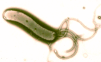


![[Frown]](frown.gif)
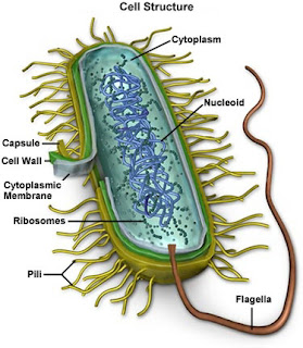

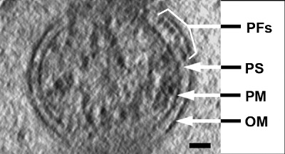
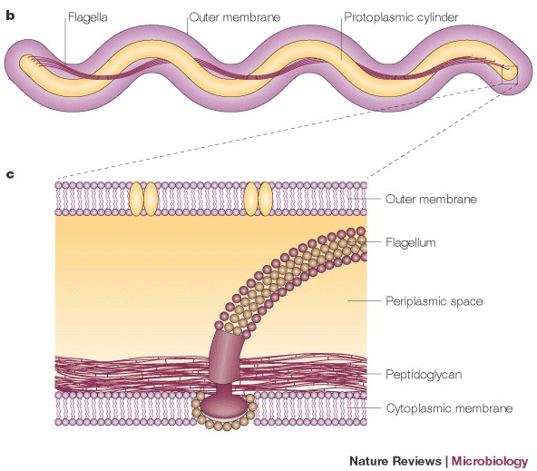

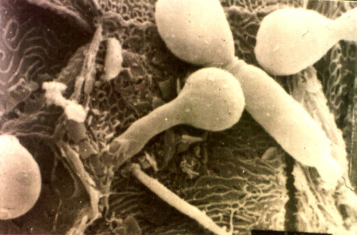

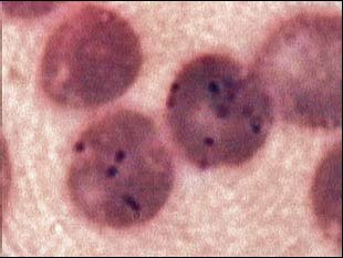


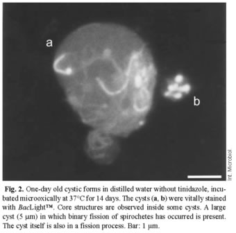
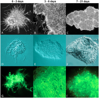


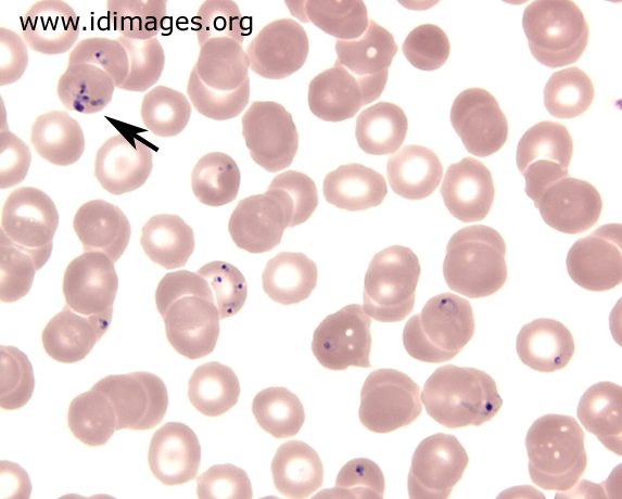


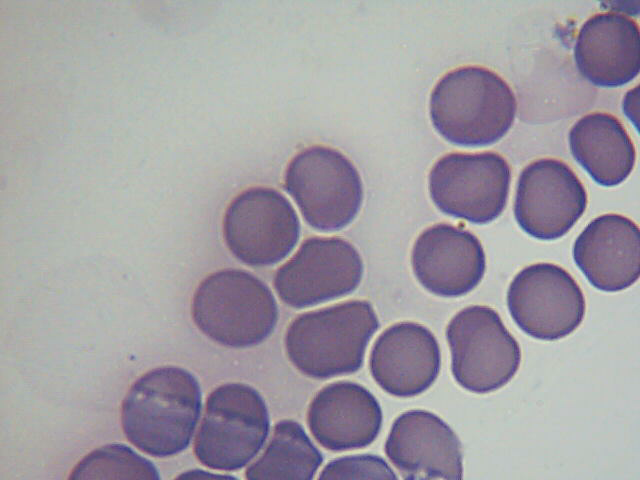
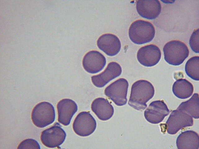
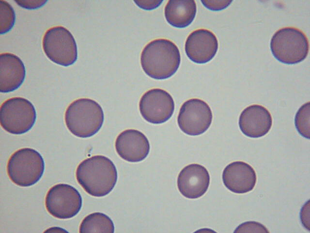
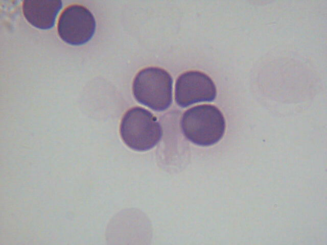
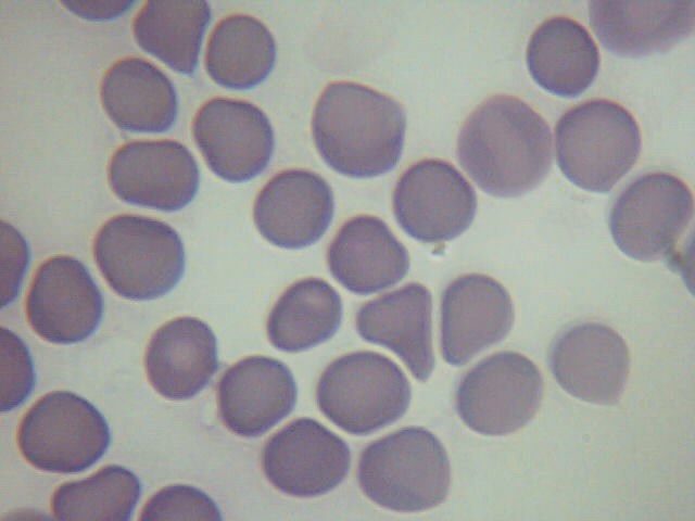
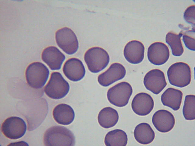


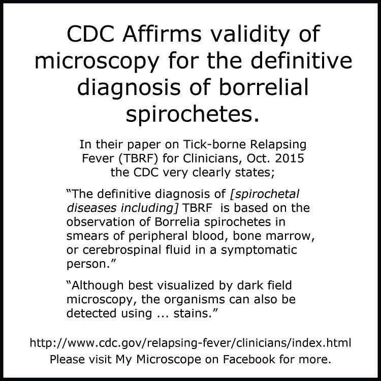


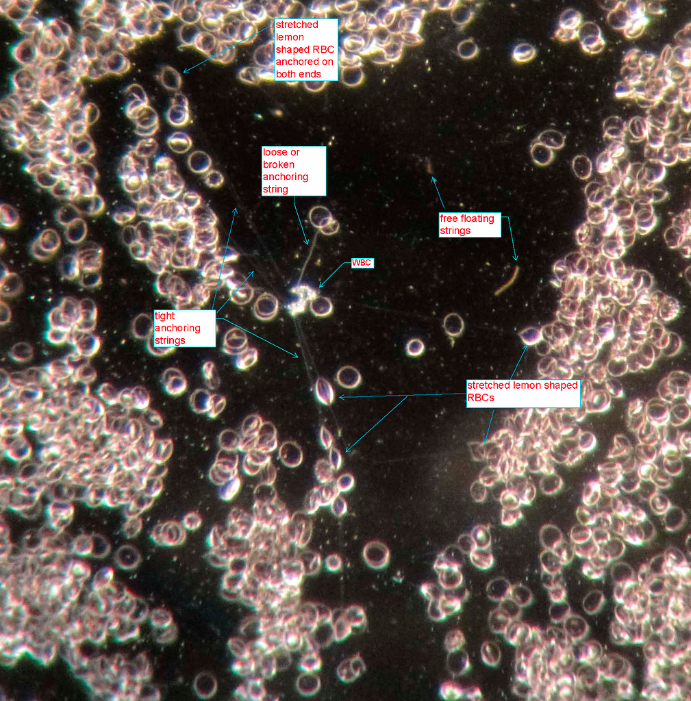


 [/IMG]
 [/IMG]
 [/IMG]


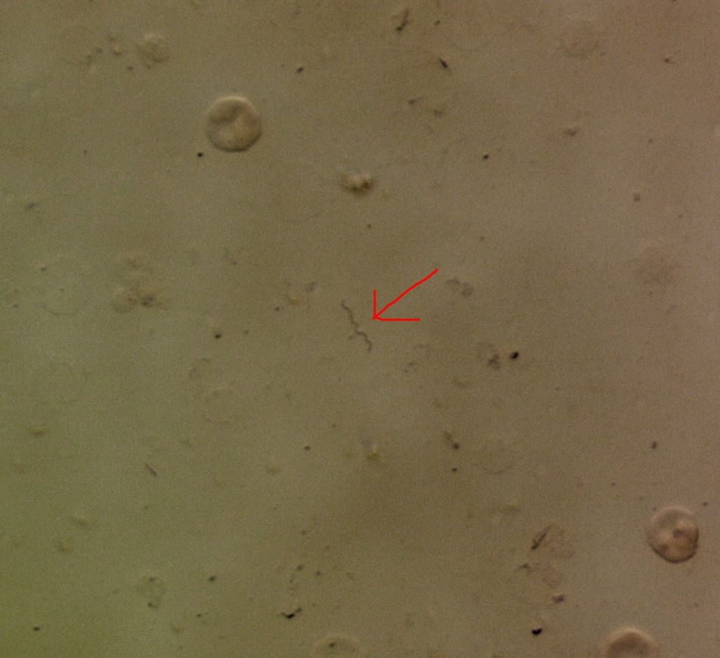
![[Razz]](tongue.gif)


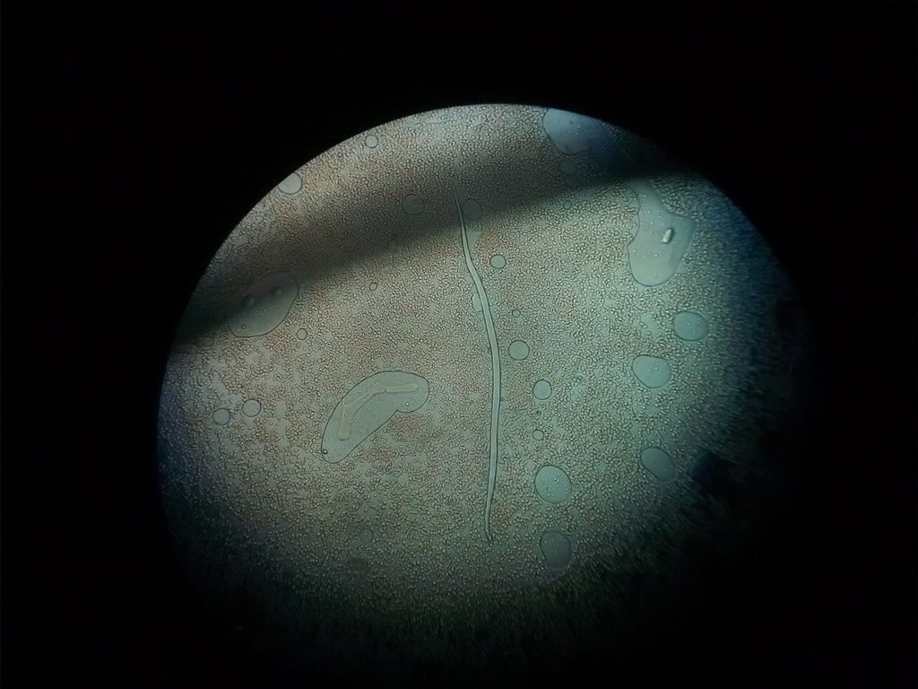


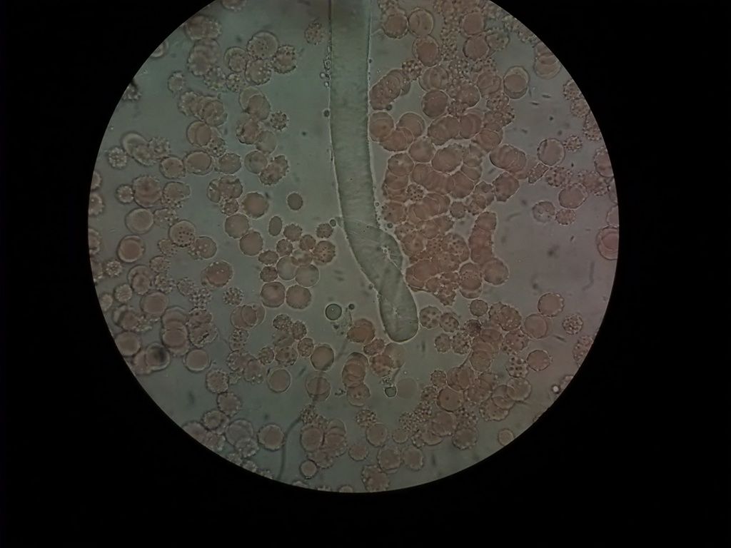


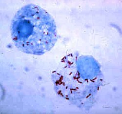


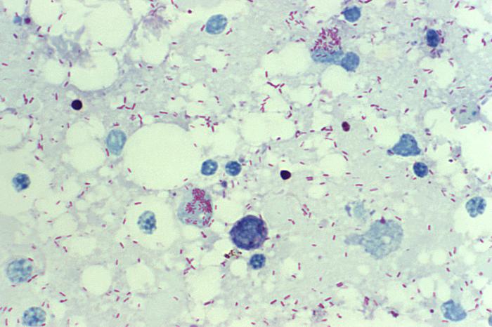

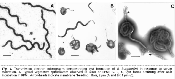


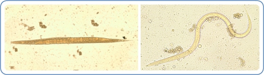

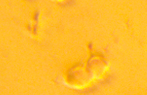
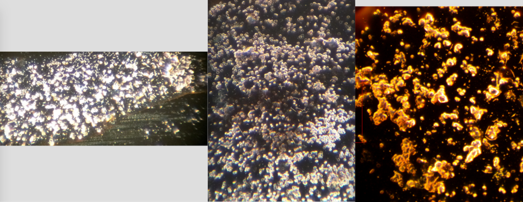
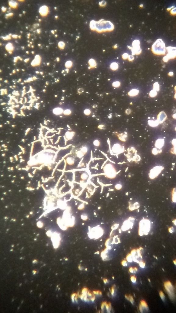
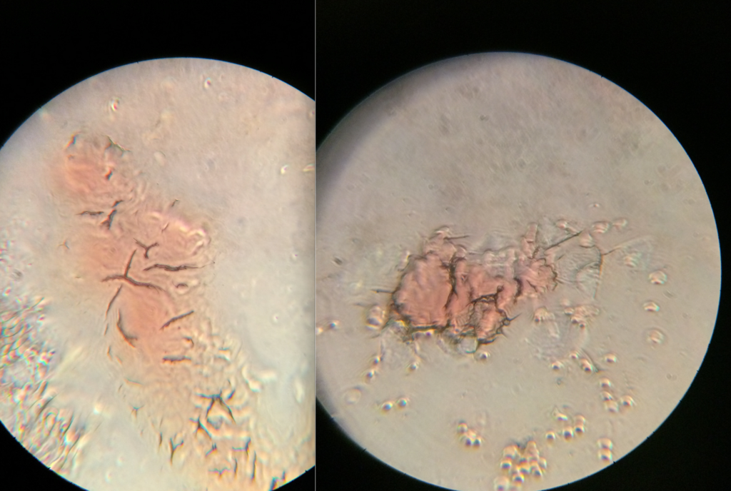
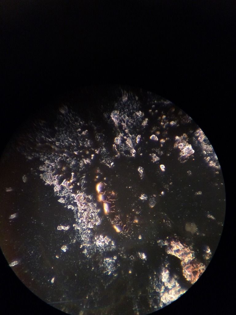
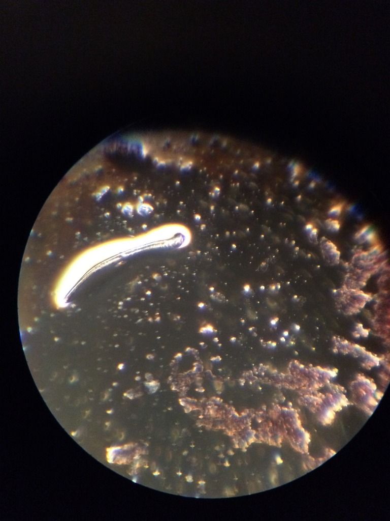
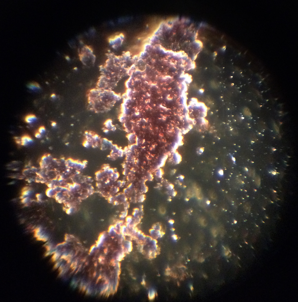

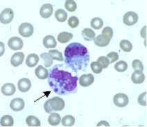


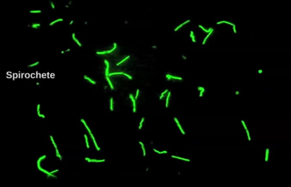
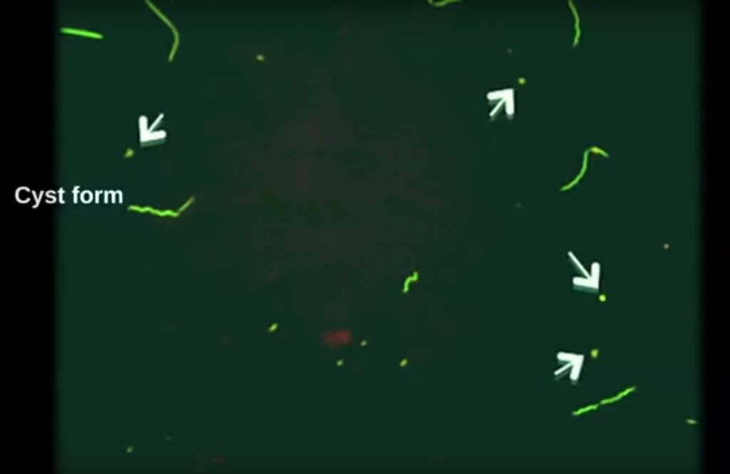
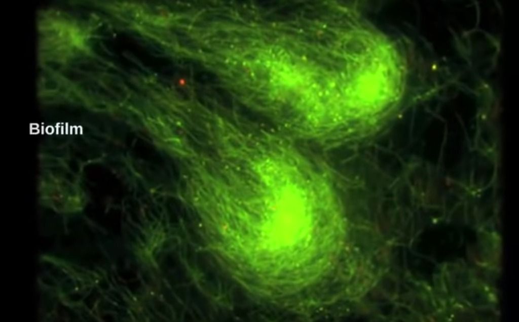


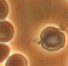
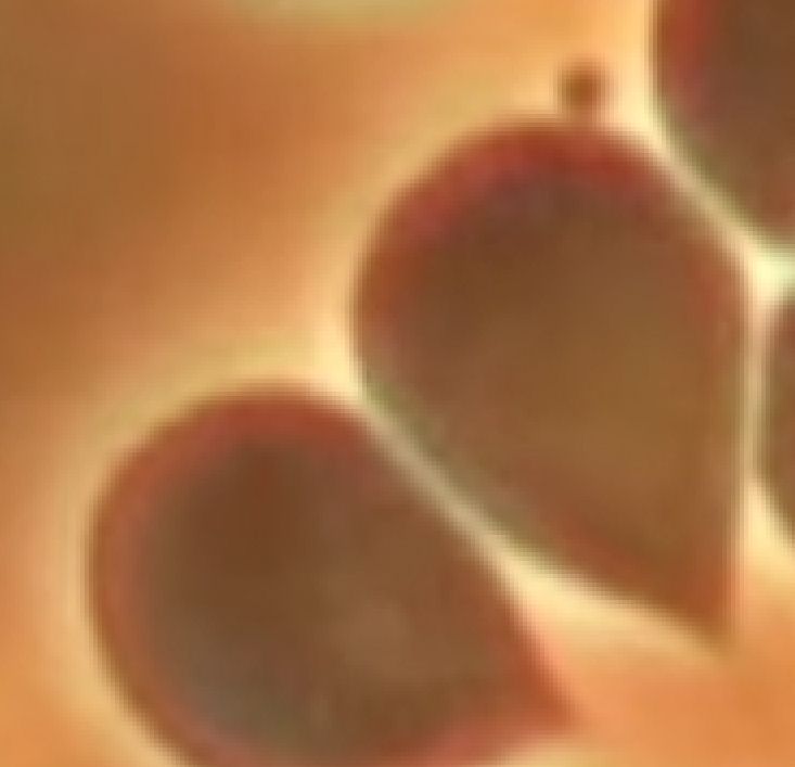
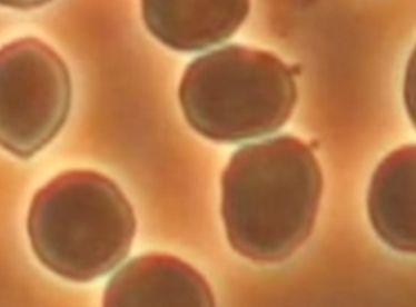
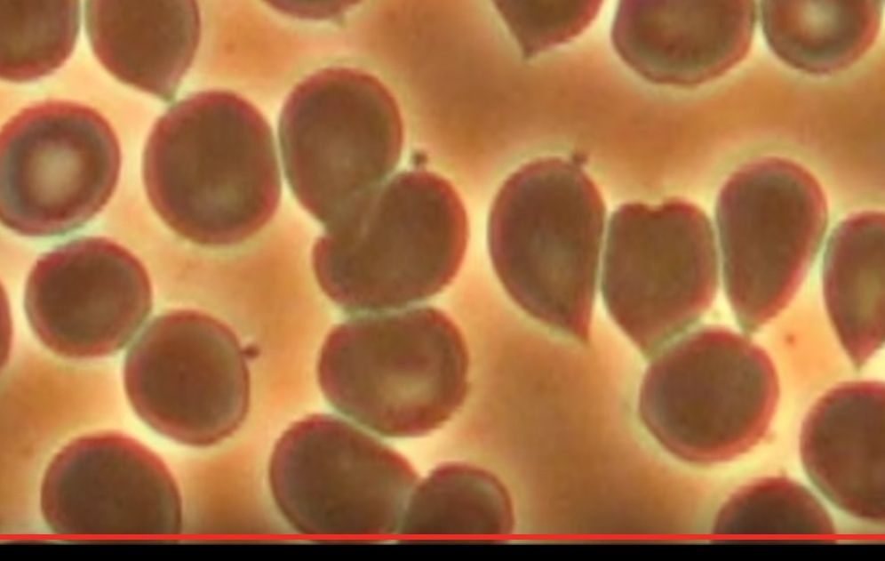
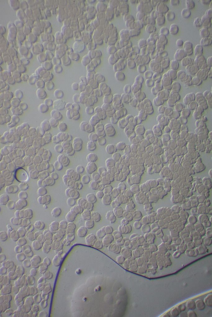


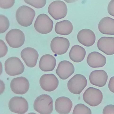


![[confused]](graemlins/confused.gif)


![[group hug]](graemlins/grouphug.gif)


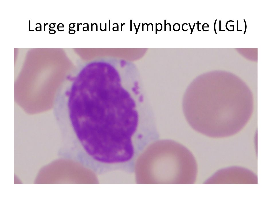

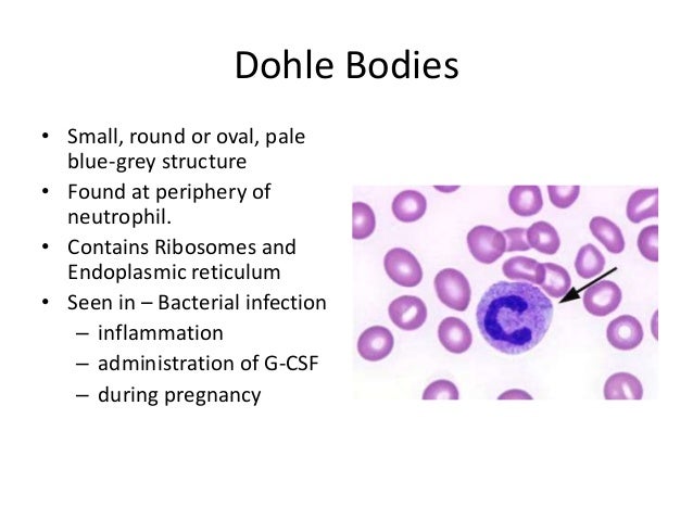
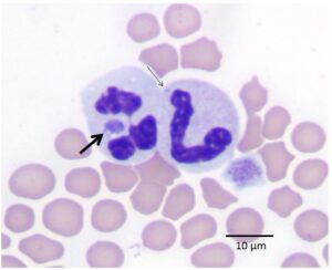


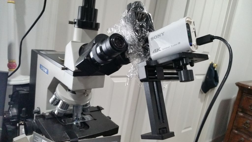


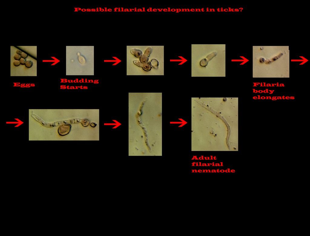


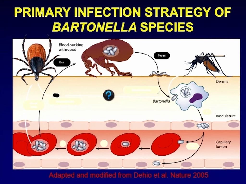

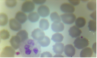


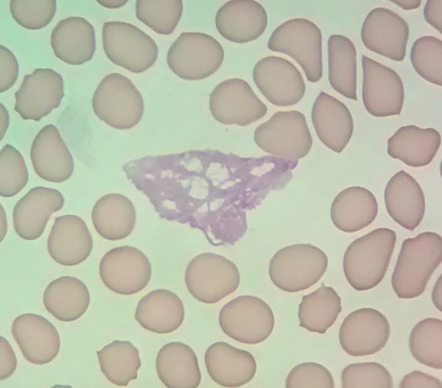
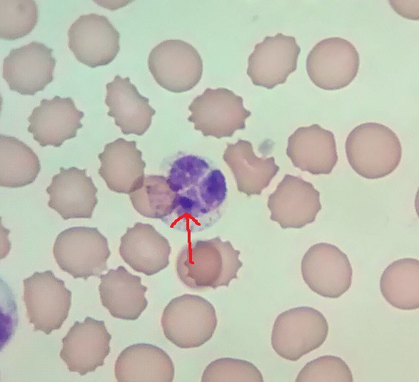


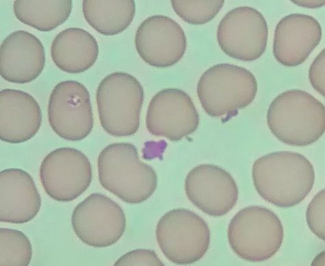

![[shake]](graemlins/shake.gif)