posted
Is there a microscope that is relatively affordable that is strong enough to see the ketes and other lovely stuff like babesia and ehrlichia.
I have done no research on this so any info. will be welcome.
I am sure a dark field microscope is extremely expensive...
Any ideas? And have any of you done this at home - checking out your blood under the micro?
Thanks!
Posts: 655 | From NC, Exit 88 on the Deer SuperHighway | Registered: Dec 2004
| IP: Logged |
SForsgren
Frequent Contributor (1K+ posts)
Member # 7686
posted
I would say no. Even with a good darkfield microscope, you really can't see ketes after a few weeks of infection according the my darkfield guy. We could see little blobs of clumped up stuff that he suspected was Borellia but it's hard to see ketes in the form I think you are hoping.
-------------------- Be well, Scott Posts: 4617 | From San Jose, CA | Registered: Jul 2005
| IP: Logged |
david1097
Frequent Contributor (1K+ posts)
Member # 3662
posted
Hi
The bottom has fallen out of the microscope market as of late. A good scope can be had for $300 to $400. You will need at least a 1000x to see babesia. Be warned though, a lot that can be seen through the microscope is the result of staining techniques. You will also have to get stain kits. I think a gram stain kit (the most common technique) sells on the retail market for about $40.
A monocular scope will do the job. You don't need PLAN objectives either (unless you have the extra money to spend and then they are nice to have). Also for dark field work, (Ie to see things that are really small in one dimension, like lyme bateria) you will need a very bright illuminator, something like 100W plus as well as a special condenser lens. This is an add on unit for most of the microscopes. Still the standard lower cost scopes will let you see babesia without any problem (although you may have to do a lot of searching!)
There are a number of companies selling microscopes here are a few,
All seem to be pretty good to deal with. Just tell them what you want to do and they will guide you in th right direction.
As implied above, dark field work is rather specialized and not too many places will sell the required equipment. There are a number of people doing dark field analysis for things like vitamin deficientcy etc. but I remain unconvinced as to the validity of what they are doing. As far as I am concerned, go with the "regular" micoscope and concentrate on learning how to do proper staining and you will much farther ahead.
As far as seeing bacteria infections, There is a VERY good veterinatian oriented web site with a lot of pathology photo's of all sorts of things (eri., bart.,bab., etc). I just don't remember its URL. If I come across it i will add it to this thread.
Finally, the lower cost scopes are made in china but they are pretty good. I feel that you are better off with one of the new but low cost units as compared to a used one of comparable cost since the new ones/companies selling them all seem to have a return option if you are not happy with it.
good hunting...
Posts: 1184 | From north america | Registered: Feb 2003
| IP: Logged |
Ann-OH
Frequent Contributor (5K+ posts)
Member # 2020
posted
What would you put on a slide under your microscope?
Blood? Spirochetes do not hang out there much, that is why blood tests are so inaccurate.
Urine? Not likely you could see a spirochete there as they would have had to pass through the kidney. Might be a possibility. The urine tests are usually done to trace the dead ones and the bits and pieces of them
Tissue? What tissue might you use? It might take years to hit the right tissue, unless you took a skin sample at just the right time, at the right spot on a rash.
We haven't even mentioned what kind of staining you would have to do.
And what might you do if you saw what you thought might be a spirochete? You would have to have verification and that might be hard to find.
This sounds like a very expensive and unsatisfying venture.
So good to read all the great info from David.
I know you probably know all of the above and know what you are doing. I just wanted to play devil's advocate as there are some people who would read this thread and say "Yeah! All I need to is get a microscope and I can find proof I have Lyme!"
david1097
Frequent Contributor (1K+ posts)
Member # 3662
posted
Hi
Lyme would be difficult but babesia is pretty easy to see if you look for a while and actually have an infection. Basically do it your self lab and pathology work is of zero clinical value, but it is interesting just to learn and discover; Just to look at your blood is interesting (and useful enough to have full courses dedicated to it in medical school)
Oh i forgot to mention; if you are real adventurous you could try you own lumbar punture using the bathroom mirror and maybe see cyst form in the CSF (this is supposed to be a joke.)
But wait, Don't laugh, in the 1600-1900's a lot of big discoveries where made by do it your self tests, even in the 1980's look at the guy that discovered h.pylori but giving it to himself.
Once you have been in the science culture for a while you learn that there are not many brave souls that will challenge the status quo. Those that do usually have to resort to unusual means to test there theories. And remember, even people that are not Dr's can discover things.
Basically just look at it as a hobby and don't get your hopes up of finding anything of use.
Posts: 1184 | From north america | Registered: Feb 2003
| IP: Logged |
posted
the most powerfull biological microscope i`ve seen is bradford variable projection microscope which magnificates 18000 times , Dr Andy Wright from UK use this. I would like to know how it creates so big magnification
david1097
Frequent Contributor (1K+ posts)
Member # 3662
posted
Be carefull of the bradford claims, or at least what you inffer from them. The magnification of a microscope is limited by physics. The claim of gazillions of times magnifications is based of the ratio of specimin size to displyed size. With a regular micro scope the eypiece has perhaps a 1/2 inch image (if that).Tthe bradford instead projects on to a screen that is perhaps 20 " across. This is 40 times larger thus the added "magnification".
You don't see anything more with the bradford, you just see it bigger. If you try to go much above 1600x with an optical microscope, the image resolution does not increase so you don;t see anything any smaller. Still the braford calin is "technically" correct. You can get the same performance froma cheap microscope and a camera.
Think of it like having a big computer screen but having a computer that set to a lower resolution. You get the same picture as a smaller screen but it is just bigger. You can't fit anything more on the screen.
At least that what I know from my physics courses. I hope I explained it in an understandable way.
Posts: 1184 | From north america | Registered: Feb 2003
| IP: Logged |
posted
David thx for explanation, so You can only get 1600 magnification with optical microscope? I thought bradrofd was using something else the light as a medium but i was wrong then.
Does binocular or monocular version has an effect on field range seen under microscope?
Posts: 636 | From Wroclaw, Poland | Registered: Mar 2004
| IP: Logged |
oxygenbabe
Frequent Contributor (1K+ posts)
Member # 5831
posted
A doctor with lyme I respect who taught herself microscopy thinks Bradford is total bull-crap.
It's not the scope so much as teaching yourself microscopy. You have to have a lot of training I think, to recognize pathogens and identify them correctly. Plus sometimes you will need certain types of agars, media, and stains to culture them and view them properly. And you already have your clinical picture so...the only reason to do this would be if you were attempting an experimental protocol like rife or herbs and wanted to try and track their effectiveness, I guess. This is not simple! Posts: 2276 | From united states | Registered: Jun 2004
| IP: Logged |
posted
Lymied, If you just want to see Borrelia burgdorferi (Bb) bacteria, as a blood test, you can use a regular microscope and use a stain instead. "Giemsa blood stain" is recommended for Bb in Bergey's Manual of Determinative Bacteriology. It is posionous, so be careful. The stain kills the bacteria, so it can only be used for establishing the presence of the bacteria. $300-500 for a scope will do for this. Make sure you have a 15X eye piece and a 100X objective lens for a total magnification of 1500X to see Bb. You'll probably have to do an oil immersion mount, but that is easy to do.
If you need to study live Bb bacteria, then you will have to go the darkfield condenser route and have a high power(100W) light source.
I order to calibrate your eye to the bacteria, then I would get some prepared slides of Bb from Wards (800-962-2660.)
Just having a lab test done is a lot easier for sure, but is not as much fun.
posted
What is the effect of phase contrast and how is done? How an effect of dark field is obtained? Is this affect of way of lightening sample or digital processing ? Imaging spirochetes with darkfield is better so ...
Posts: 636 | From Wroclaw, Poland | Registered: Mar 2004
| IP: Logged |
posted
Thanks Ernie...do you do this professionally or were you once like me, just very interested without any formal training?
Posts: 655 | From NC, Exit 88 on the Deer SuperHighway | Registered: Dec 2004
| IP: Logged |
posted
I have my own darkfield and phase contrast microscopes. They are about the only way you can see Bb in the human host, as they are mostly transparent and do not seem to stain well.
If you are expecting to see classic corkscrew shaped spirochetes though, you will be dissapointed. What you will see mostly are variant forms that you might not even recognize as Bb when you see them. Cysts and thread-like forms, but without the familiar spiral twists.
Study these pictures from very well known sources in this document to see examples of the variants.
posted
Thank You James, yes i`ve studied this dicument in fact i have translated it all. Could You please tell me what model of darkfield microscope You have ? Are they expensive?
Posts: 636 | From Wroclaw, Poland | Registered: Mar 2004
| IP: Logged |
posted
My Darkfield scope is a '10 series' American Optical equipped with an oil darkfield condenser and the #1014 Plan Achro 100x oil objective with an diaphragm in the objective.
(The 100x is the only lens I use on this, as all the magnification possible is needed to look at these organisms.)
You can often find good, serviceable older microscopes like these on eBay for a few hundred dollars, but equipped with only the standard brightfield optics. Good ones that are equipped with phase contrast or darkfield are very hard to find.
Sometimes the stuff people are selling on eBay is worn out junk that is no good to amybody, so buyer beware.
If you are very dilligent in your search and very patient, you will in time find a bargain.
I don't know about the low priced new ones that you see advertized sometimes... if they are any good or if the optics are good. I have never used one.
Posts: 714 | From San Antonio TX | Registered: Oct 2004
| IP: Logged |
posted
Very good to know...Thanks James. You have your own Dark Field Microscope...very cool...
I think I want to start out with the babesia...that is what I am most interested in at the moment since so many refuse to believe we have it here in NC.
I know there is so much I don't know and I am probably crazy to even try this but I am sure I will learn something along the way and maybe I will figure out how to stain, etc...
What do I have to lose except some time and money, and Lord knows in the past year and a half, fighting tick borne illness, I have learned how to lose these two resources like a pro.
Thanks for all the info...Thanks for that link to the adverse conditions paper...very interesting...
Posts: 655 | From NC, Exit 88 on the Deer SuperHighway | Registered: Dec 2004
| IP: Logged |
lightfoot
Frequent Contributor (5K+ posts)
Member # 2536
posted
Seems to me if it were that easy.....we would have a perfect test already!!!!!!!!!!!!
-------------------- Healing Smiles.....lightfoot Posts: 7228 | From CO | Registered: May 2002
| IP: Logged |
Just interested...I am like that kid that wants to play the guitar but wants a plastic ukalellee (sp?) for Christmas...humor me, ok?
Who knows maybe I won't find any babesia in the red blood cells but maybe I will start learning about microscopes and want to become a microbiologist in my third career change.
I posted the same thing to someone else on another board tonight...it hit me when she said she had been to a new naturopath who put her blood under the microscope and pointed on the ketes on the slide...I asked her how the woman did it and if it is this easy why is the testing so bad....so I definitely hear you.
I have just been so frustrated with a sick dog for a year and a half that I know in my bones has babesia but no one seems to think it could be true. I have this dream that I could take some of her blood from her ear with one of those tiny little straws they use to prick people's fingers and find the babesia living in the red blood cells.
I know I am dreaming...but it is fun to dream...It is what keeps me sane...
Posts: 655 | From NC, Exit 88 on the Deer SuperHighway | Registered: Dec 2004
| IP: Logged |
lightfoot
Frequent Contributor (5K+ posts)
Member # 2536
posted
Lymied,
I hear you. Dang, would that be neat or what!!!! Or what!!!!
Maybe some day!!
Healing thoughts and smiles....lightfoot
-------------------- Healing Smiles.....lightfoot Posts: 7228 | From CO | Registered: May 2002
| IP: Logged |
posted
James did You watch ketes by Your microscope ? Did u manage to make it ?
Posts: 636 | From Wroclaw, Poland | Registered: Mar 2004
| IP: Logged |
posted
James H tell me please what is the difference between oil darkfield condenser and dry darkfield condenser ? I know that oil one is more expensive. I am going to buy my own microscope too meybe Posts: 636 | From Wroclaw, Poland | Registered: Mar 2004
| IP: Logged |
david1097
Frequent Contributor (1K+ posts)
Member # 3662
posted
The reasons that microscopy is not used for routine babesia testing is because of the high manpower cost to scan the slides. Several hours and then you still donlt know if you missed anything. There are some labs that use a microscope, stage stepper and computerized camera to try to search for the buggers but even that can take several hours if there is not a high degree of parasitermia (lots of bugs).
Normally you would look for the clasic parasite form inside of the red blood cells (double or trip quadrafrom are classic), these are pretty easy to see. Also, there is also some reports of high numbers of free protozoa in the serum of infected hosts and if the sample is centrifuged then the the parasites are in higher concentration, but then you have to sort them out from other stuff than might be in the blood. (they are pretty small, perhaps a micron or 2 in diamter)
Veterinarians would typically use in office microscopy for diagnosis.
You will have to learn to do a thin blood smear to look at your blood. There ae lots of links on this ont he internet.
Posts: 1184 | From north america | Registered: Feb 2003
| IP: Logged |
posted
The oil darkfield condenser is required to use darkfield with the highest power 100x oil objective. The dry darkfield condenser can be used for the lower magnification objectives. I think the highest magnification you can get is what is needed for this application.
Here is a good tutorial on the different technologies from Olympus:
In answer to your earlier question, in our case we can see numerous organisms that resemble the photos of variant forms, and some things yet to be identified. I have almost never seen a spirochete in the classic coiled form in blood, though.
Incidentally, darkfield is used to look at live specimens in their natural state. You can do stained smears with just a standard microscope, and you can find tutorials on looking for malaria on the internet. The techniques used for Malaria can be allpied to other things.
In most of the countries where Malaria is a big problem they seldom have all the high tech lab equipment, so having an experienced microscopist look at a slide for an hour is quite common. In the US finding a lab tech that even knew what malaria looks like might be a challenge.
Borrelia do not seem to stain well, or possible do not survive the staining process intact, and are very hard to find on a dried smear. darkfield or phase contrast viewing of a live sample works better for them. They are almost transparent, so they are pretty much invisible with standard optics. Darkfield and phase contrast are 2 ways they can be given enough contrast to make them visible.
[ 31. December 2005, 09:56 PM: Message edited by: James H ]
Posts: 714 | From San Antonio TX | Registered: Oct 2004
| IP: Logged |
posted
James Olympus site is really nice, this java animations also. Does this oil used in oil darkfield condenser is the oil placed between sample covering glass and objective? Did You try leave sample overnight to see if spirochetase will leave cells and then actually can be seen? That is what Dr Andy Wright did on His slides presented with Bradford Variable System, You need time and wait for nutrients dropped in sample before spirochetes will be forced by starvation to leave cells. It takes night
Posts: 636 | From Wroclaw, Poland | Registered: Mar 2004
| IP: Logged |
posted
Yes, it is the very same oil that you would put on top of the coverslip. A drop of immersion oil on the top of the oil darkfield condenser bridges the gap so there is no distortion as light passes from glass to air and back to glass at the underside of the slide again. (The oil has the same optical properties as glass does.)
Yes, more things do come out as a sample ages. There is also alot more activity after one has had a heavy meal. It is also educational th squish a sample gently by placing pressure on the coverslip. There are often protezoan looking things inside our red blood cells, and the pressure ruptures some of the rbc's, making these visible. I am not sure what some of them are, or their medical significance (if any), but they sure are interesting.
I see pretty much the same things as in Andy Wright's videos, and there is no shortage of them. His photos that have a light colored background are done with phase contrast... the other technology that lets you see transparent living things.
I am watching for subtle and not so subtle changes caused by toe antibiotics... to know when something is working or not working.
Posts: 714 | From San Antonio TX | Registered: Oct 2004
| IP: Logged |
posted
I can buy microscope with darkfield but i would like to also buy some equipment to connect to PC and record movies/ But it looks like this is as expensive as microscope with Darkfield, i am talking about CCD cameras. Do You have any ideas how resolve this problem ? There is not much point to make work while it cannot be documented
Posts: 636 | From Wroclaw, Poland | Registered: Mar 2004
| IP: Logged |
posted
I am also at the Marshall Protocol and looking through microscope ocular with glasses would be tough Posts: 636 | From Wroclaw, Poland | Registered: Mar 2004
| IP: Logged |
posted
I bought an inexpensive ($100) camera on eBay that connects to my laptop computer and fits in place of a microscope's eyepiece. It worked, but max resolution was poor at 680 x 480 and the colors are always way to the red side of the spectrum.
Later I found out that my inexpensive HP pocket digital camera (one for regular photography) takes excellent pictures by simply holding it up to the eyepiece and letting it autofocus the image. That particular camera does video, but only at very low resolution. The stills look great though.
I think the solution to high quality photography at low cost is to get a microscope with a camera mount that will attach to a name brand camera body like a Nikon or Pentax, etc. Many of the name brand digital cameras mount their lenses the same way as their older 35mm cousins did, so an older camera mount may work on a newer camera.
Posts: 714 | From San Antonio TX | Registered: Oct 2004
| IP: Logged |
caat
Frequent Contributor (1K+ posts)
Member # 2321
posted
david1097 sd;
"Oh i forgot to mention; if you are real adventurous you could try you own lumbar punture using the bathroom mirror and maybe see cyst form in the CSF (this is supposed to be a joke.) "
OMG, LMAO!! Thanks David, I really needed that laugh today !
I just won an oldie but maybe goodie microscope for $30 on ebay thinking I could maybe see babesia at 650x... Oh well, guess I might need to get another lense for it.
Here's a couple links that might be useful;
Methods for the Optimization of Light Microscopy http://www.schwaben.de/home/mathias/english.html darkfeild wedge don't ask me- this is way over my head. All I get out of this is that you might be able to adapt a kind of sort of darkfeild with a peice of cardboard and a good plain microscope with some sort of diaphram. Maybe...
and; Microscopes to buy and to avoid http://www.absoluteclarity.com/buy&avoid.htm this is on sterio microscopes, but I went by the brand names here anyway- looked like a good source of info to me. I got an old bushnell on ebay for about $30 including shipping.
If any one comes up with any good links for babesia/microscope particulars; stains, photos, slide (blood) preservatives etc could you post them? I'm too tired to look right now but will later.
I think for anything other than babesia or tapeworms one would pretty much have to have some training in this to tell organisms apart. From the pics I've seen before on the web, some Babesia does look much easier to recognise than other things.
With babesia some say looking at blood from finger tips may be more effective than blood draws anyway for babs. No mirrors needed!
Posts: 1436 | From Humboldt county ca usa | Registered: Mar 2002
| IP: Logged |
caat
Frequent Contributor (1K+ posts)
Member # 2321
posted
oh. a friend of mine mentioned that when stabbing yourself for fingertip blood it's better to do it on the side of the fingertip below the fingernail- not the exact center. She said there are a lot of nerve endings in the center.
She mentioned a diabites tool- a spring loaded thingy for just that purpous.
Spring loaded sounds scarey to me, lol, I think I'll just use an exact-o knife with just the tip showing dipped in rubbing alcohol...
Posts: 1436 | From Humboldt county ca usa | Registered: Mar 2002
| IP: Logged |
posted
Those slides are interesting James - Very cool to look at. Took the test - didn't do too well but caught on after awhile to the different looks of different parasitic bugs...Thanks for the site!
-------------------- �Pride is concerned with who is right. Humility is concerned with what is right.� - Ezre Taft Benson Posts: 655 | From NC, Exit 88 on the Deer SuperHighway | Registered: Dec 2004
| IP: Logged |
caat
Frequent Contributor (1K+ posts)
Member # 2321
posted
Babesia....
got my microscope yesterday and today took a look at my blood cells at 600x. I can see the cells and can see an occasional round or long thing or things inside just very few of the cells, but I can't tell what the heck I'm looking at. Could even be dust on the bottom slide showing through a cell.
Everything looks clear- can see no color. Cell walls are clear but can see no cell nucleous or anything. The light is a flexible 40 watt desk lamp shone onto the mirror below the slide- seems bright enough. But I can't see the cell parts- just a wall on most cells. I guess I need to read up on stains.
I think I must have broken a lot of the cells smearing my finger on the slide... weird shapes and sizes.
This looks like an old college or high school lab microscope- good but not high end. No light condenser...
Anyone know if 900x magnification might help or not? For Babesia? I'm still searching google, but could be that I can't go higher than 900x without some kind of oil lense? Not sure if I can add a light condenser to this thing... is one absolutely needed for oil imersion?
Posts: 1436 | From Humboldt county ca usa | Registered: Mar 2002
| IP: Logged |
That is awesome...here I sit still trying to figure out what to get and you are searching the field for Babesia! WooHoo!!!!!
Keep me posted on what your seeing...occassional round or long things sound good to me ;0)
Hopefully David or James will pick up your post and give you some advice.
You have inspired me - gotta start doing some more research and find myself a scope
Rock on!!!!! (Sorry couldn't resist)
-------------------- �Pride is concerned with who is right. Humility is concerned with what is right.� - Ezre Taft Benson Posts: 655 | From NC, Exit 88 on the Deer SuperHighway | Registered: Dec 2004
| IP: Logged |
caat
Frequent Contributor (1K+ posts)
Member # 2321
posted
hi lymied
oh... BTW, there seem to be a lot of nerve endings on the *side* of the fingertip as well...
and an exacto knife tip may not be as easy as a diabetic stab thingy. Not fun... you have to kind of "saw" at it unless you really want to stab yourself deeper.
A sterilized sewing needle was easier. Might be easier on a dog too. ...the dog might not like this...
Posts: 1436 | From Humboldt county ca usa | Registered: Mar 2002
| IP: Logged |
caat
Frequent Contributor (1K+ posts)
Member # 2321
posted
Lymied,
wait on the scope! I might have gotten the wrong kind! I don't know if I can do "oil imersion" with this one- I guess you need special lenses for it. Wait till someone who knows what they are doing answers (hopefully with some advice on what to get?)
There are stain kits on ebay but I don't know what the heck is needed.
>>>>Keep me posted on what your seeing...occassional round or long things sound good to me ;0)
doesn't sound good to *me*! I am hoping NOT to do 6 months of mepron and zith followed maybe by tini...
Posts: 1436 | From Humboldt county ca usa | Registered: Mar 2002
| IP: Logged |
caat
Frequent Contributor (1K+ posts)
Member # 2321
posted
David or James- the aperture number on the objective lense is .65 is this good enough for any detail on babesia? With a stain and no light diaphram?
For others; I guess the aperture number on the objective lense is key to the "resolution" of what you see. The "objective lense" is the lense closest to the slide. The other lense is called the "eye peice". Resolution is clarity- how clear the lines are. On a basic microscope, with basic "dry" lenses the highest (best) aperture number is .95
Posts: 1436 | From Humboldt county ca usa | Registered: Mar 2002
| IP: Logged |
posted
So as well as wanting to prove my dog has babesia - I want my husband who has only been treated for 9 months to realize he may still be dealing with all this stuff...
Told him no unprotected intercourse for awhile - just freaks me out...I know I am not well yet and I am sure he is not...if I could show him on a slide his whole outlook will change and he will realize why I am being an OCD extremist about all of it...Sometimes I am sure he thinks I am
Probably sharing way too much information with you all...if I can't share it here though there is no where else I can
-------------------- �Pride is concerned with who is right. Humility is concerned with what is right.� - Ezre Taft Benson Posts: 655 | From NC, Exit 88 on the Deer SuperHighway | Registered: Dec 2004
| IP: Logged |
caat
Frequent Contributor (1K+ posts)
Member # 2321
posted
I guess your husband might not like this either... Hide that sewing needle! (just kidding)He does know that if he re-infects you you'll just reinfect him later?
Best info I've found so far for seeing babesia is;
Least expensive; Getting a basic light microscope with a light condenser(an "Abbe" Condenser keeps getting mentioned). If it has this condenser then an oil imersion objective lense can be used. The "objective lense" is the lense nearest the slide. The objective lenses can be changed on the microscope- so one can be bought seperately from the microscope but the length has to match the scope's feild. You might have to ask a good dealer for help on that.
You want 1000x magnification. 900 might be the highest a microscope without a condenser might go, that might be enough- dunno. Anyway, the total magnification is the objective lense's magnification x the magnification of the eye lense.
the appature number on the objective lense is important- see my post above.
If you are going for 1000x or higher, then an oil imersion objective lense may be what you want. It has more detail. An oil lense is made differently from a "dry" lense and neither one works well with the opposite technique. Do a google for "oil imersion technique" microscope.
There are other more expensive ways like electron microscopes...
Giemsa is the most common stain used for babesia. It's used on both thick and thin blood smears. Thin smears seem to be better from what I've read tonight. A thin smear can pick up other organisms as well.
here is a technical manual on using giemsa stain; http://www.med-chem.com/procedures/Giemsabsp.pdf I think it's much easier than it looks here- biggest problem is the technical language for those of us not familiar with it. Use a mason jar instead of lab jars, etc. Methanol is wood alcohol and can be found in a harware store. Like someone said above, it is poisonous- wear gloves (neoprene gloves?) and try not to breath any fumes. It's also very flamable. Might be better to rinse a slide with an eye dropper than to imerse in a jar- less to dispose of.
A quick search in google images will bring up magnified images of some babesia species.
Wrotec- eye glasses are no problem! You can just take them off and see things fine.
If I've misinterpreted anything somebody please correct me.
Sorry if I'm repeating anyone's info- it's late...
see one of the posts above for a link to recommended brands with good glass optics if you are buying a used microscope.
[ 08. January 2006, 04:35 AM: Message edited by: caat ]
Posts: 1436 | From Humboldt county ca usa | Registered: Mar 2002
| IP: Logged |
I am not sure my husband gets that we can just pass this back and forth. I think he chooses not to think about it and it drives me nuts!
I have dragged him to one conference with me and I thought after we left he had an epiphany when it comes to understanding lyme and coinfections but alas I think denial is a strong seductress, because he is right back to where he was before treatment...in la la land.
Thus it is left to me to be the tyrant about all this and do all the research. I am like a terrier with a bone in regards to researching every end of this I can. I have been considering RIFE at this point.
Well I am still laughing about your needle comments and sawing at your skin. Neither sound real appealing and yes, I think my dog would be less then enthusiastic.
Keep me posted on your discoveries...I hopefully will start my scope search soon...
-------------------- �Pride is concerned with who is right. Humility is concerned with what is right.� - Ezre Taft Benson Posts: 655 | From NC, Exit 88 on the Deer SuperHighway | Registered: Dec 2004
| IP: Logged |
The stain is for Babesia and not the borrilea. For Borillia, you should use dark field. More on that in a minute. The type of stain is a Giemsa Stain as someone mentioned.
Here is a website that tells you much of what to see in the blood (normally). Extremely informative.
posted
Well, I see some of you have been experimenting! I applaud your efforts and your interest.
Caat has just discovered one of the problems with looking at microscopic living things in their live state. Many of the most interesting ones are nearly transparent and almost invisible in the clear liquid in which they are found.
One approach is to spread them thin, dry them, and stain them with chemicals. This can be really useful for some purposes, but you no longer can watch them go about their business. They are 'colorful, flattened out corpses' now.
About resolution... the amount of detail you can see... This is determined by the objective lens and the condenser beneath the stage. Total magnification is expressed ad the objective's power times that of the eyepiece. A 45x objective and a 10x eyepiece yields 450x.
You can use a higher power eyepiece and an object will appear to be bigger, but the details will not be any sharper. Just bigger.
With a 40x or lower power objective the microscope can be very simple (maybe no condenser) and still give a decent image.
At the highest power, a 100x objective, subtle optical effects become very important. If you hold a pencil part way in t glass of water, it appears bent. Different density materials affect light differently.
The same thing happens as the light travels from the condenser, through some air, through the glass slide, specimen, and cover slip, through some air again, and to the objective lens. The light is refracted and the image suffers poor clarity.
That's why 100x objectives are oil immersion. A drop of a special oil is placed between the slide and the objective to bridge the gap. To the light, the oil and the glass act ecactly the same, and this distortion is eliminated. The improvement is dramatic.
Darkfield condensers designed for use at 100x usually use a drop of oil bridging the gap with the bottom of the slide too.
Why don't we see 200x, 250x, or higher power objectives? The problem is the 'size' of light waves themselves. The practical limit of total magnification for a visible light microscope is about 1200x - 1500x. Beyond that an electron microscope is required.
For observing living biological samples you really need either dark field or phase contrast. Otherwise 'jellyfish in the water' are hard to even find let alone study. The best quality optics you can find are a big help too.
This doesn't have to cost thousands if you are resourceful. Professional grade, name brand used equipment shows up all the time on eBay, and in my opinion is preferable to cheaply made new ones.
You do have to know what you are looking at to determine if it is complete with all parts, undamaged, and suitable for your needs. I am STILL looking for a phase condenser for an American Optical series 10 that I didn't look at closely enough for missing parts.
There are all kinds of different types of microscopes by the way. The low power 'stereo' ones, metalurgical, inverted ones, you name it.
'Biological' is the key word for our purposes, and the highest power 100x oil objective is needed. Phase contrast and/or darkfield are needed to view living cells.
Darkfield and phase contrast are not really 'types' of microscopes by the way... they can be added to most professional models if you can find the right accessories.
Posts: 714 | From San Antonio TX | Registered: Oct 2004
| IP: Logged |
caat
Frequent Contributor (1K+ posts)
Member # 2321
posted
thanks James, that's very clearly written.
a couple questions for James Or David or ??
I have a 15x eye peice. If I use a 60x or 63x objective I realize it won't actually magnify to 900x, but with a stain would I be able to see various cell nuclei and babesia and tell them apart? All I want to do is see if I have babesia- I don't need to know what species. Probley couldn't anyway.
Also, how can I utilise a 100x oil imersion objective without a condenser? Can I get a cheap abbe condenser and rig it to the mirror holder? Or set it below the microscope and take the mirror off? Or if I can't do that can I just direct 100 watts into the mirror and fiddle with it? I doubt the microscope I bought has any condenser built for it.
Or glue/silicon a condenser to the bottom of the stage?? Hmmm... would it work? Any idea where I could find out online?
I hate to buy another microscope. I'm only looking for babesia once, and the only other purpous this scope will have is to look at mites and maybe waterbears and moss creatures for fun.
[ 08. January 2006, 11:58 PM: Message edited by: caat ]
Posts: 1436 | From Humboldt county ca usa | Registered: Mar 2002
| IP: Logged |
posted
You should be able to see if there is something stained blue inside a red blood cell with that, but not necessarily well enough to tell what it is. A platelet lying on top of a blood cell looks an awful lot like a parasite, as they stain blue also.
I had an old American Optical Model 60 without a condenser once, and I put a 97x oil objective on it from the same brand and era. It actually worked pretty good, just not as good as a higher end instrument with parts that all matched.
I doubt it would be worth the bother of trying to retrofit a condenser though. A really good image is the combination of all precision parts designed to work together, and in perfect alignment.
Just use it for what it will do as it is. You can always sell it and get a different one later if you want to. It isn't likely to depreciate unless you drop it.
Posts: 714 | From San Antonio TX | Registered: Oct 2004
| IP: Logged |
caat
Frequent Contributor (1K+ posts)
Member # 2321
posted
Thanks James
That's very helpful. I figured out that my scope has a small "condenser lense" in the stage's light hole- so that's not so bad. And I know what length objective to get- so... first dealer I see on ebay with good deal on a 100x AND a 10x eyepeice & it'll be good to go.
Posts: 1436 | From Humboldt county ca usa | Registered: Mar 2002
| IP: Logged |
posted
If you have a 15x eyepiece in decent condition that should be just fine. While the lenses below determine the fineness of detail in the image, the eyepiece magnification makes this big enough to see. 15x is good IMO.
I have a pair of 20x eyepieces I use sometimes. While they do not make the image any sharper, they compensate for my less than perfect eyesight by making it bigger. So, technically there is no added resolution, but I am able to see more of the detail that exists.
The advantage of a widefield 10x set is you can see more area at one time... good if you are looking for something.
If you have a common digital camera, you may find it will take great pictures by just holding it right against the eyepiece once you have something in view and focused. Try it, if you happen to have one.
Posts: 714 | From San Antonio TX | Registered: Oct 2004
| IP: Logged |
caat
Frequent Contributor (1K+ posts)
Member # 2321
posted
cool! Thanks for all your help James Glad I looked here before going to ebay- just saved me $25 with shipping.
I have a digital camera but it's one of those notorious ms software abortions- only takes blue and white pictures unless you have an ms mouse...
I'll be looking for preservatives after a 100x lense. If you know offhand what to use to perserve blood slides w/giemsa stain please let me know. Otherwise- I'll find it.
My LLMD would be fine with me bringing in a preserved slide if I find anything. Right now he's set to put me on mepron anyway, but I don't want that if I don't need it. Yech.
Buying a microscope, stains etc is still cheaper than taking 3 private lab tests for babesia...
Posts: 1436 | From Humboldt county ca usa | Registered: Mar 2002
| IP: Logged |
posted
When you stain a blood smear, it is dried, fixed with Methanol, stained, and dried again. It is preserved at that point. (You don't put a coverslip on it when you prepare a slide this way.) I think they would last for years like this in a dry environment.
The Malaria testing websites warn you to cover them so flies won't eat your specimen... that tells you something about the environment those poor guys get to work in.
Of course, you put the oil directly on the dried smear, so now your slide is all oily. I've saved ones like this on end in a pill bottle so the oil would eventually run off, and more important dust would not get on it. If it is kept clean another drop of oil can be put on it if you want to look at it again later.
Babesia is not supposed to be very easy to spot by this technique unless somebody is pretty sick and you catch it at an active time, by the way. There is no harm in trying though, and it is bound to be educational. Expect them to be very scarce and require alot of careful looking.
Posts: 714 | From San Antonio TX | Registered: Oct 2004
| IP: Logged |
david1097
Frequent Contributor (1K+ posts)
Member # 3662
posted
A couple of things on babesia (and malaria) you might see dots in/on the RBC but a lot of things can do this(even playing with the focus knobs). A more definative indication is the double bumps in the RBC than are sort of welded together. This is pretty definative as being a parasite. Of course there also the cross form which is talked about a lot, but not seen that often. If this is seen this is rather conclusive (I think).
Another interesting point is that babesia infection makes the RBC walls weak and they wil break during a thin smear. You might see clusters of 4 dots and a stringy looking thing. I have seen this before (they are all in a cluster between other RBC's) and suspect it is the shell of the fractured RBC with 4 parasites. I have also seen the RBC just as it has fractured and the "parasites" coming out, likly from the surface shear during the smear.
If the blood is dried you can just put the oil directly in the slide but the oil lens is designed for use with a cover slip (in most cases). With a cover slip the image is a bit better defined.
I have a question Someone here mentioned "something:" growing out the RBC's after a period of time, Does anyone have a more detailed description of what they see? the RBC's in wet smears will develop funny bumps over time and not look like RBC's at all but this as far as I can see is normal.
Posts: 1184 | From north america | Registered: Feb 2003
| IP: Logged |
quote:Originally posted by david1097: I have a question Someone here mentioned "something:" growing out the RBC's after a period of time, Does anyone have a more detailed description of what they see? the RBC's in wet smears will develop funny bumps over time and not look like RBC's at all but this as far as I can see is normal.
I believe that this was without the staining and done with the dark field filter. What they are referring to is the borrelia hiding in a RBC (from the ABX) comes out when the nutrients in that RBC get low. Apparently there is a llmd that does this I think.
Then you can see the Bb when it comes out. I don't think it was to do with babs because you definitely need a stain for that which kills anything alive on the slide.
This has just sparked off another question of my own. Why does the Bb go and hide in the RBC? or more to the point - How? If we can stop it from hiding, perhaps we would be in a better position.
If it just lands up there through no choice of it's own, there is probably nothing much we can do to stop it. If it actively seeks out the RBC's to hide then perhaps there is a way of stopping that from happening. - another crazy idea? Posts: 27 | From Mars | Registered: Jan 2006
| IP: Logged |
posted
Supposedly Bb is not found inside RBC's, but it seems like it does alot of things it is 'not supposed to'. Maybe they should have explained the rules to it.
If you look at the bottom of page 20 (page 23 of the pdf) of the 'survival in adverse conditions' paper you can see a drawing of a more complex life cycle for syphilis that was proposed in 1913 by McDonagh. (Read his text too.)
The spider like forms in his drawing I have seen many times in by own blood using darkfield and a live specimen. They appear to be inside the RBC's, and are usually only visible in that form if the RBC gets ruptured. Sometimes a wormlike projection or two are seen hanging out of an otherwise intact RBC. Other RBC's are sometimes seen trembling and with distorted shapes.
I had tried lysing the RBC's with distilled water and other chemicals to see better what was inside, bit found the organisms inside would also lyse and be destroyed almost as fast as their rupturing RBC home made them visible.
A better technique was to simply apply firm pressure on the coverslip over the fresh blood sample using a clean piece of lens paper. With a little practice alot of the cells are ruptured, leaving their occupants exposed but still intact. They are very numerous.
Leaving a sample overnight also causes wormlike things to become visible, like the ones Andy Wright filmed.
Note that the familiar spiral appearance is lacking in the organisms seen. This is also true of many filmed by people like Marie Kroun, Andy Wright, and Mr. Burgdorfer himself.
Alot of the spirally ones filmed are in cultures, from ticks, or are different species. For whatever reason the perfect spiral form is hard to find in a human.
So what are these things? Are they all Bb? I believe at least some of them are. Do some research and you will find there is controversy.
Some of them could be of species yet to be identified, and of unknown medical significance, if any. Or, they could just be pieces of protein, as some propose. The 'old guys' from the early part of the last century came to associate them with disease in their human hosts, though.
The technology now exists to answer these questions, but unfortunately there is little interest in researching cures for disease anymore. Everyone is too busy looking for the next blockbuster symptomatic drug for erectile dysfunction, or their latest insult, restless leg syndrome. Posts: 714 | From San Antonio TX | Registered: Oct 2004
| IP: Logged |
posted
This is such an interesting thread...Thanks for all the information and links to excellent papers! I wished my background was in the field of microbiology.
I simply can't believe more scientists are not researching Bb and other coinfections - seems pretty fascinating to me...but I do think that the human spirit is one of wanting to conquer...these little bugs are simply humbling...they are an enigma and their behaviour is still a mystery.
A scientist is not left feeling God like in the presence of these bugs...for many there is no fun in that
Keep the great info. coming. Still haven't gotten a microscope or shopped for one. I am in a major transition in life...time is at a minimum...money at a minimum too but I will continue to learn and hopefully be able to get a microscope sometime soon.
-------------------- �Pride is concerned with who is right. Humility is concerned with what is right.� - Ezre Taft Benson Posts: 655 | From NC, Exit 88 on the Deer SuperHighway | Registered: Dec 2004
| IP: Logged |
david1097
Frequent Contributor (1K+ posts)
Member # 3662
posted
Do you have a lionk to any of the pictures? I would really like to see them.
I had often wondered about RBC. WBC infection myself. This would be the ONLY way that a insect could pick up the disease since there are so few organisms free flowing in the blood. The only issue I suppose is that Bb are longer than a RBC diameter (I think), so it would be a tight fit, ie i would have to roll up or something.
Has anybody tried to culture the RBC or better yet inject it into an immunocomrpmized mouse to see what if any infection is passed on. Since the resouvoir for all this nasty stuff are rodents, what ever it is it must be viable in them.
Posts: 1184 | From north america | Registered: Feb 2003
| IP: Logged |
posted
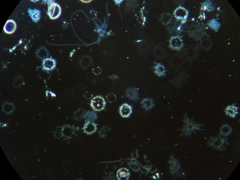
OK, I have my scope and I saw some keets in the blood today!
The blood was from a small finger prick last night, smeared over the slide and half of it was covered by a slip. The other half was left to dry.
It took allot of looking and a "make your own" dark field patch stop filter but interesting to see the keets in the end.
I couldn't get a good picture from the digital camera, so this is from a digital video camera.
Keets in the middle!
There are Three in the middle. The lower one is quite long and the ones just above that have just divided - I saw them do it, but didn't manage to capture it
There also seems to be allot of small (very) granules about (not in this pic though). More so than the keets. The granules are about 1/8th (maybe smaller) the size of a RBC.
I have seen some of the black spidery looking things about as well. These stretch across many RBC's and I think it is a bit of candidia. Not too much thank god.
No stain used, just a small disc under the condenser for the dark field contrast.
Next - staining for the babs...
edited for JPG picture
[ 16. January 2006, 01:17 PM: Message edited by: Starship Trooper ]
Posts: 27 | From Mars | Registered: Jan 2006
| IP: Logged |
posted
WooW, this is not dark field right ? How big magnification is this ??? The best way is to have CCD camera and record movie
Posts: 636 | From Wroclaw, Poland | Registered: Mar 2004
| IP: Logged |
quote:Originally posted by wrotek: WooW, this is not dark field right ? How big magnification is this ??? The best way is to have CCD camera and record movie
As I understand it, it is dark field. It may not be the best in the world because it was a home made filter. I have got very good dark field images using this technique with a lower magnification (400X) but the filter has to be smaller on 1000X for some reason.
This is at 1000X - 100 oil objective and a 10x wide field eye lens.
Here is the science bit (see one of my posts above):
Here is the filter I made. Yes it is stuck on with tape and yes it was cut out with scissors and no I don't really know what I am doing. The blue filter helps the contrast a bit as well. Posts: 27 | From Mars | Registered: Jan 2006
| IP: Logged |
posted
Yes i am familiar how dark field condenser works, Interesting u did it . IF this forms u have seen were spirochere than congratulations cause laboratories have problem with visualisation this bugs i guess.
U should make a movie., what scope u have?
Posts: 636 | From Wroclaw, Poland | Registered: Mar 2004
| IP: Logged |
Starship... if you save your files as .JPG type instead of .BMP they will be about 1/10th the size and will load faster for anyone with a slow connection. Good work!
--------
The darkfield photos are from my 30 year old American Optical series 10.
You do not have to spend huge amounts of money if you are resourceful. I found that one on eBay.
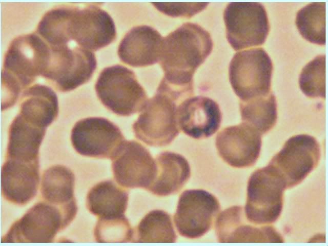
Blood cells on a slide so the same thing blood cells do when you cut your finger... they clot.
Seeing them clump up on a slide after it sits awhile is pretty normal, but is used to scare people and sell people various nutritional supplements by some.
If you prepare a very thin film so the cells have to lay flat you will not see this. If the film is thicher they will tend to stack like a roll of Lifesaver candies. Blood spread out on a glass slide is not 'happy' blood anymore... it is blood that is dying.
Starship, I understood that you were looking at a live specimen. Actually I thought you were pretty creative with that little darkfield stop you constructed. You might try enlarging the dark spot gradually until the light barely shows at the edges of the field. On some scopes you might not be able to get it to work, but there is no harm in trying.
Those could well be a form of the spirochetes you saw, as divsion like that has been described in alot of the older scientist's writings. They will be more worm-like and you may not see the classic coiled look.
That's the problem (and the addiction) with these instruments... we always see things we wish we could see a little bit more clearly!
[ 16. January 2006, 02:56 PM: Message edited by: James H ]
Posts: 714 | From San Antonio TX | Registered: Oct 2004
| IP: Logged |
posted
James did You do this dark field photos? They are beautifil. How do u pick blood ?
Posts: 636 | From Wroclaw, Poland | Registered: Mar 2004
| IP: Logged |
posted
Wrotek, it is a 516B-II BIO-microscope 40X-1600X from Digilens.
James, the blue filter might make it look like a stain, but I haven't used one - honest. I thought I was getting a dark field effect because what look like the keets (not so sure now) are brighter than it's background.
I honestly did see them divide - which took about 30 seconds from start to finish and then they were still for the best part of an hour when I left them alone. This is what made me think it was bacteria - a keet at that.
Tell you what, I will repeat the experiment and try a better disk on the filter. Don't know what I'm going to cut it with though.
Posts: 27 | From Mars | Registered: Jan 2006
| IP: Logged |
A have question about this photo, what do You think about these clumps of red cells? I mean bacteria is causing red cell to gather together what do You think ?
Posts: 636 | From Wroclaw, Poland | Registered: Mar 2004
| IP: Logged |
A have question about this photo, what do You think about these clumps of red cells? I mean bacteria is causing red cell to gather together what do You think ?
"CAUSE: protein linkage. Often poor protein digestion. The pancreas may be off. Excess dietary protein, poor assimilation. Eating too much animal protein. Blood too toxic (altered blood pH-zeta potential down) from stress, coffee, cigarettes, meat, etc. Dehydration, not drinking enough water (which by the way, is one of the top undiagnosed causes of many ailments). Eating the wrong foods for the blood type, e.g. wheat consumption by type O's, beef consumption by type A's, etc.
SIGNS: Fatigue, shortness of breath - RBC's cannot carry oxygen; stress on heart. Cold hands/feet - poor circulation."
Posts: 27 | From Mars | Registered: Jan 2006
| IP: Logged |
I am no good at web sites (as you can probably see) but I have managed to do a video. The size of the file is just over 1.7mb.
This was done with a 10x objective and a 10x eyepiece and the camera set to another 8x making a total of 800x. I could only manage to get this dark field to work at such a low objective. Don't know why?
The smear it allot thinner than the last one and I now also have a larger disk on the filter - still stuck on with tape though!
[ 04. April 2006, 10:02 AM: Message edited by: Starship Trooper ]
Posts: 27 | From Mars | Registered: Jan 2006
| IP: Logged |
posted
The darkfield photos are from my 30 year old American Optical series 10. You do not have to spend huge amounts of money if you are resourceful. I found that one on eBay.
Blood cells on a slide do the same thing blood cells do when you cut your finger... they clot.
Seeing them clump up on a slide after it sits awhile is pretty normal, but is used to scare people and sell people various nutritional supplements by some.
If you prepare a very thin film so the cells have to lay flat you will not see this. If the film is thicher they will tend to stack like a roll of Lifesaver candies. Blood spread out on a glass slide is not 'happy' blood anymore... it is blood that is dying.
Watching the fibrinogen strands form as it sits awhile is also normal, and is part of the clotting process that dying blood goes through. If it didn't, you would bleed to death from a papercut.
Fibrinogen strands look like spider webs that glue everything in place, whereas the 'spidery' things, also called the 'darkfield objects' are free floating objects with waving tentacles. They are very different.
Unfortunately live blood microscopy has in my opinion been taken over by snake oil salesmen using it to sell vitamins. Not all of what you will find on the subject is good science. Go to the sites where they sell the microscopes packaged with a video monitor. Often they will say straight out that it isn't for diagnosis, but is a sales tool to get patients to agree to a practitioner's chosen protocol. That is a disgrace in my opinion.
Starship: yes, I understood that you were looking at a live specimen. Actually I thought you were pretty creative with that little darkfield stop you constructed. You might try enlarging the dark spot gradually until the light barely shows at the edges of the field. On some scopes you might not be able to get it to work, but there is no harm in trying.
Those could well be a form of the spirochetes you saw, as divsion like that has been described in alot of the older scientist's writings. They will be more worm-like and you may not see the classic coiled look. Identity can be a problem.
That's the problem (and the addiction) with these instruments... we always see things we wish we could see a little bit more clearly!
Posts: 714 | From San Antonio TX | Registered: Oct 2004
| IP: Logged |
treepatrol
Honored Contributor (10K+ posts)
Member # 4117
posted
I have a question What abx's are you guys on when you took these pics????
-------------------- Do unto others as you would have them do unto you. Remember Iam not a Doctor Just someone struggling like you with Tick Borne Diseases.
posted
Excellent! You got the darkfield working really good for having to improvise. You might not be able to get the geometry right for it to work on a higher power, but I think that is pretty good.
Could you see movement of those spirochete looking things? Slides usually have microscopic contaminants on them that can trick us, but contaminants don't move.
Good work! It is interesting, isn't it.
James
Posts: 714 | From San Antonio TX | Registered: Oct 2004
| IP: Logged |
quote:Originally posted by James H: Excellent! You got the darkfield working really good for having to improvise. You might not be able to get the geometry right for it to work on a higher power, but I think that is pretty good.
Thanks..
quote:Originally posted by James H:
Could you see movement of those spirochete looking things? Slides usually have microscopic contaminants on them that can trick us, but contaminants don't move.
Unfortunately I couldn't see any movement. I did on the slide before, but that was before I sorted out the darkfield. The one before was also a much thicker smear.
This one was quite thin and was left over night. Could they be dead ones? They look kind of spiral shaped. Don't know if they are the right size. Does anyone have a size comparison with some of the work that Andy W. has done?
Posts: 27 | From Mars | Registered: Jan 2006
| IP: Logged |
treepatrol
Honored Contributor (10K+ posts)
Member # 4117
posted
quote:Originally posted by treepatrol: I have a question What abx's are you guys on when you took these pics????
up for comments
-------------------- Do unto others as you would have them do unto you. Remember Iam not a Doctor Just someone struggling like you with Tick Borne Diseases.
You have hit on the reason behind this little microbiology exercise. It is really difficult to tell sometimes if a particular treatment or medicine is helping. Response is always slow, and many times a beneficial response means that you feel worse. I am looking for evidence of effectiveness. Interpretation is very subjective of course, but you can see any CHANGE. It has to be weighed with how you feel too.
The photos I posted cover a period of a year and a half, and some are from different people.
We've cycled through quite a few of the common antibiotics, and unfortunately none have the kind of effectiveness we'd like to see.
Doxycycline starts to visibly clean things up at about 600mg, but few can stand that for long.
Macrolides and Ketek do not have any effects that are VISIBLE as far as I have been able to determine, but some report feeling alot better with these.
Penicillins and the Cephalosporins have widely known effect of forcing the Borrelia into cyst form, but seem to fall short of eliminating them.
Fluconazole in combination with a penicillin shows some interesting activity... tentatively.
Posts: 714 | From San Antonio TX | Registered: Oct 2004
| IP: Logged |
They look right. If it was a dried smear without a coverslip on it you wouldn't see movement anymore.
Look for some more if you want.
My point in all of this is that much of what is going on with this is observable, and without huge amounts of money.
Interpreting what is seen is maybe another matter.
These are the only ones I have seen on this slide. The other one had more. I know I have lyme because of a positive test from Igenex, but I have never been tested for Babs which is a far more definitive test by the looks of things - if you see any.
So - Ill get back once I have stained the slide and maybe some pics too. Thanks for your help. Very interesting!
Posts: 27 | From Mars | Registered: Jan 2006
| IP: Logged |
The Lyme Disease Network is a non-profit organization funded by individual donations. If you would like to support the Network and the LymeNet system of Web services, please send your donations to:
The
Lyme Disease Network of New Jersey 907 Pebble Creek Court,
Pennington,
NJ08534USA http://www.lymenet.org/


 UBBFriend: Email this page to someone!


![[Wink]](wink.gif)
![[Eek!]](eek.gif)
![[Smile]](smile.gif)
![[loco]](graemlins/loco.gif)
![[group hug]](graemlins/grouphug.gif)
![[Frown]](frown.gif)

![[bonk]](graemlins/bonk.gif)


 Printer-friendly view of this topic