posted
My understanding of blebs and cysts comes from Stephen Buhner's book Healing Lyme.
Blebs are fragments of genetic material which the spirochetes shed. These fragments attract the immune response. As a result blebs are one of the ways Bb eludes the immune system.
Cysts are the entire spirochete which has metamorphosed into a different form when exposed to an unfavorable environment. And when the environment is safe they convert back to spirochetes
Flagyl, Tindamax, and GSE are cysts busters.
So not only do we have to contend with cysts turning back into spirochete, blebs also can become spirochetes.
I was so astonished by this that I sent the link to Buhner. It would thrill me to provide him with new information.
Sigh - I sent a retraction to Buhner. I don't feel it was ever answered.
So - Is there new research which show that these fragments of genetic material (blebs) can turn into spirochetes ?
Or - Should everyone be saying cyst not bleb ?
I don't feel I'm splitting hairs. The answer is important. Is there new research about blebs ?
-------------------- You won't know how sick I was until you see me when I'm well ! Posts: 123 | From Colorado | Registered: Sep 2012
| IP: Logged |
GretaM
Frequent Contributor (1K+ posts)
Member # 40917
posted
LuluBelle-my memory is the pits for specifics nowadays.
But Alan McDonald has 2 youtube videos.
In the second video, around 48 minutes (I think) it explains blebs.
Something about strings of pearls. Breaking apart. Each pearl is a bleb. Just rna or simple DNA.
Two Norwegian (?) scientists founded the term string of pearls I think.
Whereas a cyst has many spirochetes in it.
It explains it in the video.
Sorry I can't help more. I took notes. But I'll be danged if I can find them. Haha.
Dr. Alan M. calls them round bodies and found round bodies in the neurons of ALS MS and other neurodegenerative brain tissue.
Posts: 4358 | From British Columbia, Canada | Registered: Jun 2013
| IP: Logged |
susank
Frequent Contributor (1K+ posts)
Member # 22150
posted
I have been researching this very thing this afternoon.
Ended up at Lymenet.europe and under the topic, "Persisting Atypical and Cystic Forms of Borrelia Burgdorferi" dd May 4, 2012.
Saw photos showing, "cyst with abundant bleb forms", and "blebs and cyst forms".
Inferring they are two separate forms.
Also a mention about blebs turning into filaments that might turn into spirochetes.
Cystic forms and blebs are two wholly distinct entities. The blebs are extremely tiny, contain some DNA, and surrounded by a fatty envelope. This makes them resemble liposomes, the artificial vesicles which scientists use to get genetic information into a cell.
What is horrifying is that in vitro work has raised the spectre that such a bleb could not only penetrate the cell membrane of a human cell , but even gain access to the nucleus, with its cargo of Borrelial DNA.
Cysts are very variable in size, have bacterial cell wall material (peptidoglycan) forming their outside boundary (as opposed to the blebs which do not), and may contain one or many whole spirochaetes.
Spicohaetes that have converted to cystic form can divide inside the cyst, but it seems the cyst itself can divide quite independently of any spiros within it.
Another thing many people are often confused about is the role of a cyst. The Borrelia cyst does form as a response to bad (from the spiro's point of view) environmental conditions, such as presence of antibiotics. But it should not be thought of as merely a dormant form or spore.
It can actively invade human cells - Dr. Macdonald has micrographs of cysts invading human nerve cells on his website.
Hope this helps, Elena
-------------------- Justice will be ours. Posts: 786 | From UK | Registered: Oct 2007
| IP: Logged |
GretaM
Frequent Contributor (1K+ posts)
Member # 40917
posted
Thank you Elena
That was exactly the video I took notes on!
If only I could find the dang notes. Haha.
Posts: 4358 | From British Columbia, Canada | Registered: Jun 2013
| IP: Logged |
susank
Frequent Contributor (1K+ posts)
Member # 22150
posted
"Bleb" where others have used the term "cyst"?
Using Tini as a cyst buster is nothing new.
It brought forth a discussion that blebs and cysts are two different forms.
And that blebs are forms that also cause havoc with the immune system and can cause symptoms? and apparently when not dealt with can hinder recovery.?
Blebs as immune system decoys etc is one thing. Blebs that could possibly spin off more spirochetes is another.
I have never heard or read about doctors treating the bleb form.
And the links from susank mention Tindi reducing cysts. Since blebs come from cysts reducing blebs would result in fewer blebs being produced. To me the research does not say Tindi has a direct affect on blebs.
But I did find interesting in the link from susank to learn that Tindi penetrates the nervous system better. That is very useful information. Thanks !
posted
Also, if you are taking an non-prescription substitute for tindamax, that artemisinin also kills the blebs and crosses the blood-brain barrier.
(artemesinin has some precautions)
I do not know if tindamax also kills the cysts, but someone listed some things that "pop" the cyst form, including GSE which I take for that.
[ 03-04-2014, 08:36 PM: Message edited by: faithful777 ]
Posts: 254 | From North Carolina | Registered: Nov 2013
| IP: Logged |
Abxnomore
Frequent Contributor (5K+ posts)
Member # 18936
posted
Yes, Tindamax/ Tinidazole kills the cyst form as well as other pathogens. It's traditionally used to treat intestinal parasites.
Posts: 5191 | From Lyme Zone | Registered: Jan 2009
| IP: Logged |
sixgoofykids
Honored Contributor (10K+ posts)
Member # 11141
posted
*** People who keep bringing up banned members or past conflict will be banned. Please keep the discussion on topic. If you want to discuss things the moderators have asked you not to discuss publicly, do it by PM only. Thanks. ***
-------------------- sixgoofykids.blogspot.com Posts: 13449 | From Ohio | Registered: Feb 2007
| IP: Logged |
susank
Frequent Contributor (1K+ posts)
Member # 22150
posted
Lulu - don't understand your post re: blebs and cysts. Typos?
Also - re: CNS and Tindi. I may have missed that myself. Or do you mean Mino?
-------------------- Pos.Bb culture 2012 Labcorp - no bands ever Igenex - Neg. 4 times With overall bands: IGM 18,28,41,66 IND: 23-25,34,39 IGG 41,58 IND: 39 Bart H IGG 40 Posts: 1613 | From Texas | Registered: Aug 2009
| IP: Logged |
'Kete-tracker
Frequent Contributor (1K+ posts)
Member # 17189
posted
As I've understood it- There's the [wild] spirochete, the L-form OR spheroplast OR "cell wall deficient" (which it can convert to in a hostile environment such as the presence of a "cell-wall antibiotic")
& the "cyst", the most mis-understood form that some claim is dead, but many now believe is the mechanism by which Lyme symptoms can re-occur months or years later (Lyme relapse).
susank
Frequent Contributor (1K+ posts)
Member # 22150
posted
I wish that Dr. Sapi had included Mino in her study. Wonder how similar the Tetras are in regards to forming cysts? and blebs?
Those of you that look at your blood under microscopes - can the transformations be seen?
Have you been able to see differences when on Doxy and Mino?
Are these the dumbest questions I have ever asked?
To add: Oh Geez - Cell wall deficient and L-forms? Of course I have read about those forms - but now don't have the foggiest idea where they play into all of this. I need brain meds obviously..
This is the second to last sentence of the second research link you posted
Another advantage of TZ compared to MZ is its higher accumulation in the cerebrospinal fluid [16].
-------------------- You won't know how sick I was until you see me when I'm well ! Posts: 123 | From Colorado | Registered: Sep 2012
| IP: Logged |
Lymedin2010
Frequent Contributor (1K+ posts)
Member # 34322
posted
"Those of you that look at your blood under microscopes - can the transformations be seen?
Have you been able to see differences when on Doxy and Mino?"
With my blood what I have seen is that NOTHING & I mean NOTHING kills these things within your Red Blood Cells (RBC). At least the particular ones I have, which look exactly like the ones Dr. Alan MacDonald shows on his youtube video.
I do herx with GSE & have never checked my blood while taking GSE. I am on a LLMD protocol & my doc does not call for GSE. It is interesting how the cheapest natural they do not promote, because there is no money for them to make on it.
If we cannot get them to die within the RBC's then how can we expect to stop the infection. I have taken so many different kind of ABX already. Actually with continued ABX, the only thing I see is more & more & more spirochetes.
There was another member on here years ago who did his own blood studies & also discovered that nothing can kill them. For some reason they persist. I think the reason they persists is that certain cells will simply not allow outside chemicals to easily pass their cell walls/membranes, if they allowed foreign chemicals to easily pass then it would defeat it's highly specialized task.
For instance the heart & brain (neurons). Visually I would say that blood cells fall into this category. Allowing foreign substances into the red blood cell would be detrimental to your body & not allow for proper oxygenation. Why would the cell easily allow outside particulates into the bodies most important function (hemoglobin for oxygen transport), if it's primary function fails then all else fails.
I have never had the patience to look at something long term to see any transformation. It can easily be seen though if you had a camera that can perform time lapse video or a PC that can do time lapse video.
Posts: 2094 | From NY | Registered: Oct 2011
| IP: Logged |
posted
Lymedin2010, thanks for sharing your microscope observations. Simultaneously fascinating and horrifying to think that antibiotics can't touch the bacteria in the RBC.
One would have to wait until the the spirochetes decide to come out or until the RBC completes its life cycle?
Posts: 96 | From USA | Registered: Sep 2013
| IP: Logged |
Lymedin2010
Frequent Contributor (1K+ posts)
Member # 34322
posted
Check this video out. The more symptoms you have the more your blood will look like this.
You can see cysts & the blebs (that look like dancing plateletes). The bigger & darker & more hollow ones are cysts.
This is a great dark field video. I cannot see these WITHIN my cells in my lightfield, but I know there is something within the cells. Some RBC's are irregular & dance around. The RBC moves funny & looks irregular, that is when I get very suspicious there are spirochetes in there.
Given time with blood smear left out, they burrow through the cell & you can readily see them in the blood plasma.
Here is another youtuber's video & has amazingly clear video.
Lymedin2010
Frequent Contributor (1K+ posts)
Member # 34322
posted
"One would have to wait until the the spirochetes decide to come out or until the RBC completes its life cycle? "
When proper conditions are met (temp/ph...etc) then the spirochetes come out on their own. The more time the blood is left out, the more spirochetes you will see. Within 24 hours you should see plenty.
If you search efficiently enough, then you will see them in your blood right away & that is DESPITE abx.
If you only have a few symptoms then it is harder to find them & you have to search harder. When I first got sick I searched for HOURS & could not find anything. One day I left the blood out for over a week & I was shocked that my RBC's looked rather healthy & not damaged.
I noticed that I had poured more oil immersion on that particular slide & that it covered the whole peripheral of the slide. It created a vaccum & seal, preserving my cells better. To my SHOCK I saw so many spirochetes out in the open & some in the process of coming out of RBC's.
Posts: 2094 | From NY | Registered: Oct 2011
| IP: Logged |
susank
Frequent Contributor (1K+ posts)
Member # 22150
posted
Thanks - but Geez.....
I don't know what kind of scope you have to see these things.
I asked the tech at my hematologist office to look at my cells with her scope. She said she did not see anything unusual. Other than what would be expected - I guess - with one who has macrocyctic anemia.
I often see her looking at slides - dunno - cancer cells? What would be the difference between trying to see Bb in cells vs other blood cell abnormalities?
-------------------- Pos.Bb culture 2012 Labcorp - no bands ever Igenex - Neg. 4 times With overall bands: IGM 18,28,41,66 IND: 23-25,34,39 IGG 41,58 IND: 39 Bart H IGG 40 Posts: 1613 | From Texas | Registered: Aug 2009
| IP: Logged |
Lymedin2010
Frequent Contributor (1K+ posts)
Member # 34322
posted
Here is the problem if you only have a few symptoms, then most of the spirochetes are sequestered to the area in question & very few of them are in your cells most likely.
You will have "preserve" the slide, by placing the oil around the slip cover & all around. Then check the slide in 24, 48, & 72 hrs.
My scope is cheap & I cannot see as clearly as in those videos, but I see all of what that video shows. Only difference is that I have to wait for them to come out of the RBC. As far as any person looking at the blood initially they would think it looked like normal.
The spirochetes are INVISIBLE in the RBC under lightfield & phase contrast microscopy. Unless someone comes up with a dye that we can see them while the blood is kept alive.
Posts: 2094 | From NY | Registered: Oct 2011
| IP: Logged |
posted
My understanding from research is that the Tindamax AND artemisinin will kill the blebs, and cross the blood-brain barrier so they kill the blebs in the brain, too.
I originally thought the Bb inside the red-blood and other host cells had to wait until the host cells died a natural death to come out and be vulnerable to antibiotics and drugs.
Now I understand that they can be killed while INSIDE the host cells and the biofilms.
I can't remember which, the tindamax and artemisinin, or the amygdalin foods, does this. I remember the mechanism, though.
Will check & see if I can cut/paste that research and the source. If correct, we would not have to wait months to kill the Bb inside host cells.
I also read that the reason you see red-blood cells clumped together in bunches in Bb infected people (by dark field microscopy), is that the Bb are in there or on the outside of those cells causing the clumping. They are attracted to the iron carried by the red-blood cells, as they need it.
Posts: 254 | From North Carolina | Registered: Nov 2013
| IP: Logged |
"Tinidazole (as a very effective co-treatment together with Tetracyclines and Macrolides). Very recent studies (Profs E.S.and A.M., Conn, 2011) have shown its high efficacy against INTRACELLULAR persister forms and BIOFILMS as well as against the spirochetal Borrelia." (my caps)
Hey! Some good news for a change!
Posts: 254 | From North Carolina | Registered: Nov 2013
| IP: Logged |
posted
Hey Nancy That is good news about Tinidazole. Thanks for continuing to share your research!!
Lymedin2010.... I was wondering if you ever got to examine your blood while treating with Tinidazole?
Posts: 96 | From USA | Registered: Sep 2013
| IP: Logged |
Lymedin2010
Frequent Contributor (1K+ posts)
Member # 34322
posted
I did check with Tinidazole & I saw spirochetes in my blood. At that time I did not know clearly what the blebs & cysts looked like exactly. Tinidazole had no herx effect on me, nor did it do anything for me.
Flagyl made me feel a bit more energetic after 4 months, but during that time I never checked my blood. I just tried to enjoy life a bit more & try to forget some of this horror.
It looks like I hear the scientist & others realizing it is in the blood & that is where they have to focus.
It is easy to test out someones blood, since it can survive for weeks outside you & you can perform actual real world tests. No need for animal studies. If we cannot eliminate the infection from the blood, at best, then there is no hope for improving otherwise.
Here is what Dr. Sapi had to say about the subject matter:
" About your question it is an excellent question and we just started to set up experiment to see what happens if Borrelia in the blood- we will label Borrelia and monitor it in human and mouse blood cells with all of our microscope. We are also setting up experiment to see what happens to Borrelia if it is the skin using fibroblast model.
I think you are absolutely right understanding the interaction between Borrela and human cells are crucial to better understand how to treat this bug. Thank you very bests Eva"
Posts: 2094 | From NY | Registered: Oct 2011
| IP: Logged |
posted
There is some new research on how the blebs turn into cysts within 48 hours with all the DNA in them, and then (unless threatened to stay as cysts probably) after 9 days, they open and new spirochetes come out.
Also that tinidazole (Tindamax a newer form I think) stays in the system over 48 hours.
Did you have your blood checked after Tinidazole treatment? How long did you take it? every day?
Posts: 254 | From North Carolina | Registered: Nov 2013
| IP: Logged |
Abxnomore
Frequent Contributor (5K+ posts)
Member # 18936
posted
Nancy, Tinidazole is the same thing as Tindamax not a newer form. In the old days Tinidazole (generic name) was manufactured by Pfizer internationally but not domestically in the U.S. under the name Fasigyn (brand name).
During those times, Lyme patients in the U.S. could only get Tinidazole from abroad or from a compounding pharmacy that was willing to make it.
Eventually in 2004, Mission Pharmacal started to market Tindamax in the U.S. I guess because it was an old drug probably having long lost it's patent, Pfizer could not contest or block the manufacture of it by Mission Pharmacal.
Using Tinidazole for the treatment of Lyme is not new and has always been used by more cutting edge LLMD's to kill the cyst form and blebs, most especially because it's much easier for patients to tolerate and the earlier work of the Broron's from Norway showed it to be more effective than Flagyl.
posted
Thanks for the explanation Abx and the research cites. I will read when I can on the effectiveness.
I guess the new German study says "tinidazole" because it is still that name in Germany, tho here it is Tindamax.
Posts: 254 | From North Carolina | Registered: Nov 2013
| IP: Logged |
Abxnomore
Frequent Contributor (5K+ posts)
Member # 18936
posted
You're welcome. That's correct that in Europe and abroad it is referred to as Tinidazole but it is the same medication as Tindamax.
Posts: 5191 | From Lyme Zone | Registered: Jan 2009
| IP: Logged |
posted
In studying the German paper I found this saying that tindamax also kills the blebs. And that minocycline is better for chronic lyme than doxy:
Pg18: Antiprotozoal medication such as Metronidazole (e.g. Clont®, Arilin®, Flagyl®) or Tinidazole(e.g. Trimonase®, Fasigyn®, Tindamax®) is also effective intracellularily and thus can enhance macrolides or tetracyclines antibiotic therapy, especially since an undetected parasite (especially e.g. giardia/ lamblia or trichomonas) can prolong and intensify chronic Borrelia infection.
Also the persister forms of Borrelia (e.g. cysts, blebs, granula, biofilms) can be treated with Metronidazole as well as Tinidazole.
From p20-21: Of all the above-mentioned antibiotics, Minocycline is the most effective in crossing the blood-brain-barrier (40% can enter the CFS).
Thus, in my experience, Minocycline should be preferred to Doxycycline with regard to neurological, psychiatric, cognitive and vegetative symptoms in the chronic persistent stage of LD.
posted
I am really enjoying all the info my thread has generated.
Thanks !
But to get back to my original question.
The current research indicates that blebs, which are incomplete pieces of genetic material are -
#1 Capable of becoming complete spirochetes ?
#2 Tinidazole kills blebs in addition to cysts ?
Yes to both #1 and #2 ?
-------------------- You won't know how sick I was until you see me when I'm well ! Posts: 123 | From Colorado | Registered: Sep 2012
| IP: Logged |
Lymedin2010
Frequent Contributor (1K+ posts)
Member # 34322
posted
#1 Capable of becoming complete spirochetes ?
YES, that is the whole point!!!
#2 Tinidazole kills blebs in addition to cysts ?
Nobody knows for sure.
From a very prominent scientist, that is what they are looking into & studying. I think there is more unknown about the blebs & the scientists looking at them thought them to be insignificant at first and now they are getting more attention.
Some say indestructible, while others say 104 deg for 20 min will kill it. That is why sauna & hot baths work for some & they are able to eliminate infection just that way.
Also, some proof that borrelia persists in various tissue, including MACROPHAGES (White Blood Cells, WBC's). I have seen them clearly in Red Blood Cells & bursting out of White Blood Cells (WBC's).
Most none of the studies account for genetic variation within the same class of spirochete & I think that is where the difference is between getting treatment & being successful & seeing borrelia still persist in the blood for example.
I may have a genetically different spirochete that is more resistant not to mention cellular penetration of various ABX.
*****researchers conclude that the intracellular location may enable the spirochete to remain inaccessible to (such) antibiotics (that do not enter into cells,*****
"B. burgdorferi has been shown to be capable of persisting in human hosts despite extensive antibiotic treatment (17, 85 - 88). Because in vitro studies demonstrate that B. burgdorferi
can be recovered from antibiotic-treated fibroblast monolayers (89) and because B. burgdorferi has been shown to lodge inside human fibroblasts (89), mouse macrophages (90), and human endothelial cells (91),
researchers conclude that the intracellular location may enable the spirochete to remain inaccessible to (such) antibiotics (that do not enter into cells, see Brouqui et al. 1996, parenthesis by J. Gruber) and normal immune surveillance. Sequestration in other antibiotic- and immunologically priviledged sites
(e.g.
CNS, joints, anterior chamber of the eye)
may also account for persistent illness despite antibiotic treatment (20). "
Posts: 2094 | From NY | Registered: Oct 2011
| IP: Logged |
posted
First, I do not understand all of this discussion, but I ran across the following while doing other research and now I have some questions.
Wondering about Transfection and what is a biological multiplier mentioned in this article that says blebs are liposomes.
Seems like it's talking about human and pathogen DNA combining to mess us up with mutations. Are we MUTANTS! Well no wonder my poor body is more and more confused as to how to react to food, scents or anything else I come in contact with.
Maybe this isn't what you all are talking about. But if someone can do a "plain english" recap it would be helpful. This seems to be trying to explain the reason we have autoimmune responses or allergy attacks as I call them?? Am I close? ----------------------------------------------
This seems to be related to the MacDonald info posted in other comments.
"LIPOSOMES -- THE CORRECT SCIENTIFIC NAME FOR "BLEBS"
Liposome is the scientific name for the "blebs" which emanate from the outer surface membrane of Borrelia species as the spiral microbes move through tissues and body fluids.
It is , at first glance, astonishing, that any DNA or MRna at all would be found in the space between the outer surface membrane region and the cylindrical cell wall.
One would predict that DNA "Belongs" INSIDE of the CELL Wall Cylindrical region of the spirochete.
After all, the Cell Wall Cylinder region of the spirochete is Rigid, protective, and persists even after the Outer Surface Membrane "Outer cylinder" has been stripped away by washing the spirochetes in phosphate buffered saline.
The Outer surface membrane is "slime" , moveable, and relatively fragile, when Compared with the Cell wall invested Inner cylinder compartment. The Cell Wall is the Protective Wrapper for all of the Guts of the spirochete.
Be that as it may, Nature has its own set of rules. The Outer surface membrane "cylinder region" contains its own DNA and RNA.
Bleb like bodies [Liposomes] regularly "pinch off" from the outer surface membrane cylinder and these Liposomes contain DNA and Rna as demonstrated by Dr Claude Garon by Transmission Electronmicroscopy study.
Individual moleclues of actual Circular DNA and Linear DNA were photographed in his classcial article in Scanning Electron Microscopy journal ( supplement 0 in 1988.
There appears to be NO LIMIT to the number of blebs which can be shed by a living borrelia during its lifetime, and no decrement in the total DNA composition of the bleb shedding Borrelia as they "shed their blebs" as they move through tissues.
This Bleb shedding during lifetime of the spirochete is a Biological Multiplier.
The Free blebs have been photographed in high density at the tick bite site {Beerman et al} and have been tracked as the Bleb/liposomes fuse with the cell membranes of living human lymphocytes, neurons, and other non-phagocytic cells.
After fusion of the Bleb/ liposomes with the mammalian cell membrane, the Bleb/liposomes enter the mammalian cytoplasm.
The Internalized cytoplasmic bleb/liposomes then move across the cytoplasm to fuse with the Nuclear membrane. Penetration of the mammalian cell nucleoplasm by 'borrelia bleb/liposomes enables the release of Borrelia DNA and Rna contained within the blebs with the Nucleoplasm and direct access to Mammalian DNa.
Liposomes are a tool for the TRANSFECTION of Mammalian DNA.[Commercially available kits available for purchase to induce laboratory TRANSFECTION of mammalian cells.
Internalized Bleb DNa is free within the nucleoplasm to TRANSFECT Human DNA. Transfection is a Permanent Chemical incorporation of DNA of one species {ie Borrelia}with the DNA of another species { ie Human}.
Borrelia DNA contains Covalently linked ends. Human DNA has Covalently linked ends . After TRANSFECTION, the Borrelia DNA has a permanent place in the TRANSFECTED cell DNA content.
Consequences of such a transfection are enormous. Mutation of human DNA, Borrelia DNA transcription of its DNA, now resident inside a human cell, and movement of borrelia directed proteins to the cell surface of the Transfected human cells.
Borrelia proteins which are chemically incorporated into the cell membranes of human cells would provoke an immune response against the Transfected human cells.
Auto-immunity is thereby explained. Autoimmunity in Borrelia infections has long been noted by Dr. Allen Steere. But Dr. Allen Steere has no scientific explanation for the reason that Autoimmunity would actually develop as a result of borrelia infection.
Bleb [liposome] mediated Transfection provides the answer to Autoimmune events in borreliosis.
Alan B.MacDonald
Posts: 590 | From Canada | Registered: Oct 2007
| IP: Logged |
posted
Do you have a link to this new info, Looking?
My understanding (but may be older info) was that the Bb matched the proteins of the host cell from their own cache of proteins (they have many) and put them on their outer cell membrane, so our immune cells would not think they were foreign, and they are then safe from the immune cells.
And that the auto-immune part was that when some immune cells did somehow recognize them as foreign and attack them, then they also attack the actual host cells with that same natural proteins on them, thus auto-immune.
Could that be the meaning of the technical explanation?
LuluBelle, From that German paper, Yes, Tindamax kills the blebe and cysts. Mino and doxy only kill the spirochetes.
And yes, the blebs become complete spirochetes. There is new research that shows this: the blebs actually become cysts, develop into complete spirochetes within the cysts, then after about 9 days, the cysts break open and the spirochete, now doubled into two chetes, comes out and feeds.
Lovely, huh? At least they are slow to replicate compared to most bacteria.
Posts: 254 | From North Carolina | Registered: Nov 2013
| IP: Logged |
Lymedin2010
Frequent Contributor (1K+ posts)
Member # 34322
posted
Liposomes are basically bilipid layers & they are actually very common in nature & with animals. It was reported that some algae in the oceans shed them by the billions & billions and that scientist suspect they play a vital role in the ocean's ecology.
They can be spontaneously formed in nature & in the lab. So you basically add some lipid in water & they will naturally & automatically form a cell wall due to the nature of the lipid. It has a hydrophilic head & a hydrophobic tail.
Now biologically organisms use them to carry various substances to & fro. Some carry enzymes, others carry DNA/RNA, borrelia carries the latter.
Because they are lipids they can fuse with other lipid layers, like your cells (which also have a cell wall made of lipids). With certain cells they cannot simply fuse, they need a surface protein to open up the lock & be able to fuse. Once they fuse they can deposit their content within the cell.
In this manner they can exchange DNA with your cells if they make it to your nucleus & they can exchange DNA with viruses & other bacteria. Most of the time the fusion means nothing & borrelia can spare millions of losses due to the billions of blebs it creates. Once in a while something special comes along, like a new strain or a strain with human surface proteins that becomes more resistant & difficult to kill.
The significance in borrelia is that it has DNA & RNA in the outer surface membrane. Normally you would expect DNA/RNA to be in a membrane & within another membrane (nucleus), but borrelia needs to multiply quickly & rapidly so it has found a way to explode in numbers quickly.
These blebs also grow up to be tiny borrelia that continue the cycle.
Posts: 2094 | From NY | Registered: Oct 2011
| IP: Logged |
posted
Please excuse brief answer - I've not had time yet to read all of this interesting thread.
nancy, Dr Macdonald's info quoted here re blebs behaving like a genetic engineer's liposomes is a completely distinct explanation of "autoimmunity" from the theories re Borrelial proteins being similar to human ones. Elena
-------------------- Justice will be ours. Posts: 786 | From UK | Registered: Oct 2007
| IP: Logged |
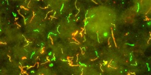
Diverse morphologies of borrelia demonstrated in this Superb Microscopic Image;
Spiral forms
Knob endforms
uncoiled undulating forms
Dot like granlar forms
Cystic (round body forms)
Fat forms (? intertwined pairs of borrelia)
Tiny spiral forms
Axial bulbosity forms
Speckled fluorescence forms
Orange staining forms
Green staining forms
Straightened forms (Bacillus-like forms)
Tennis racket forms
Respectfully,
Alan B. MacDonald, MD Fellow College of American Pathologists'
January 6,2014
Note: These diverse borrelia Morphologies Contradict the recent article by Lantos, P. et al, in Clin Infectious Diseases, 2013,DECEMBER epub, which alleges that so called "morphologic variants" of borrelia are non-contributory to the true pathology of Chronic Borreliosis infections in the Human Host.
{ Clin Infect Dis is an Official Publication of the Infectious Disease Society of American (IDSA) with Corporate Headquaters in Washington D.C.}
In addition, vague rounded areas of blush pale green staining are probably Extruded DNA/Rna of once living now dead Borrelia.
Posts: 590 | From Canada | Registered: Oct 2007
| IP: Logged |
posted
Just wanted to add, from what I've observed, it seems the descriptions of various forms of borrelia are often referred to by more than one name, so maybe the terminology isn't written in stone.
Cysts, blebs, round forms, etc. Are there universal scientific designations to these terms or are they just descriptive. And do all the researchers use the same terminology? Is it important, does it really matter?
Kinda confusing to me.
What have others found?
Posts: 590 | From Canada | Registered: Oct 2007
| IP: Logged |
posted
I have found that blebs and cysts are separate forms, though blebs turn into cysts by late info.
Spirochetes are the spiral form, but all the rest seems to be any number of various shapes and forms.
Thanks so much Looking and you other guys for all the good info & websites Am saving this thread.
Posts: 254 | From North Carolina | Registered: Nov 2013
| IP: Logged |
Lymedin2010
Frequent Contributor (1K+ posts)
Member # 34322
posted
There is a BEAUTIFUL video of Borrelia releasing a BLEB on one of it's end. So it looks like the bulbous ends might have two functions.
1) Burrowing into cells 2) Bleb formation
Don't forget blebs can also be formed throughout the length of the spirochete in the "string of pearls" formation.
Look at 6:40 on this video, the link below will take you right into the scene:
At 4:12 you can see the one of many, many, many forms of the cyst. So many & depends on how big the spirochete is when it folds in on itself & how it folds in on itself. The longer it is in cyst form I believe the longer it develops an outer protective layer & fortifies itself. Looking more bean or slipper like.
The Lyme Disease Network is a non-profit organization funded by individual donations. If you would like to support the Network and the LymeNet system of Web services, please send your donations to:
The
Lyme Disease Network of New Jersey 907 Pebble Creek Court,
Pennington,
NJ08534USA http://www.lymenet.org/


 UBBFriend: Email this page to someone!


![[Smile]](smile.gif)


 Printer-friendly view of this topic