posted
I am wondering if there is any controversy or uncertainty regarding whether the cyst form of borellia exists. My LLMD told me that the cell wall deficient form exists but not the cyst form or that he doesn't treat the cyst form for some reason?
He is a well regarded LLMD and has been recommended on this board. He has been treating lyme for about 20 yrs and is very experienced.
He mentioned something about new electron microscopes being developed but I am not sure what he meant by that. Can anybody shed light on this or what he may have meant?
If you have any info about the cyst form of borellia that I can bring to him can you please share? I am very concerned right now because I thought there was not controversy about whether the cyst form needed to be treated.
-------------------- Do unto others as you would have them do unto you. Remember Iam not a Doctor Just someone struggling like you with Tick Borne Diseases.
treepatrol
Honored Contributor (10K+ posts)
Member # 4117
posted
Whoops thats a yes
-------------------- Do unto others as you would have them do unto you. Remember Iam not a Doctor Just someone struggling like you with Tick Borne Diseases.
posted
Perhaps the LLMD is thinking that the spherical forms that look like cysts are actually the L-form/cell wall deficient forms, which he says doxy is effective against? If the L-form looks like a sphere, that may be that may be the cyst form...I'd appreciate any thoughts on this.
The article by Tom Grier on the wildernetwork refers to something like this.
CELL WALL DEFICIENT SPIROCHETES: The nice clearly definable spiral shape of the Lyme spirochete is formed by the presence of the bacteria's cell wall. Without this cell wall to determine its shape, the bacteria only has a thin pliable membrane to hold its structure together. When the bacteria turns off its genes that govern cell wall synthesis, the bacteria can change from a spiral shape, to a sphere. These spherical forms are known as L-forms or Cell Wall Deficient (CWD) forms. They represent a new hazard in the diagnosis and treatment of common diseases.
Many doctors and microbiologists have a hard time understanding that spirochetes can actually be fuzzy blobby looking spheres, but they are a reality. As a defense mechanism to mammalian immune systems, the Lyme spirochete has learned to turn off its group of genes that result in cell wall formation. This also eliminates all associated cell wall antigens.
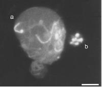
How does a microbiologist recognize Borrelia burgdorferi once it is no longer a spirochetea? In order to prove that these spheres floating around in human blood are actually spirochetes is difficult because they do not culture well. Instead a microbiologist will make a wet mount of the infected blood on a slide, and then add acrodine orange stain to stain the nucleic acids. Then a second stain that is specific for Borrelia burgdorferi is added. It is prepared by first making a monoclonal antibody specific to a Borrelia membrane antigen. Then that monoclonal antibody is tagged with a fluorescent dye. When this dye is added to the wet mount slide it stains only the Borrelia species of bacteria. Suddenly what was once totally invisible in blood by normal staining techniques, becomes visible. Spirochetes can in fact become cell wall deficient spheres.
Another technique to identify spheroplasts as being Borrelia bacteria is to take antibodies unique against Borrelia and tag them with inert gold spheres. Then culture the L-forms with the antibody-tagged-gold . When these cultures are viewed under electron-microscopes, the gold can clearly be seen attached to the membrane indicating that the antibody has attached to a protein specific to Borrelia. Therefore the spheroplasts must have once been spirochetes.
Posts: 163 | From Cleveland, OH | Registered: Sep 2007
| IP: Logged |
Tracy9
Frequent Contributor (1K+ posts)
Member # 7521
posted
Dr. Sam Donta has published on this as well.
13 years Lyme & Co.; Small Fiber Neuropathy; Myasthenia Gravis, Adrenal Insufficiency. On chemo for 2 1/2 years as experimental treatment for MG. Posts: 4480 | From Northeastern Connecticut | Registered: Jun 2005
| IP: Logged |
posted
Thanks Keebler- my caveat is that I think this doctor likely knows much more than I do about lyme disease. He is in ILADs, treated lyme since the late 80s, and has lyme and coinfections himself so he has a ton of motivation to do the research. His practice is pretty much only lyme and he is highly recommended.
On one hand, I would feel strange arguing with him about this because I feel there must be a theory or reason why he does not think the cyst form needs to be addressed. On the other hand, if there is a cyst form and I need some other med, I'm worried b/c I want to get back to my life.
But of course, every LLMD has his own approach and since there are so many uncertainties with this, maybe this is what he found in his practice. I am so unsure and confused right now. If people continue to post info/theories of these cysts I'd be grateful.
Here are just two titles from MacDonald/Borrelia search:
3: MacDonald AB.
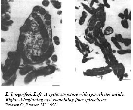
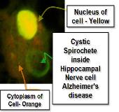
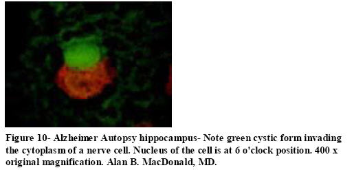
Spirochetal cyst forms in neurodegenerative disorders,...hiding in plain sight.
Med Hypotheses. 2006;67(4):819-32. Epub 2006 Jul 7. PMID: 16828236 [PubMed - indexed for MEDLINE]
===================
4: MacDonald AB.
A life cycle for Borrelia spirochetes?
Med Hypotheses. 2006;67(4):810-8. Epub 2006 May 22. PMID: 16716532 [PubMed - indexed for MEDLINE]
excerpt:
. . . Stereotypes of what a spirochete "should " look like, have actually produced a state of "perseveration" in spirochetal pathobiology. We have been "stuck" like a broken record, on the corkscrew form, and have failed to see the rest of the life cycle.
Cystic, granular, and cell wall deficient spirochetal profiles, which were well known in the 19th and 20th centuries by such titans as Schaudinn, Hoffman, Noguchi, Delamater, Steiner, and Mattman, have been repudiated by professional microbiologists, and by pathologists who practice and who confer the status of 21st century truths in microbiology matters.
Proper microscopic study, as is required by Dr. Robert Koch's second postulate, for establishing links between microbes and disease, presupposes that the microscopist be aware of the complete array of morphologic repertoires of the alleged pathogen. (Morphologies, which are herein introduced.).
LYME DISEASE PRESENTING AS POPLITEAL CYST IN CHILDREN.
Magee TH, Segal LS, Ostrov B, Groh B, Vanderhave KL.
Department of Orthopaedics and Rehabilitation, Milton S. Hershey Medical Center, The Pennsylvania State University College of Medicine, Hershey, PA
Lyme disease is the most common tick-borne disease in North America. Our review of the literature found few reports of Lyme disease presented in the orthopaedic literature. However, Lyme disease presenting as a popliteal cyst, with or without rupture, is rarely reported.
We present 4 cases of Lyme disease that initially presented to our pediatric orthopaedic clinic for treatment of a popliteal cyst.
The early diagnosis and treatment of Lyme disease may help prevent the often-devastating long-term sequelae of Lyme disease. The goal of this article is to increase the awareness of Lyme disease presenting in children as a popliteal cyst.
PMID: 17065933 [PubMed - indexed for MEDLINE]
--
A popliteal cyst is simply a sac filled with normal joint fluid. A cyst will vary in size within a given patient, depending on the quantity of fluid present ...
D Bergy
Frequent Contributor (1K+ posts)
Member # 9984
posted
I am not sure why he thinks they need a new Electron microscope to see the cyst form. There are light microscopes that go beyond the Abbe limit that should be able to do the job better as they do not kill the specimen. Much like Rife's universal microscope.
However,they are not very cheap.
D Bergy
Posts: 2924 | From Minnesota | Registered: Aug 2006
| IP: Logged |
posted
The LLMD does not think he needs a new microscope to see the cyst form I don't think. I was not sure what exactly he was getting at- but it is something along the lines that the cyst form is really the same as the L-form (or cell wall deficient) bacteria.
Posts: 163 | From Cleveland, OH | Registered: Sep 2007
| IP: Logged |
The Lyme Disease Network is a non-profit organization funded by individual donations. If you would like to support the Network and the LymeNet system of Web services, please send your donations to:
The
Lyme Disease Network of New Jersey 907 Pebble Creek Court,
Pennington,
NJ08534USA http://www.lymenet.org/


 UBBFriend: Email this page to someone!


![[Big Grin]](biggrin.gif)


 Printer-friendly view of this topic