david1097
Frequent Contributor (1K+ posts)
Member # 3662
posted
A couple of things on babesia (and malaria) you might see dots in/on the RBC but a lot of things can do this(even playing with the focus knobs). A more definative indication is the double bumps in the RBC than are sort of welded together. This is pretty definative as being a parasite. Of course there also the cross form which is talked about a lot, but not seen that often. If this is seen this is rather conclusive (I think).
Another interesting point is that babesia infection makes the RBC walls weak and they wil break during a thin smear. You might see clusters of 4 dots and a stringy looking thing. I have seen this before (they are all in a cluster between other RBC's) and suspect it is the shell of the fractured RBC with 4 parasites. I have also seen the RBC just as it has fractured and the "parasites" coming out, likly from the surface shear during the smear.
If the blood is dried you can just put the oil directly in the slide but the oil lens is designed for use with a cover slip (in most cases). With a cover slip the image is a bit better defined.
I have a question Someone here mentioned "something:" growing out the RBC's after a period of time, Does anyone have a more detailed description of what they see? the RBC's in wet smears will develop funny bumps over time and not look like RBC's at all but this as far as I can see is normal.
Posts: 1184 | From north america | Registered: Feb 2003
| IP: Logged |
quote:Originally posted by david1097: I have a question Someone here mentioned "something:" growing out the RBC's after a period of time, Does anyone have a more detailed description of what they see? the RBC's in wet smears will develop funny bumps over time and not look like RBC's at all but this as far as I can see is normal.
I believe that this was without the staining and done with the dark field filter. What they are referring to is the borrelia hiding in a RBC (from the ABX) comes out when the nutrients in that RBC get low. Apparently there is a llmd that does this I think.
Then you can see the Bb when it comes out. I don't think it was to do with babs because you definitely need a stain for that which kills anything alive on the slide.
This has just sparked off another question of my own. Why does the Bb go and hide in the RBC? or more to the point - How? If we can stop it from hiding, perhaps we would be in a better position.
If it just lands up there through no choice of it's own, there is probably nothing much we can do to stop it. If it actively seeks out the RBC's to hide then perhaps there is a way of stopping that from happening. - another crazy idea? Posts: 27 | From Mars | Registered: Jan 2006
| IP: Logged |
posted
Supposedly Bb is not found inside RBC's, but it seems like it does alot of things it is 'not supposed to'. Maybe they should have explained the rules to it.
If you look at the bottom of page 20 (page 23 of the pdf) of the 'survival in adverse conditions' paper you can see a drawing of a more complex life cycle for syphilis that was proposed in 1913 by McDonagh. (Read his text too.)
The spider like forms in his drawing I have seen many times in by own blood using darkfield and a live specimen. They appear to be inside the RBC's, and are usually only visible in that form if the RBC gets ruptured. Sometimes a wormlike projection or two are seen hanging out of an otherwise intact RBC. Other RBC's are sometimes seen trembling and with distorted shapes.
I had tried lysing the RBC's with distilled water and other chemicals to see better what was inside, bit found the organisms inside would also lyse and be destroyed almost as fast as their rupturing RBC home made them visible.
A better technique was to simply apply firm pressure on the coverslip over the fresh blood sample using a clean piece of lens paper. With a little practice alot of the cells are ruptured, leaving their occupants exposed but still intact. They are very numerous.
Leaving a sample overnight also causes wormlike things to become visible, like the ones Andy Wright filmed.
Note that the familiar spiral appearance is lacking in the organisms seen. This is also true of many filmed by people like Marie Kroun, Andy Wright, and Mr. Burgdorfer himself.
Alot of the spirally ones filmed are in cultures, from ticks, or are different species. For whatever reason the perfect spiral form is hard to find in a human.
So what are these things? Are they all Bb? I believe at least some of them are. Do some research and you will find there is controversy.
Some of them could be of species yet to be identified, and of unknown medical significance, if any. Or, they could just be pieces of protein, as some propose. The 'old guys' from the early part of the last century came to associate them with disease in their human hosts, though.
The technology now exists to answer these questions, but unfortunately there is little interest in researching cures for disease anymore. Everyone is too busy looking for the next blockbuster symptomatic drug for erectile dysfunction, or their latest insult, restless leg syndrome. Posts: 714 | From San Antonio TX | Registered: Oct 2004
| IP: Logged |
posted
This is such an interesting thread...Thanks for all the information and links to excellent papers! I wished my background was in the field of microbiology.
I simply can't believe more scientists are not researching Bb and other coinfections - seems pretty fascinating to me...but I do think that the human spirit is one of wanting to conquer...these little bugs are simply humbling...they are an enigma and their behaviour is still a mystery.
A scientist is not left feeling God like in the presence of these bugs...for many there is no fun in that
Keep the great info. coming. Still haven't gotten a microscope or shopped for one. I am in a major transition in life...time is at a minimum...money at a minimum too but I will continue to learn and hopefully be able to get a microscope sometime soon.
-------------------- �Pride is concerned with who is right. Humility is concerned with what is right.� - Ezre Taft Benson Posts: 655 | From NC, Exit 88 on the Deer SuperHighway | Registered: Dec 2004
| IP: Logged |
david1097
Frequent Contributor (1K+ posts)
Member # 3662
posted
Do you have a lionk to any of the pictures? I would really like to see them.
I had often wondered about RBC. WBC infection myself. This would be the ONLY way that a insect could pick up the disease since there are so few organisms free flowing in the blood. The only issue I suppose is that Bb are longer than a RBC diameter (I think), so it would be a tight fit, ie i would have to roll up or something.
Has anybody tried to culture the RBC or better yet inject it into an immunocomrpmized mouse to see what if any infection is passed on. Since the resouvoir for all this nasty stuff are rodents, what ever it is it must be viable in them.
Posts: 1184 | From north america | Registered: Feb 2003
| IP: Logged |
posted
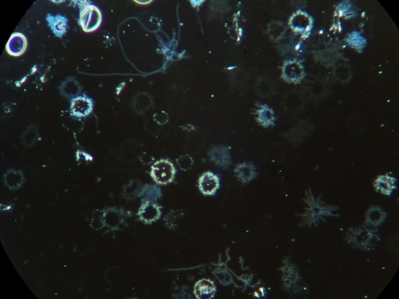
OK, I have my scope and I saw some keets in the blood today!
The blood was from a small finger prick last night, smeared over the slide and half of it was covered by a slip. The other half was left to dry.
It took allot of looking and a "make your own" dark field patch stop filter but interesting to see the keets in the end.
I couldn't get a good picture from the digital camera, so this is from a digital video camera.
Keets in the middle!
There are Three in the middle. The lower one is quite long and the ones just above that have just divided - I saw them do it, but didn't manage to capture it
There also seems to be allot of small (very) granules about (not in this pic though). More so than the keets. The granules are about 1/8th (maybe smaller) the size of a RBC.
I have seen some of the black spidery looking things about as well. These stretch across many RBC's and I think it is a bit of candidia. Not too much thank god.
No stain used, just a small disc under the condenser for the dark field contrast.
Next - staining for the babs...
edited for JPG picture
[ 16. January 2006, 01:17 PM: Message edited by: Starship Trooper ]
Posts: 27 | From Mars | Registered: Jan 2006
| IP: Logged |
posted
WooW, this is not dark field right ? How big magnification is this ??? The best way is to have CCD camera and record movie
Posts: 641 | From Wroclaw, Poland | Registered: Mar 2004
| IP: Logged |
quote:Originally posted by wrotek: WooW, this is not dark field right ? How big magnification is this ??? The best way is to have CCD camera and record movie
As I understand it, it is dark field. It may not be the best in the world because it was a home made filter. I have got very good dark field images using this technique with a lower magnification (400X) but the filter has to be smaller on 1000X for some reason.
This is at 1000X - 100 oil objective and a 10x wide field eye lens.
Here is the science bit (see one of my posts above):
Here is the filter I made. Yes it is stuck on with tape and yes it was cut out with scissors and no I don't really know what I am doing. The blue filter helps the contrast a bit as well. Posts: 27 | From Mars | Registered: Jan 2006
| IP: Logged |
posted
Yes i am familiar how dark field condenser works, Interesting u did it . IF this forms u have seen were spirochere than congratulations cause laboratories have problem with visualisation this bugs i guess.
U should make a movie., what scope u have?
Posts: 641 | From Wroclaw, Poland | Registered: Mar 2004
| IP: Logged |
Starship... if you save your files as .JPG type instead of .BMP they will be about 1/10th the size and will load faster for anyone with a slow connection. Good work!
--------
The darkfield photos are from my 30 year old American Optical series 10.
You do not have to spend huge amounts of money if you are resourceful. I found that one on eBay.
Blood cells on a slide so the same thing blood cells do when you cut your finger... they clot.
Seeing them clump up on a slide after it sits awhile is pretty normal, but is used to scare people and sell people various nutritional supplements by some.
If you prepare a very thin film so the cells have to lay flat you will not see this. If the film is thicher they will tend to stack like a roll of Lifesaver candies. Blood spread out on a glass slide is not 'happy' blood anymore... it is blood that is dying.
Starship, I understood that you were looking at a live specimen. Actually I thought you were pretty creative with that little darkfield stop you constructed. You might try enlarging the dark spot gradually until the light barely shows at the edges of the field. On some scopes you might not be able to get it to work, but there is no harm in trying.
Those could well be a form of the spirochetes you saw, as divsion like that has been described in alot of the older scientist's writings. They will be more worm-like and you may not see the classic coiled look.
That's the problem (and the addiction) with these instruments... we always see things we wish we could see a little bit more clearly!
[ 16. January 2006, 02:56 PM: Message edited by: James H ]
Posts: 714 | From San Antonio TX | Registered: Oct 2004
| IP: Logged |
posted
James did You do this dark field photos? They are beautifil. How do u pick blood ?
Posts: 641 | From Wroclaw, Poland | Registered: Mar 2004
| IP: Logged |
posted
Wrotek, it is a 516B-II BIO-microscope 40X-1600X from Digilens.
James, the blue filter might make it look like a stain, but I haven't used one - honest. I thought I was getting a dark field effect because what look like the keets (not so sure now) are brighter than it's background.
I honestly did see them divide - which took about 30 seconds from start to finish and then they were still for the best part of an hour when I left them alone. This is what made me think it was bacteria - a keet at that.
Tell you what, I will repeat the experiment and try a better disk on the filter. Don't know what I'm going to cut it with though.
Posts: 27 | From Mars | Registered: Jan 2006
| IP: Logged |
A have question about this photo, what do You think about these clumps of red cells? I mean bacteria is causing red cell to gather together what do You think ?
Posts: 641 | From Wroclaw, Poland | Registered: Mar 2004
| IP: Logged |
A have question about this photo, what do You think about these clumps of red cells? I mean bacteria is causing red cell to gather together what do You think ?
"CAUSE: protein linkage. Often poor protein digestion. The pancreas may be off. Excess dietary protein, poor assimilation. Eating too much animal protein. Blood too toxic (altered blood pH-zeta potential down) from stress, coffee, cigarettes, meat, etc. Dehydration, not drinking enough water (which by the way, is one of the top undiagnosed causes of many ailments). Eating the wrong foods for the blood type, e.g. wheat consumption by type O's, beef consumption by type A's, etc.
SIGNS: Fatigue, shortness of breath - RBC's cannot carry oxygen; stress on heart. Cold hands/feet - poor circulation."
Posts: 27 | From Mars | Registered: Jan 2006
| IP: Logged |
I am no good at web sites (as you can probably see) but I have managed to do a video. The size of the file is just over 1.7mb.
This was done with a 10x objective and a 10x eyepiece and the camera set to another 8x making a total of 800x. I could only manage to get this dark field to work at such a low objective. Don't know why?
The smear it allot thinner than the last one and I now also have a larger disk on the filter - still stuck on with tape though!
[ 04. April 2006, 10:02 AM: Message edited by: Starship Trooper ]
Posts: 27 | From Mars | Registered: Jan 2006
| IP: Logged |
posted
The darkfield photos are from my 30 year old American Optical series 10. You do not have to spend huge amounts of money if you are resourceful. I found that one on eBay.
Blood cells on a slide do the same thing blood cells do when you cut your finger... they clot.
Seeing them clump up on a slide after it sits awhile is pretty normal, but is used to scare people and sell people various nutritional supplements by some.
If you prepare a very thin film so the cells have to lay flat you will not see this. If the film is thicher they will tend to stack like a roll of Lifesaver candies. Blood spread out on a glass slide is not 'happy' blood anymore... it is blood that is dying.
Watching the fibrinogen strands form as it sits awhile is also normal, and is part of the clotting process that dying blood goes through. If it didn't, you would bleed to death from a papercut.
Fibrinogen strands look like spider webs that glue everything in place, whereas the 'spidery' things, also called the 'darkfield objects' are free floating objects with waving tentacles. They are very different.
Unfortunately live blood microscopy has in my opinion been taken over by snake oil salesmen using it to sell vitamins. Not all of what you will find on the subject is good science. Go to the sites where they sell the microscopes packaged with a video monitor. Often they will say straight out that it isn't for diagnosis, but is a sales tool to get patients to agree to a practitioner's chosen protocol. That is a disgrace in my opinion.
Starship: yes, I understood that you were looking at a live specimen. Actually I thought you were pretty creative with that little darkfield stop you constructed. You might try enlarging the dark spot gradually until the light barely shows at the edges of the field. On some scopes you might not be able to get it to work, but there is no harm in trying.
Those could well be a form of the spirochetes you saw, as divsion like that has been described in alot of the older scientist's writings. They will be more worm-like and you may not see the classic coiled look. Identity can be a problem.
That's the problem (and the addiction) with these instruments... we always see things we wish we could see a little bit more clearly!
Posts: 714 | From San Antonio TX | Registered: Oct 2004
| IP: Logged |
treepatrol
Honored Contributor (10K+ posts)
Member # 4117
posted
I have a question What abx's are you guys on when you took these pics????
-------------------- Do unto others as you would have them do unto you. Remember Iam not a Doctor Just someone struggling like you with Tick Borne Diseases.
posted
Excellent! You got the darkfield working really good for having to improvise. You might not be able to get the geometry right for it to work on a higher power, but I think that is pretty good.
Could you see movement of those spirochete looking things? Slides usually have microscopic contaminants on them that can trick us, but contaminants don't move.
Good work! It is interesting, isn't it.
James
Posts: 714 | From San Antonio TX | Registered: Oct 2004
| IP: Logged |
quote:Originally posted by James H: Excellent! You got the darkfield working really good for having to improvise. You might not be able to get the geometry right for it to work on a higher power, but I think that is pretty good.
Thanks..
quote:Originally posted by James H:
Could you see movement of those spirochete looking things? Slides usually have microscopic contaminants on them that can trick us, but contaminants don't move.
Unfortunately I couldn't see any movement. I did on the slide before, but that was before I sorted out the darkfield. The one before was also a much thicker smear.
This one was quite thin and was left over night. Could they be dead ones? They look kind of spiral shaped. Don't know if they are the right size. Does anyone have a size comparison with some of the work that Andy W. has done?
Posts: 27 | From Mars | Registered: Jan 2006
| IP: Logged |
treepatrol
Honored Contributor (10K+ posts)
Member # 4117
posted
quote:Originally posted by treepatrol: I have a question What abx's are you guys on when you took these pics????
up for comments
-------------------- Do unto others as you would have them do unto you. Remember Iam not a Doctor Just someone struggling like you with Tick Borne Diseases.
You have hit on the reason behind this little microbiology exercise. It is really difficult to tell sometimes if a particular treatment or medicine is helping. Response is always slow, and many times a beneficial response means that you feel worse. I am looking for evidence of effectiveness. Interpretation is very subjective of course, but you can see any CHANGE. It has to be weighed with how you feel too.
The photos I posted cover a period of a year and a half, and some are from different people.
We've cycled through quite a few of the common antibiotics, and unfortunately none have the kind of effectiveness we'd like to see.
Doxycycline starts to visibly clean things up at about 600mg, but few can stand that for long.
Macrolides and Ketek do not have any effects that are VISIBLE as far as I have been able to determine, but some report feeling alot better with these.
Penicillins and the Cephalosporins have widely known effect of forcing the Borrelia into cyst form, but seem to fall short of eliminating them.
Fluconazole in combination with a penicillin shows some interesting activity... tentatively.
Posts: 714 | From San Antonio TX | Registered: Oct 2004
| IP: Logged |
They look right. If it was a dried smear without a coverslip on it you wouldn't see movement anymore.
Look for some more if you want.
My point in all of this is that much of what is going on with this is observable, and without huge amounts of money.
Interpreting what is seen is maybe another matter.
These are the only ones I have seen on this slide. The other one had more. I know I have lyme because of a positive test from Igenex, but I have never been tested for Babs which is a far more definitive test by the looks of things - if you see any.
So - Ill get back once I have stained the slide and maybe some pics too. Thanks for your help. Very interesting!
Posts: 27 | From Mars | Registered: Jan 2006
| IP: Logged |
treepatrol
Honored Contributor (10K+ posts)
Member # 4117
posted
Thanks James maybe everybody who are checking blood for Bb should do it like this.
ABX on = Date started=
ABX first of month blood check
ABX end of month blood check
or three sldes at first of month and spirochetes any?
or three slides at end of month and spirochetes any? ::::::::: ___________________ ------------------- __________________
Or the ABX your on= Date started=
And do the blood check every 10 days = 3 x in 30 days.
That way you catch cycles of Bb and significant abx exposure too.
-------------------- Do unto others as you would have them do unto you. Remember Iam not a Doctor Just someone struggling like you with Tick Borne Diseases.
posted
If only it would lend itself to such neat, orderly, and quantifiable analysis!
The problem is that what can be seen in the blood at a given moment does not represent the whole body. In the morning after a good night's rest the blood can appear almost sterile... yet appear totally septic again by that evening. I don't think the infection fluctuates that much, but what is seen in the blood does. The blood is maybe not the focal point of the infection, but it is a convenient thing to examine.
I think daily samples taken at the same time of day could give a general idea of quantity, especially for the same person. There is alot of variance to deal with though.
I'm looking for changes in appearance and condition, and take lots of photos to compare. If membranes look damaged or deformed, or the organisms appear sluggish, that is of interest.
Posts: 714 | From San Antonio TX | Registered: Oct 2004
| IP: Logged |
"CAUSE: protein linkage. Often poor protein digestion. The pancreas may be off. Excess dietary protein, poor assimilation. Eating too much animal protein. Blood too toxic (altered blood pH-zeta potential down) from stress, coffee, cigarettes, meat, etc. Dehydration, not drinking enough water (which by the way, is one of the top undiagnosed causes of many ailments). Eating the wrong foods for the blood type, e.g. wheat consumption by type O's, beef consumption by type A's, etc.
SIGNS: Fatigue, shortness of breath - RBC's cannot carry oxygen; stress on heart. Cold hands/feet - poor circulation."
I drink coffee and i deffinetely notice difference In my hands and feet, they become very cold.
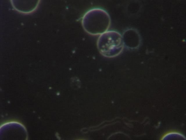
This clothing explenation as a dying blood(thrombosis) and bad diet (stress) is very convincing but i also have seen in Dr Lida Mattman book Stealth Pathogens a photograph where young spirochetes created hemagglutinin that makes erythrocytes rosseting around spirochete. This is this photo http://www.borelioza.tivi.pl/photogallery.php?photo=15 signature of photo is "Young colonies of B.Butgdorferi may make hemagglutinin which causes erythrocytes to rosette around them", any ideas they do it? Easier cross from one cell to another without risk meeting immune system cell ?
Posts: 641 | From Wroclaw, Poland | Registered: Mar 2004
| IP: Logged |
posted
Interesting pictures. This one looks alot like what Starship Trooper was showing us:
That picture you linked is definitely not normal.
It is just that some people will point to any kind of normal appearance or normal degradation of an old sample and use it to scare people so they buy products. I am sure you know what I mean.
Eastern Europe is WAY ahead of the US in studying this desease and looking for practical ways to deal with it. If anyone finds a cure, it will be them, using 50 year old equipment, not our 'head in the sand' US CDC geniuses.
Posts: 714 | From San Antonio TX | Registered: Oct 2004
| IP: Logged |
posted
This pictured You showed is multiple sclerosis spirochete also from Dr Lida Mattman Book-Stealth Pathogens. Dr Lida Mattman is not selling anything, she is microbiologist nominated to nobel prize in medicine
Posts: 641 | From Wroclaw, Poland | Registered: Mar 2004
| IP: Logged |
posted
I was not referring to Dr. Lida Mattman in that way... she is of the kind I greatly admire. Posts: 714 | From San Antonio TX | Registered: Oct 2004
| IP: Logged |
david1097
Frequent Contributor (1K+ posts)
Member # 3662
posted
OK guys... for your viewing (dis) pleasure try these links:
And James, what you have seen in the RBC's has been looked in some detail and PCR'd to confirm that they are some type of bacteria.... even in "healthy" people. It looks like it has been reported several times and once in the journal "Nature" so it appears to be able stand the assualt of peer naysayers...... Still it looks like nobody listens (or does not want the added complication in their lives... or maybe know something that everyone else does not.)
Heres the link to recent paper with references. They apparently stumbled across this using dark field trying to spot spirocettes in some degerenative brain disporders (sounds familiar?)
posted
Thanks, David. That last link was interesting. It seems they isolated Pseudomonas maltophilia from some people's blood.
From the pictures I have seen elsewhere of Pseudomonas maltophilia I don't think that particular bacteria is the same as the darkfield forms I am seeing, though some of the rod forms look similar.
It does show that they CAN identify an unknown bacterial organism in blood fairly easily if they want to, and that blood is anything but sterile.
These strange looking forms have been noticed as early as 80 or 90 years ago, and they really have not been studied much in all that time.
I don't automatically assume that everything seen is a pathogen, they may just be interesting 'things'.
How about this guy?
[ 18. January 2006, 01:03 PM: Message edited by: James H ]
Posts: 714 | From San Antonio TX | Registered: Oct 2004
| IP: Logged |
caat
Frequent Contributor (1K+ posts)
Member # 2321
posted
Wow. I missed a lot this week. So much great information! Thanks James and thanks David for posting that atlas etc.
Congradulations on that home made darkfeild filter Starship Trooper, that's impressive! Do you have a photo of it? I can't understand it from the diagram but might understand a photo. Where are the glass lenses underneath the stage from? Or is that the condenser and you tape cardboard under the stage? Maybe it's not that simple...
I still haven't ordered stain yet, I think my liver is rebeling and I can only do about 3 things per day right now. I feel like I drank a case of whiskey last night...(It's OK though- I'm ramping down and ending this protocol and should start feeling a little better in several days.)
Posts: 1436 | From Humboldt county ca usa | Registered: Mar 2002
| IP: Logged |
posted
What a fascinating post! I'm going to have to come back and read it properly later on. I have just looked at a couple of the sites that were linked and somehow I really don't fancy my breakfast now! Yuk!!!
Posts: 229 | From United Kingdom | Registered: Jul 2005
| IP: Logged |
david1097
Frequent Contributor (1K+ posts)
Member # 3662
posted
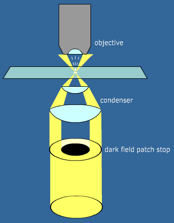
OK, Here's a detailed explanation of how I did it.
First a little about the microscope. You will notice from the pics that the objective is quite big. In fact the whole thing was bigger than I expected. I guess that the bigger the lenses, the better the pictures but the size shown here is a standard in microscopes called the DIN standard.
This means that you can go and buy an objective from any local shop and it will fit. This is the objective I used.
Just under my finger is what you place the slide on and that is called the "stage". There is adjustment underneath the scope so that you can move the slide around without touching it yourself.
Under the stage is the condenser. This is responsible for focusing the light onto the slide you are looking at. It is important to have an adjustment on this for the dark field, but a little hard to explain why..
This is the condenser under the stage. I am pointing to a filter holder where we will be putting our filter. This just slides out.
Sliding out the filter holder, you see this.
In fact, this is a bright field condenser with a filter holder. What I am going to explain below uses this condenser. I have asked about a dark field condenser and they are available. Because this Microscope is standardised, I am able to just buy one off the shelf. This could be a decision if you decide to get your own.
Here is the condenser removed. Note that I have inserted the filter in the holder in this photo.
OK, that said, lets get on to making the filter. The filter is a device to block the light from going into the objective. The condenser focuses a ring of light onto the slide all into one point, so the slide is well lit.
When the objective sees it, the only light it sees is from the objects on the slide. This is why you see light objects on a dark background.
These were the disks I made to produce the hole in the light.
Next I laminated them and cut them out so that they would fit into the filter holder.
The disks need to be central within the laminate and they need to be as round as you can cut them.
Place them in the filter holder and look at a slide through the scope. I found it best to use a 10x objective and a 10x eyepiece lens. Yep that is just 100x total. This is to get your dark field working.
You will have to alter the adjustment for the focus of the condenser so that you see bright blood cells on a black background. If you do not see this, change the disk for a larger one and repeat until you do.
Once you have the darkfield sorted at 100x, connect your camera or video camera to the eye piece as shown and zoom to about 8 times. This takes a little setting up as you have to the lenses straight.
You may have to slightly refocus on the microscope for the camera to see it better.
Once you have got this far, looking is all there is left to do. Instead of looking through hte eyepiece, you can now look on the screen of your camera. Move the slide about using only the adjustment for the stage on the scope.
One thing that crossed my mind was that my pics are the classic spirochete form, where by others are rolled up into cysts. This may be because I am on doxy and they are not. Either way, another interesting fact is that my blood was taken late in the evening. I don't think they are in the blood so much in the morning. One ref to back this up is at http://www.canlyme.com/lymeconf04.html
Skip down to the part on Nick Harris and lyme testing.
Another interesting fact about my logs is that I have a +ve PCR - first time. Don't know if this was good luck or that there is something in it, but out of the two smears that I have done, I have seen Keet looking bodies in both of them.
If you have any further questions, I will gladly answer them.
Posts: 27 | From Mars | Registered: Jan 2006
| IP: Logged |
Edited - Actually saying that they do look a bit pricey
Posts: 27 | From Mars | Registered: Jan 2006
| IP: Logged |
caat
Frequent Contributor (1K+ posts)
Member # 2321
posted
Starship, that was very clear. Thank you for taking the time to make a DIY lesson! I've saved this web page to look at later if I get a scope with a condenser. And it helps to know what condenser to look for (w/filter holder).
[ 24. January 2006, 04:19 PM: Message edited by: caat ]
Posts: 1436 | From Humboldt county ca usa | Registered: Mar 2002
| IP: Logged |
caat
Frequent Contributor (1K+ posts)
Member # 2321
posted
I'm starting to look to order giemsa stain and what others might be useful later.
One of the manuals says to dilute giemsa with ph buffered water.
Does anyone know if I can just use baking soda or vinegar to buffer with or would those interact with the stain? Do I need something special? maybe citric acid & ???
And if giemsa has any other name? So far all the ones that carry it seem to be professional's companies. I know I had a problem ordering orchid sowing materials before from these- many want a title or company to send to.
Posts: 1436 | From Humboldt county ca usa | Registered: Mar 2002
| IP: Logged |
quote:Originally posted by caat: I'm starting to look to order giemsa stain and what others might be useful later.
One of the manuals says to dilute giemsa with ph buffered water.
Does anyone know if I can just use baking soda or vinegar to buffer with or would those interact with the stain? Do I need something special? maybe citric acid & ???
And if giemsa has any other name? So far all the ones that carry it seem to be professional's companies. I know I had a problem ordering orchid sowing materials before from these- many want a title or company to send to.
Hi Caat,
I ordered mine in a pre made solution from www.ralamb.net Their part number was LAMB/120-D. I am not sure if they do a ready-made solution in your part of the world, but they will sell you the powder to make it up yourself. It might be worth asking them for this part number, as they are the same company just a different country.
The buffer solution can be deionised water. You should be able to get this in most super markets as people use in irons to prevent them from scaling up. Alternatively use distilled water. Mix 9 parts stain to 1 part water.
The problems I have run into (not big ones) is that the stain seems to have microscopic hair like things in it and other debris. It needs to be filtered just before it is put on the slide.
Another thing you need to have is ethanol to stop the RBC's from bursting when the stain is applied. I tried it with some vodka for 5 minutes and it seemed to be OK! - Don't drink it afterwards though...
When I get a better procedure together I will post it here. Also, just in case people don't know how a smear is done, I can probably do a small example video.
ST.
Posts: 27 | From Mars | Registered: Jan 2006
| IP: Logged |
caat
Frequent Contributor (1K+ posts)
Member # 2321
posted
ST,
Thanks!
I think distilled water has a ph of 5.something. From what I've read the water should be buffered to between pH 6.8 & 7.2 for diluting giemsa.
They usually use phosphates as a buffer; a mix of sodium phosphate and potassium phosphate, although it seems you could use one or the other. I found no mention of baking soda, although it was mentioned as a buffer for other stains. The phosphate mix goes for around $6 in the US.
Of course we probley don't need to be as particular...
I'm going to try Wright's stain first. Haven't read the manual on it yet.
Thanks for the tip about vodka!! I was going to buy wood alcohol in the hardware store today, but wasn't sure if it should be straight methenol or if it could be what they sold- methenol ket-something.
Vodka is a lot less toxic and I can dispose of any left over to some of my neighbors who will be very happy about that. I will just have to listen to them fight later... LOL
Posts: 1436 | From Humboldt county ca usa | Registered: Mar 2002
| IP: Logged |
posted
OK, still trying slightly different techniques for the staining, but thought you might like to see some results.. Not absolutely sure what I am looking at!
Caat, pure water has a ph of 7. If you use the wright stain, I believe it has a fixer in it so you can skip the ethanol bit. 90% ethanol is pretty easy to get hold of though.
I have also found that the pictures come out better if the eyepiece on the scope is removed.
posted
We've seen videos of the spirochetes on the Canadmian Lyme site. They were actually very frightening to see, burrowing into the B cells. So I wonder what kind of technology was used in finding those and I wonder why tests for Lyme aren't finding the spirochetes on this kind of technology.
I saw my blood many times on the bradford microscope. I have a severe case of Lyme, reinforced by positive Western Blot. The spirochete could not be seen on this microscope. All we saw were signs of infected B cells, and the cyst form -- both indirect speculations.
-------------------- Jeff Posts: 533 | From CA | Registered: Mar 2006
| IP: Logged |
Dave6002
Frequent Contributor (1K+ posts)
Member # 9064
posted
I think the video on that website was in vitro culture not from in vivo.
A European scientist suggest that looking for "apple" instead of "snake" when you check your blood under a microscope.
Because the spirochetes have become granuated. Still a puzzle that why PCR cannot detect the granuated Bbs, he suggested that the granuated Bbs have RNA not DNA. Of course this is a wild thinking. I doubt it.
Posts: 1078 | From Fairland | Registered: Apr 2006
| IP: Logged |
posted
hi, Just thought I'd mention that I have a crappy little microscope.. So I wouldn't be able to see the bb anyway. But you can look at other things like yeast. But I was using it the other day and I have so many floaters in my eyes that I can't really even see anything. I'm assuming that since this is a common lyme symptom that others will have the same problem. So before you blow a bunch of money on a microscope, you might want to check out the number of floaters in your eyes.
I did the biology thing back in college and the floaters weren't there then... so it was a bit of a surprise.
Posts: 207 | From san francisco, ca | Registered: Mar 2005
| IP: Logged |
posted
Ive been watching the PBS special Malaria:Fever Wars and I noticed that the scientist at Oxford quick-dry their blood samples with hair dryers.I assume the parasites are much easier to identify,because of their size. I am excited about this thread and the Knock-Out clinical trials thread and I hope they stay active! Im going to keep mentioning the fact that we dont compile personal treatment data(like on remedyfind.com)untill the administrators of this site create a format or easy links ...for that reason! Alan
-------------------- Charter member of the ~ Delux Toasting Club ~ Our Moto: "Take No Prisoners" Posts: 95 | From San Diego | Registered: Nov 2005
| IP: Logged |
The Lyme Disease Network is a non-profit organization funded by individual donations. If you would like to support the Network and the LymeNet system of Web services, please send your donations to:
The
Lyme Disease Network of New Jersey 907 Pebble Creek Court,
Pennington,
NJ08534USA http://www.lymenet.org/


 UBBFriend: Email this page to someone!


![[loco]](graemlins/loco.gif)
![[Frown]](frown.gif)
![[Wink]](wink.gif)

![[bonk]](graemlins/bonk.gif)

![[Eek!]](eek.gif)

![[Smile]](smile.gif)


 Printer-friendly view of this topic