posted
It's a stain ya, but I've never seen a malaria ring look quite like that before so I'm not sure.
Posts: 71 | From Canada | Registered: May 2016
| IP: Logged |
Lymedin2010
Frequent Contributor (1K+ posts)
Member # 34322
posted
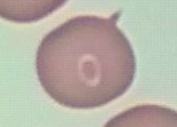
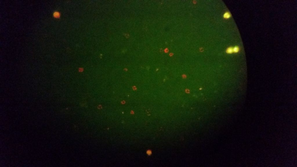
It sure looks like the shape of a ring form, but it did not stain & the circle around it did not stain as well. The center of the circle is pink like hemoglobin and the circle is clear like a halo. As a result I don't think this is Babs ring form. The spike or protrusion of the rbc is just a deformity in the rbc I think.
The rings around stains that do have them stain blue to purple & are clear stained rings AROUND the halo.
Check out these forms here & you can see a halo around them too. I still have not done anything else to figure out if they are platelets or rbc crenation burrs for sure. Maybe even some of the darker ones might be pathogens, around the rbc, but I am still not sure on this video until I do more live viewing with the filters & compare it to normal blood.
I was ready to call it off as a random artifact, but in a stain I did tonight I found two more identical to the one I posted! It's very weird. I'm going to have to dig a little deeper into this...
I tend to think those are platelets, but it's obviously hard to tell. Have you done any work with the quick dip?
Posts: 71 | From Canada | Registered: May 2016
| IP: Logged |
Lymedin2010
Frequent Contributor (1K+ posts)
Member # 34322
posted
Some, but not a whole lot & I never got to refining my technique yet. Before 2017 is over I will get to that.
Posts: 2094 | From NY | Registered: Oct 2011
| IP: Logged |
Lymedin2010
Frequent Contributor (1K+ posts)
Member # 34322
posted
DIRECTLY FROM THE CDC!!!!!
"Although no cases of Lyme disease have been linked to blood transfusion, scientists have found that the Lyme disease bacteria can live in blood that is stored for donation. Individuals being treated for Lyme disease with an antibiotic should not donate blood. Individuals who have completed antibiotic treatment for Lyme disease may be considered as potential blood donors. Information on the current criteria for blood donation is available on the Red Cross website"
If anyone of these anti-Lymers gives you grief, just tell them to put their science where their big anti-Lyme rhetoric mouths are & offer them an infusion of 1 pint of your blood. Then see how quickly they reveal their spineless inconsistencies in the deplorable act of terrorizing the sick.
If they are too worried about receiving blood, then just offer to inject them with 1 CC of Borrelia burgdorferi colonies & tell them don't worry it is easy to treat with 2 weeks of Doxycyline.
Lymedin2010
Frequent Contributor (1K+ posts)
Member # 34322
posted
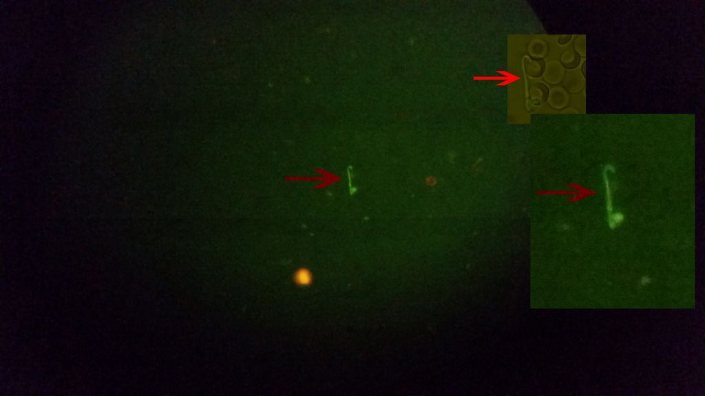
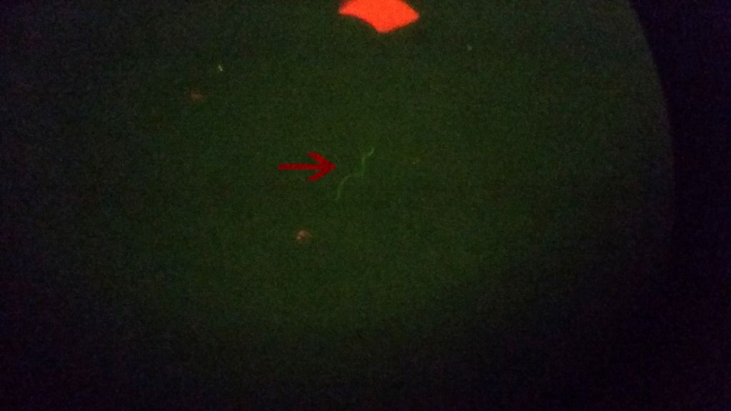
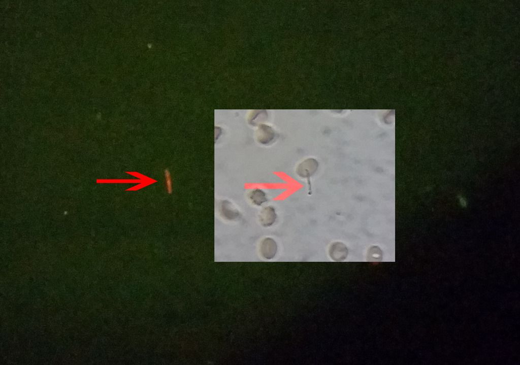
So I have only found 2 string-like subjects in my blood so far out of 7-8 slides, and not all slides were ideally made. One of them did not fluoresce & I have video of it and I will post it up eventually. And this one, which did fluoresce.
I do see a lot of smaller particles fluoresce though, but it is too hard to determine if they are DNA from the WBC's or spiro cysts and/or blebs. Since I am not seeing many string-like objects, be it actual spiros or false spiros, it must be that the AO solution is causing them to get destroyed or cyst up. Once they cyst up, it is hard to tell them from other components in the blood. I have not had the energy to check with much aggression though & the attention/time that it deserves.
Spirochetes are also delicate subjects & can lyse/die easily. If they are not given ample time to cyst up, they can get destroyed too and their delicate nature is known by the people who handle them...hardy but delicate.
In the pic below I also provide a zoomed version of it to the right & one with some light to see the rbc's that it is next to. Also, to the right of the subject in the center is another what might be infected rbc, similar to my last co-infection video.
Lymedin2010
Frequent Contributor (1K+ posts)
Member # 34322
posted
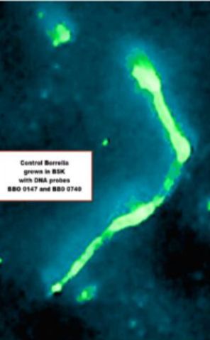
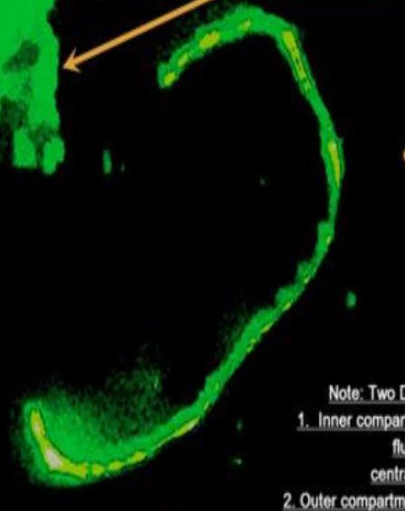
Here are some of Alan MacDonald's PanDNA and DNA Probe stains. The stains are not always ideal & what we would normally expect and they don't look very typical of lab grown spiros.
Lymedin2010
Frequent Contributor (1K+ posts)
Member # 34322
posted
"Figure 3. Spirochetes are present within xenodiagnostic ticks that fed upon saline- or antibiotic-treated mice at 12 months after treatment.
Indirect immunofluorescent staining of B. burgdorferi in the midguts of ticks that fed upon saline-treated (A) or ceftriaxone-treated mice (B) at 12 months after treatment."
posted
Keep up the good work with the fluro lymedin2010. Looks very promising.
Posts: 71 | From Canada | Registered: May 2016
| IP: Logged |
Lymedin2010
Frequent Contributor (1K+ posts)
Member # 34322
posted
Thanks mustard!
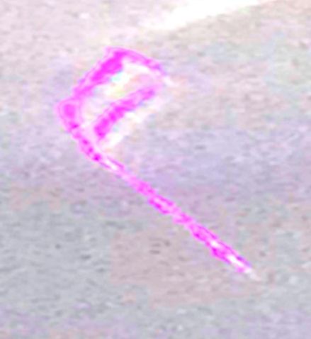
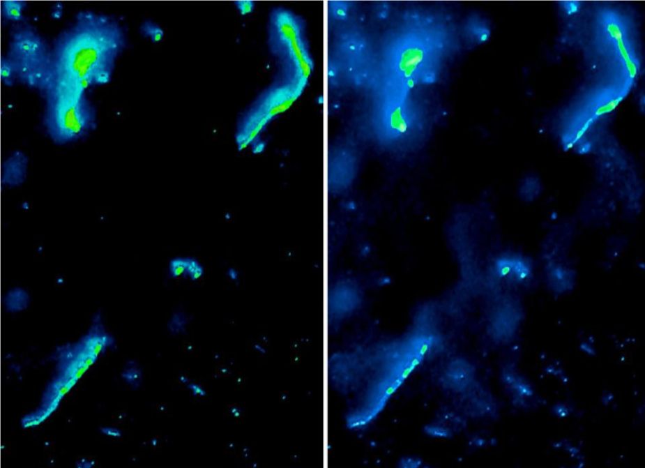
A new video from Ann, who grows confirmed laboratory grown Borrelia cultures. Interesting, as at time 3:25 the Borrelia has a slight bend, just like the one that I captured in my lyme blood via fluorescent staining.
I will also attest to the variation in spiro length & size & shapes, as that became obvious with my tick studies. In this one tick I found ridiculously super massive spirochetes of the same unusual type I had been finding. I couldn't believe just how big they were. And it was not just one, ALL OF THEM were big... a DNA lineage of massive spiros.
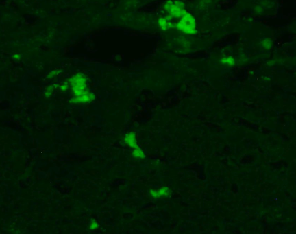
"Examples of fluorescent antibody staining from one donor showing fluorescence filtered image on the left of each matched pair and normal darkfield image on the right.:"
Lymedin2010
Frequent Contributor (1K+ posts)
Member # 34322
posted
I am finding that a lot of my rbc's have DNA on them at times & they are scattered throughout the sample. The fuller & brighter objects are WBC's & the speckled ones with DNA are rbc's.
________________________________________
This is fluorescence in the plasma. It might be components from WBC's that have lysed, as the WBC's have DNA in them & that is why they fluoresce in AO stains.
Also, this is that vacuole video I was talking about a while back & I finally got to uploading it.Vacuole in the rbc or biconcave lighting tricks. I would probably just dismiss this as lighting tricks until further evidence, although it looks tempting to say it is a vacuole.
Lymedin2010
Frequent Contributor (1K+ posts)
Member # 34322
posted
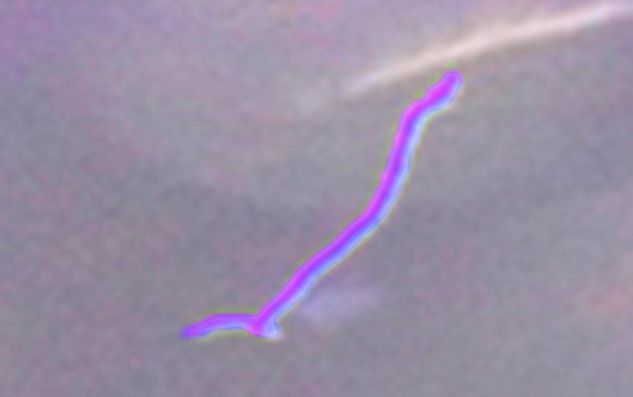
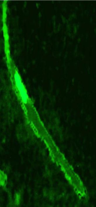
Another spirochete that fluoresced with Acridine Orange in my blood. Green means it is something with DNA, but who knows what species it might be.
Lymedin2010
Frequent Contributor (1K+ posts)
Member # 34322
posted
So with refinement of my technique of diluting & spreading the prep thinner I am finding spiros that fluoresce, but they are few & scattered. Another spiro in my blood pic below. I now also have other string-like subjects & even SOP in my preps & they do not fluoresce, which means they do not have DNA/RNA and are probably false spiros. I will make a video of the false spiros eventually.
Lymedin2010
Frequent Contributor (1K+ posts)
Member # 34322
posted
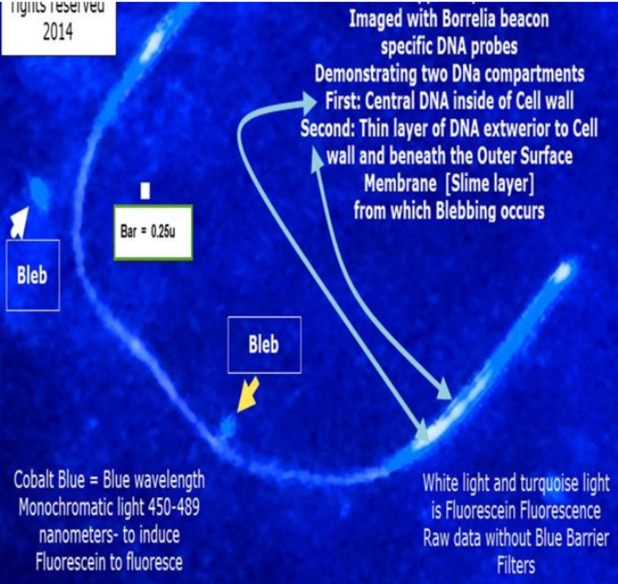
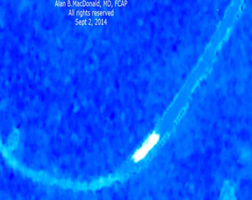
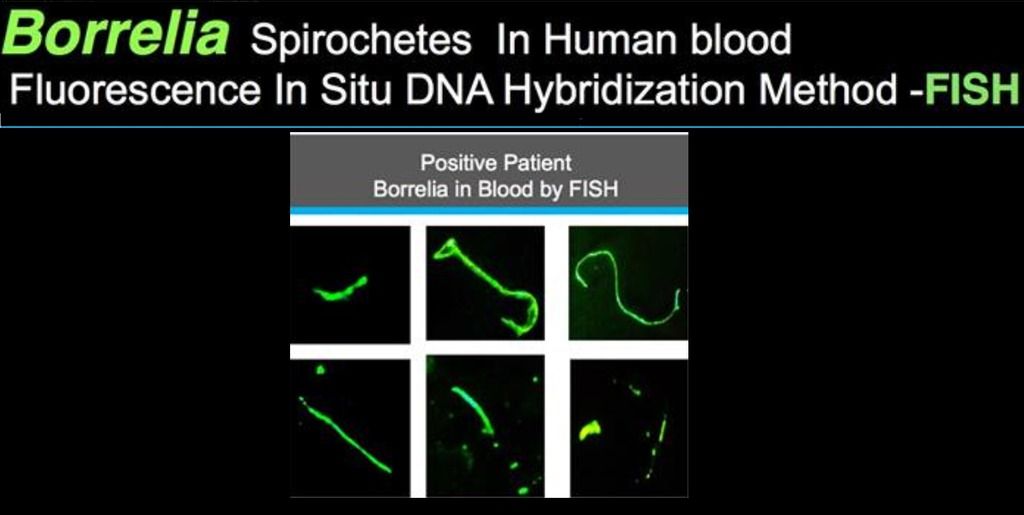
I like to remind myself of the odd & unusual shapes of Borrelia in the human body at times. Alan MacDonald's DNA FISH probes only bind to Borrelia DNA & makes for a good reference. Here is his result from probing in human blood.
bluelyme
Frequent Contributor (1K+ posts)
Member # 47170
posted
Wow what great work ...hard proof that it is indeed very much intercellular ...maybe why dr h loves double and triple intercellular abx .thanks lymed
-------------------- Blue Posts: 1539 | From southwest | Registered: Dec 2015
| IP: Logged |
Lymedin2010
Frequent Contributor (1K+ posts)
Member # 34322
posted
Thanks.
I have found SOP's that fluoresce in my blood now. I also found spiro-like ones that do not fluoresce & segmented ones that do no fluoresce & I will put them in a future false spiro video.
Lymedin2010
Frequent Contributor (1K+ posts)
Member # 34322
posted
HUGE discovery & further proof for all of us!!!!!!
Spirochetes, SOP's, blebs, 2 daughter splitting in my Lyme blood by fluorescent microscopy...so that explains all the symptoms & the grappling fatigue and shortness of breath...these things are all over my blood just as we see them in regular light microscopy! Only now they pop & stand out more with fluorescent stains and this adds proof that they have DNA & they are not just artifacts!
Plenty of info in the description & since it was too long, I had to add some more info in the comments section as well.
I also figured out what those dumbbells are that we have been seeing since the beginning of our microscopy. On regular lightfield they look like dumbbells & when I switch to fluorescence they reveal as 2 daughter splitting and so the segment between the 2 splitting spiros looks like the handle to the dumbbell and since the dumbbells typically move fast in live blood preps with Brownian motion it distorts it and makes the tips look thicker then what they really are and it gives us the illusion of dumbbells.
You can see the dumbbells & the other many forms from the breakup of SOP in this video I made back in 2014. And now we get to see them really stand out!
Lymedin2010
Frequent Contributor (1K+ posts)
Member # 34322
posted
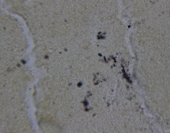
"Results
Bb Culture Dieterle staining of the blood culture pellet demonstrated both long slender spirochetes ranging from approximately 0.1 to 0.5 μm in width and approximately 2 to 6 μm in length, some with visible helices, and round morphological variants ranging from approximately 0.5 to 2 μm in diameter that looked much like some of the morphological variants seen in the liver section (Figure 1). " " Anti-Bb immunostaining of the blood culture was strongly positive, exhibiting a bright red cherry color (Figure 2). There was some positive staining of cellular debris, possibly because of the antigens released from lysed spirochetes or secreted by Bb. Strongly positive staining was not observed in the culture pellets of control microorganisms. Molecular beacon staining of the culture pellet sections was strongly positive throughout (Figure 3). Culture pellets contained both cultured bacteria and human cellular debris (intact red blood cells and lysed blood cells) indicating that Bb nucleic acid sequences were present throughout the cellular debris and within intact RBCs."
Figure 3. Blood culture beacon stain, Probe Fla B. 400X magnification. Posts: 2094 | From NY | Registered: Oct 2011
| IP: Logged |
Lymedin2010
Frequent Contributor (1K+ posts)
Member # 34322
posted
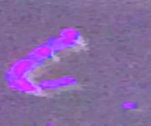
This one is super special as it is the first stained SOP that I know of from one of our members, Sylvie. Great contrast between the rbc's & the stained SOP to show it is not a false SOP that is generated from the rbc, but a true SOP. So might be from spirochete and others will be adamant that it is from yet another organism.
-------------------- Blue Posts: 1539 | From southwest | Registered: Dec 2015
| IP: Logged |
Lymedin2010
Frequent Contributor (1K+ posts)
Member # 34322
posted
Yea, I have to transfer those pics over to another site one day.
I am not familiar with that scope, but the Nikon's & Zeiss's have some nice turrets that show up from time to time. Some times they show up cheaper & without the filters, and so one can DIY the filters & place them in the turret. It is valuable to have a swivel for the condenser lens, as sometimes a slight angle on the light can make a world of difference, as it does on my Zeiss when I am in the mood for images that stand out. My Reichert Oblique Illumination is outstanding when it comes to impressive views & outperforms my Zeiss in this respect, even with worst optics. The view can appear very 3D & DIC like.
________________________________ After combing through my videos of False Spirochete proofs, I decided to put out a video before I gather more evidence via 4K video & other methods. I think there is enough proof in this video & it shouldn't be too far of a stretch to believe this?
So I think both real & False Spirochetes (FS) can exist in our Lyme blood and sometimes they will be hard to tell apart. There is more info in the video & in the video description.
__________________________________________ CYST OR WBC COMPONENTS???
I tried live staining with Parker's blue/black ink to see if I can buy more clues. In this video the objects did stain blue/black/purplish, as spirochetes do stain that color, but the wbc's also stained aquablue, blue, & purplish as well, so it was hard to differentiate whether these were spiro cysts or wbc components. I time lapsed them for 1.5 days here & no sign of uncysting.
That's a really good Bartonella FISH video. Interestingly, my fluorescent "Bart" organisms look almost identical.
It's too bad I cannot get some of this DNA/RNA-specific fluorescent stain. I'd likely have to have a license (a certified lab) to purchase this stuff once they find a distributor.
I'm still having trouble with my AO-stained dry smears. I've done some experimenting--even got some AO from a different company--but my smears keep washing off when I rinse the AO off the specimen. The only other thing I think it could possibly be is perhaps my methanol is not good anymore. I'm not sure why that would be though, unless perhaps it has absorbed some moisture somehow. I can't see how that's possible since I store it in the capped plastic bottle it came in. Just in case, I have some more methanol coming. I hope that's the fix, because I have been really bummed out about microscopy because of this.
Posts: 1308 | From Eastern USA | Registered: Oct 2013
| IP: Logged |
Lymedin2010
Frequent Contributor (1K+ posts)
Member # 34322
posted
Hey TNT. Yea, I would love to get my hands on some DNA Probes.
TNT, it is better to dilute the AO & do live staining, that is how I saw the spirochete SOP's & 2 daughter splitting, blebs, & developing juvenile spiros. Before that I was using pure 0.2% AO & it was too strong & cytotoxic and just destroys the spiros. I spoke to someone in a lab & they said that when they exposed sperm to pure AO it too destroyed the sperm cells & so I confirmed the toxicity. I gave the solution in MY HORRIFIC LYME BLOOD P2 video, it is 2 step dilution to mimic the exact one that I used. You then add a drop of that to a drop of your blood, sometimes a little less than a drop so you can get better dispersion of the rbc's. You seal the cover slip with vaseline or immersion oil so that it does not dry up & you wait for the spiros to take up the dye & fluoresce. Usually some start right away & more will pick it up in 12-24 hrs & sometimes ever more at 36 hrs later.
It seems like when the spiros are not active their double bound cell membranes are hard to penetrate at this low concentration, but if they undergo change, such as SOP formation then the AO is taken up & they light up.
Instead of washing out the AO from your slide, have you tried NOT to wash it out & just simply let it dry out naturally. BUT you must place a smaller amount of blood & disperse them really well so you don't get as much clumping when the AO evaporates. Also, evaporate in a well ventilated areas & better to do it in a garage but enclosed in a box & then open the garage door to vent...any stain fumes are not good for the Lyme body.
Posts: 2094 | From NY | Registered: Oct 2011
| IP: Logged |
TNT
Frequent Contributor (1K+ posts)
Member # 42349
posted
Thanks for the tips Lymedin. I've got to say, you are doing some EXCELLENT work with spirochetes and live blood! I've really been impressed! And, your "schizont" looks more like one than the one in ID-Fish's video. Keep up the great work. I honestly think your channel will eventually go viral as your microscopy work with Borrelia is definitely on the cutting edge.
Personally, I guess I'm just not as interested in doing live blood as much anymore since I've seen the conversion and I've never doubted from the beginning that I was seeing spirochetes. I'm more intent on catching pyriforms/ringforms or morulas in their distinct morphologies under fluorescence because those infections (COMBINED with the Borrelia) are what I believe have made and kept me sick. Trouble is, I have less and less a chance to see this as I keep improving and can't get my game on with the dried smears.
I will probably do more live blood under fluorescence once I have mastered the fluoro dried smears though.
Posts: 1308 | From Eastern USA | Registered: Oct 2013
| IP: Logged |
Lymedin2010
Frequent Contributor (1K+ posts)
Member # 34322
posted
Thanks TNT! I think collectively we did & learned a lot together.
Looking forward to seeing some new stuff from you.
Posts: 2094 | From NY | Registered: Oct 2011
| IP: Logged |
Lymedin2010
Frequent Contributor (1K+ posts)
Member # 34322
posted
Lymedin2010/TNT and all - this is a wonderful thread. Thanks for sharing all of the amazing information. Been reading on and off for a while. First time posting.
I am interested in getting started with some dark field analysis. What do you recommend for a microscope/camera to get started without going with a full pro setup (Olympus/Zeiss etc) in September 2017? More specifically, can I get the job done well with a cheap microscope (Amscope etc)?FWIW - I am good with electronics so I am able to hack/mod anything if necessary if that helps.
Posts: 65 | From minneapolis, san francisco | Registered: Nov 2004
| IP: Logged |
quote:Originally posted by electric: Lymedin2010/TNT and all - this is a wonderful thread. Thanks for sharing all of the amazing information. Been reading on and off for a while. First time posting.
I am interested in getting started with some dark field analysis. What do you recommend for a microscope/camera to get started without going with a full pro setup (Olympus/Zeiss etc) in September 2017? More specifically, can I get the job done well with a cheap microscope (Amscope etc)?FWIW - I am good with electronics so I am able to hack/mod anything if necessary if that helps.
I don't have a specific microscope recommendation, but I just started doing darkfield and here's what I recommend as far as the type of equipment:
Darkfield at 40x isn't all that impressive. I've done some at 63x which is pretty decent, but 100x is the best.
You'll want to make sure that the NA of your objective is always less than the NA of your condenser. For instance, for 100x darkfield, I use a darkfield oil condenser with an NA of 1.25, so my 100x objective (which also has an NA of 1.25) has an adjustable iris to close down the NA (otherwise the image isn't dark enough).
You'll also want a fairly strong light source. My 18 watt incandescent bulb on my microscope is slightly under-powered for 100x darkfield, but it's not terrible either. A high powered LED is probably the way to go.
Check out older microscopes on eBay, or a new Amscope (but make sure it has both an oil condenser and 100x objective with iris; often the Amscopes are sold as "Darkfield" but only come with a dry condenser and 100x objective without adjustable NA, basically limiting darkfield work to 40x).
Posts: 71 | From Canada | Registered: May 2016
| IP: Logged |
Lymedin2010
Frequent Contributor (1K+ posts)
Member # 34322
posted
Personally, I don't own a darkfield & have limited experience. I would def ask & listen to the owners of DF...thanks MS2!
posted
mustardseed2 - awesome information! Thank you, it really helped me understand exactly what I need to look for shopping around.
Also, how valuable is adding phase contrast ability to the setup? Would it be valuable for trying to view lyme/co-infections in blood samples etc?
Lymedin2010 - Thanks for the link to My Microscope group. Will check it out.
Posts: 65 | From minneapolis, san francisco | Registered: Nov 2004
| IP: Logged |
Lymedin2010
Frequent Contributor (1K+ posts)
Member # 34322
posted
Sometimes it helps & other times it can also hinder, depends on how the slides are prepared and how busy the slide is. You don't need it really, but only if you want more impressive images on a clean slide/prep.
Posts: 2094 | From NY | Registered: Oct 2011
| IP: Logged |
Lymedin2010
Frequent Contributor (1K+ posts)
Member # 34322
posted
My new venture on high contrast staining. WBC's & platelets are a dramatic blue contrast to the golden/yellow rbc's. This makes things easier to spot & distinguish.
This is on one of my bad delaminated objectives though, better images in the future.
Even in high clutter, spot things easily. Posts: 2094 | From NY | Registered: Oct 2011
| IP: Logged |
Lymedin2010
Frequent Contributor (1K+ posts)
Member # 34322
posted
Another true spiro in my blood using a proprietary technique other than the freezing method.
posted
Lymedin2010 -Thanks for sharing. What magnification levels are the images above?
Posts: 65 | From minneapolis, san francisco | Registered: Nov 2004
| IP: Logged |
Lymedin2010
Frequent Contributor (1K+ posts)
Member # 34322
posted
They are all w/ 40x objective + 15x eyepiece, so 600x & then I can zoom in & out with my Samsung S5 phone cam.
You can see them better with a 100x oil objective, but I did not use it with the above pics.
Posts: 2094 | From NY | Registered: Oct 2011
| IP: Logged |
Great video, Lymedin! At 7:11 in the video shows classic acridine orange coloring. The green background of cellular components and debris with the bright orange pathogenic (Borrelia) material is awesome classic presentation. Makes me want to do a few smears, HA HA!
Are you still using your RGB LED light hack (particularly on that clip of the video)??? If so, that is great because that is plenty enough intensity.
Posts: 1308 | From Eastern USA | Registered: Oct 2013
| IP: Logged |
Lymedin2010
Frequent Contributor (1K+ posts)
Member # 34322
posted
Thanks. Yes, still using the same bulb. I bought 2 more rgb bulbs, including the one you linked to & another white color flash light. They all work, but the 1st bulb I bought has 3 of 3w blue LED's & they overlap for increased intensity & so it produces the best and brightest images.
Sometimes the stain sample does not take the stain right away or looses the stain over time & that is why it is dimmer in the other videos. Timing is important & I don't have the energy to monitor perfect stained times & so sometimes they come out better than others. Plus my AO solution mix has seen too much light & I should have stored it in a dark container and so I will have to make a new batch eventually.
Been waiting forever to see your stains man, hopefully soon?
Posts: 2094 | From NY | Registered: Oct 2011
| IP: Logged |
TNT
Frequent Contributor (1K+ posts)
Member # 42349
posted
quote:Originally posted by Lymedin2010: Been waiting forever to see your stains man, hopefully soon?
Yeah, I'm working on it, but not too diligently
I got some new methanol a few weeks ago, and, like I said, the color at 7:11 in your video got me fired up again about trying some more stains. Unfortunately, the specimens on the first two slides washed right off again. A third slide I dunked in the methanol instead of flooding the top, and I may have a winner. I still need to look at it, as it was too late to get out the scope last night.
But, maybe you are hoping for some wet mount stains?...
I just might be inspired enough to do one of those again before long. Though I have some Giemsa smears that are pressing at the moment unless the AO is working out. If that is the case, I will be doing some more fixed AO smears.
Posts: 1308 | From Eastern USA | Registered: Oct 2013
| IP: Logged |
TNT
Frequent Contributor (1K+ posts)
Member # 42349
posted
Also, that's great that your hack is working so well!!
I proposed on another forum that using a RGB lamp directed right onto the slide and stage would be a usable brightfield scope hack. When I personally tried it with my 5 watt RGB bulb I was able to see absolutely nothing. So, maybe the theory doesn't work out for real world application. Or, maybe I didn't have enough wattage for the smear to pick up any fluorescence.
Maybe something like this light with much more wattage would work for such a hack:
TNT
Frequent Contributor (1K+ posts)
Member # 42349
posted
quote:Originally posted by TNT: I proposed on another forum that using a RGB lamp directed right onto the slide and stage would be a usable brightfield scope hack.
What I meant was that you could turn a brightfield scope into a fluorescent microscope simply by shining one of these RGB LED lights onto the specimen plane directly. But when I tried it with a low-power bulb I saw nothing. But, again, the problem could have been wattage....
Posts: 1308 | From Eastern USA | Registered: Oct 2013
| IP: Logged |
Lymedin2010
Frequent Contributor (1K+ posts)
Member # 34322
posted
Yes, I think one can do a complete hack of the fluoro scope & all one needs is the yellow filter to place between the specimen & camera or eyes.
I think a bulb like this will work too, as it has a collimator right on it.
Quite honestly I think you don't even need a RGB bulb & you could use a white LED light bulb & place a blue filter on top of it, but it has to be the right hue & one can get a filter match book set.
Posts: 2094 | From NY | Registered: Oct 2011
| IP: Logged |
The Lyme Disease Network is a non-profit organization funded by individual donations. If you would like to support the Network and the LymeNet system of Web services, please send your donations to:
The
Lyme Disease Network of New Jersey 907 Pebble Creek Court,
Pennington,
NJ08534USA http://www.lymenet.org/


 UBBFriend: Email this page to someone!


![[Frown]](frown.gif)


 Printer-friendly view of this topic