TNT
Frequent Contributor (1K+ posts)
Member # 42349
posted
quote:Originally posted by WakeUp: The reason Im interested is because Alan McDonald has shown that borrelia is harbored inside these worms-- so one needs to get rid of the worms FIRST if there is any hope of curing the borrelia.
I personally really caution against killing "foundational" pathogens (or larger pathogens/organisms) first as Dr. K in Washington does. I believe it just makes a bad situation MUCH MUCH worse because you are opening up the Pandora's Box of microbes and releasing them into the body immediately. That can make a person extremely sick, disabled, and can even be deadly.
I personally feel it is advisable to KILL WHAT'S INSIDE those filarial worms AT THE SAME TIME we kill the worms. This poses the least amount of risk.
Remember the "Heartworm infection model" in dogs!
But, I agree with you! You must address the larger organisms/worms in order to fully address the smaller or "symbiotic" pathogens harbored inside them.
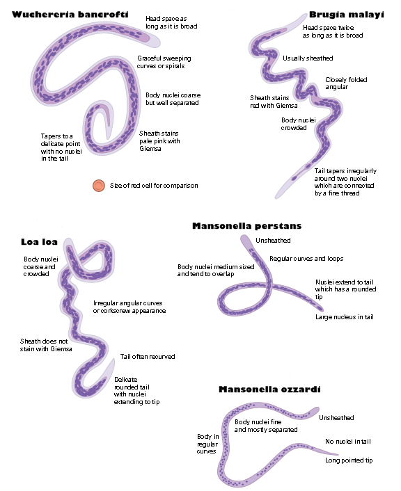
And, YUCK, those DO look like filarial worms in Lymedin's tick juice video!!!
(Again) Great work, Lymedin! But, yuck! Posts: 1308 | From Eastern USA | Registered: Oct 2013
| IP: Logged |
Lymedin2010
Frequent Contributor (1K+ posts)
Member # 34322
posted
We are in trouble guys, as I think resistance is a real thing too & even with Borrelia. On one sample I used my blood on tick juice & what happened was that the Borrelia just disappeared in 24 hrs. Hard to say if they went into cyst or just died altogether. I will have to try sheeps or rabbits blood on a future round or maybe saliva + the blood.
TNT, how much longer are you going to do the BVT for...you can't keep on doing it forever?
I have a video of one of those filarial objects in a tick & all the surrounding spirochetes have been attracted to the object & are trying to burrow inside of it. I found the Borrelia gathering after I had taken my phone & contraption apart already & was just too crispy to record anything. I recorded 24 hrs later though, but there were fewer Borrelia & I am assuming some most have gotten inside. I will post the video in a few.
Posts: 2094 | From NY | Registered: Oct 2011
| IP: Logged |
Lymedin2010
Frequent Contributor (1K+ posts)
Member # 34322
bluelyme
Frequent Contributor (1K+ posts)
Member # 47170
posted
quote:Originally posted by TNT: And, wow, very nice scope, Lymedin! Definitely a research grade Reichert! Especially with the 6 lens turret.
Great job on the experiments with saliva and tick "juice."
I hope to have some things to share before long. But, not concerning tick juice, ha ha.
Oh the suspense ... on bvt im at 1 yr , bvters say 2 to 3 ...my nurse friend is in yr 6 after lyfe of lyme..how long have you been at full dosing ? I heard of a dr h client switching to do 75 sting a day making strides ..
I have some liquid venom i will put on a slide of ketes soon?!
-------------------- Blue Posts: 1539 | From southwest | Registered: Dec 2015
| IP: Logged |
Lymedin2010
Frequent Contributor (1K+ posts)
Member # 34322
posted
I betcha, just like the Westernback Lizard, some snakes will be immune to Borrelia & their immune system will not let it grow in their bodies. I wish someone would do Borrelia studies on reptiles.
quote:Originally posted by WakeUp: The reason Im interested is because Alan McDonald has shown that borrelia is harbored inside these worms-- so one needs to get rid of the worms FIRST if there is any hope of curing the borrelia.
I personally really caution against killing "foundational" pathogens (or larger pathogens/organisms) first as Dr. K in Washington does. I believe it just makes a bad situation MUCH MUCH worse because you are opening up the Pandora's Box of microbes and releasing them into the body immediately. That can make a person extremely sick, disabled, and can even be deadly.
I personally feel it is advisable to KILL WHAT'S INSIDE those filarial worms AT THE SAME TIME we kill the worms. This poses the least amount of risk.
Remember the "Heartworm infection model" in dogs!
But, I agree with you! You must address the larger organisms/worms in order to fully address the smaller or "symbiotic" pathogens harbored inside them.
And, YUCK, those DO look like filarial worms in Lymedin's tick juice video!!!
(Again) Great work, Lymedin! But, yuck!
The heartworm analogy is possibly, but not necessarily true in the case of filaria- but remember that worms also have chemicals that disable the immune system-- meaning they make the victim weaker, and less able to fight other pathogens as time goes on. If given the choice, Id get rid of any filarial worm infestation first if at all possible, while continuing to take anti borrelia herbs. I know its possibly risky-- i.e. like removing asbestos from a house without encapsulating it first-- you risk breathing it all in-- I think I would take that risk.
Filarial worms are a perfect "payload" delivery vehicle in binary biological warfare. The less filaria you have, the smaller the dangerous payload is over time inside your body. All you then have to do is get rid of biofilm-- and you're cured.. LOL just kidding. We are all screwed royally.
Posts: 696 | From New York | Registered: Aug 2006
| IP: Logged |
quote:Originally posted by Lymedin2010: I betcha, just like the Westernback Lizard, some snakes will be immune to Borrelia & their immune system will not let it grow in their bodies. I wish someone would do Borrelia studies on reptiles.
LOL-- When I win the mega millions lottery -- and create my Lyme foundation-- this will be one of the first areas funded (aside from my list of promising herbal compounds...) LOL Thanks for all you do !!
Posts: 696 | From New York | Registered: Aug 2006
| IP: Logged |
Lymed-- thanks again for your incredible video work!! This video, plus McDonald's PCR work pretty much proves that the spirochetes attach to, feed off and burrow into the filarial worms, and are thus harbored inside the worm--- in a similar way to how they attach to , and are harbored inside our red blood cells and bone marrow. FUBAR for us.
The corkscrew burrows into everything--- is like an archimedes killing machine-- a perfect biological weapon..
Posts: 696 | From New York | Registered: Aug 2006
| IP: Logged |
quote:Originally posted by TNT: For those of you/us who have and use Rife machines, here is an amazing video of live blood showing the destruction of a spirochete with a TrueRife machine.
I would really like to know what frequency they were using for this demonstration.
Yea-- I wonder what frequency he was using-- theres a phone number for him if someone wants to call him. I think he was using this PLASMA rife machine which is very powerful: https://www.youtube.com/watch?v=GvJGZM908gE
It would be great to do more microscopy using this machine.
Posts: 696 | From New York | Registered: Aug 2006
| IP: Logged |
TNT
Frequent Contributor (1K+ posts)
Member # 42349
posted
quote:Originally posted by Lymedin2010:
TNT, how much longer are you going to do the BVT for...you can't keep on doing it forever?
I sure will!!! As long as it's helping me, I will continue them. It's much cheaper and easier than ABX and other modalities such as mHBOT!
That's interesting blue that some people are doing that many stings. I stick with 10-12 per time.
Posts: 1308 | From Eastern USA | Registered: Oct 2013
| IP: Logged |
posted
Here's another "filaria" or I hope just a fiber----- it has a pointy end, and is about the length of 60 red blood cells, I think-- I'll try to upload these images smaller -- still working on all this tech stuff: Posts: 696 | From New York | Registered: Aug 2006
| IP: Logged |
Lymedin2010
Frequent Contributor (1K+ posts)
Member # 34322
posted
I've been told that when there are sharp bends then it is not a worm, but then again I see sharp bends sometimes in the tick filarial & you can see them in my last video where there are quite a few worms. So I think everything has to be taken with a grain of salt.
I don't think yours are nematodes & I think they are fibers. The first pic has a synthetic fanned end on one end. They are about the right size for a worm though & it could be, as sometimes it is hard to tell for sure. They always say staining is best for the worms.
I've also told people to leave out a glass slide in the open for 48 to 60 hours & then to add some water & a cover slip. Then you can compare the potentials of real fibers to the known nematode pictures & videos. I've been meaning to do this myself for quite some time, but never got to it as of yet.
Great pics with your iPhone, what is that the iPhone 6?
Posts: 2094 | From NY | Registered: Oct 2011
| IP: Logged |
posted
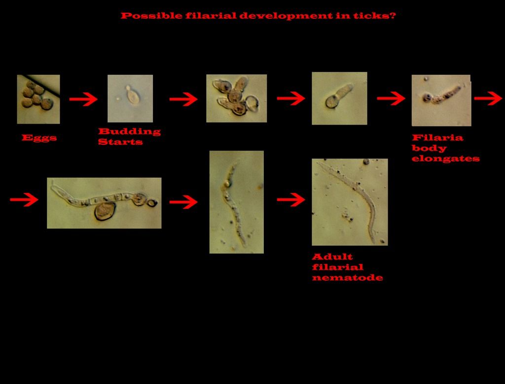
Thanks ! Yes-- its an iphone 6-- -- its really hard to get the lens lined up to the eyepiece-- my pics above come from blood from an open morgellons skin lesion. I'm a microscope newbie -- so much to learn but its a fun hobby and hopefully useful to other sick people and even scientists who come here. I Hope "they" are fibers but I have a feeling they might not be. My Amscope camera software isnt working hardly at all on my mac -- so Im stuck with the handheld phone for now. Here's circopithifilaria in 3 stages I came across online ( from a squashed tick). Circopithifilaria is a filarial skin infection in dogs, I believe.
Posts: 696 | From New York | Registered: Aug 2006
| IP: Logged |
[ 03-18-2017, 02:01 AM: Message edited by: Lymedin2010 ]
Posts: 2094 | From NY | Registered: Oct 2011
| IP: Logged |
Lymedin2010
Frequent Contributor (1K+ posts)
Member # 34322
posted
Tick juice in BSK-H @ 1000x + cam zoom. The spirochete zig-zags back & forth & then at times becomes limber and just gyrates & wriggles just like in our Lyme Diseased blood.
Other times I have seen them become straightened out & very rigid, what they call "needle form."
Lymedin2010
Frequent Contributor (1K+ posts)
Member # 34322
posted
This sample is from 2 months later post prep.
I see these occasionally & they are usually much smaller. This one is fairly larger & worth sharing.
To me it is more like segmentation, which is what I see more of toward the end life of the spirochetes...when the nutrients run out.
https://www.youtube.com/watch?v=nvKrN0ggnwc ___________________________________________ Dr. Alan MacDonald explains the granular forms of the SOP in this video, with a nice pic.
The Bart's are small & in a vacuole, which was most likely inside a rbc at one point that then ruptured to release the vacuole + Barts inside the vacuole. When the vacuole is inside the rbc, the vacuole can rupture too & release the Bart's so that they freely move WITHIN the rbc. So one can see multiple variations & conditions, and even the Bart plantonic (free-moving/existing) in the plasma.
Remember that the organisms are in an environment & the environment can change very rapidly & their "behavior" can change as well. It depends on how long ago the blood was stored in vacutainers before slide sample was taken , how long ago sample was taken and video recorded, whether the slip cover was sealed so as not to dry out the sample, ambient temp & temp from illumination source...etc...many possible situations.
For instance, in ticks when I first look at the slide there are no spirochetes, because they were shocked by the water or water+ some other mixture addition. Then when they slowly emerge & appear to seemingly pop out of thin air. When they first emerge they spiral (zig-zag) back & forth violently & with much fervor and energy and they also start to divide and multiply. Then over time as the nutrients run out, they become stagnant & with little mobility just like in our blood...they just gyrate & move in limited motion. I will make a video eventually & it will be crystal clear and the motion lessons here can be applied to all microscopic organisms.
Borrelia do not need to form the vacuoles, since they have the more messier & random biofilm formations. Babesia does not form the vacuoles either. Bartonella is the only one that I know of out of the trio (Borrelia, Babs, Bart) that form the vacuole. Others that I have spoken to also believe that visually this is probably Bart, but of course they want testing as we all do. For me this is 100% bart visually. I have seen the Bart in another persons blood & I have seen the vacuoles once before, but at that time it was earlier in my microscopy ventures & I did not know what it was...I cannot find that video now as it was online somewhere.
Posts: 2094 | From NY | Registered: Oct 2011
| IP: Logged |
TNT
Frequent Contributor (1K+ posts)
Member # 42349
posted
quote:Originally posted by Lymedin2010: At time 35s in this video...looks like that is more Bartonella in the vacuole & in the rbc.
The Bart's are small & in a vacuole, which was most likely inside a rbc at one point that then ruptured to release the vacuole + Barts inside the vacuole. When the vacuole is inside the rbc, the vacuole can rupture too & release the Bart's so that they freely move WITHIN the rbc. So one can see multiple variations & conditions, and even the Bart plantonic (free-moving/existing) in the plasma.
It's very possible that is an example of Bart organisms inside a vacuole or even a ghosted RBC. It is just so hard to tell many times (with live blood). I have definitely seen this in my live blood, but have chalked it up to lysosomes getting trapped in the remains of a RBC. I don't see this "phenomena" demonstrated in my giemsa smears.
Great job and finds, especially your own nematodes-in-ticks videos! I think those are particularly interesting, and CONFIRMING.
I've actually considered doing some giemsa smears of the blood of biting flies and ticks, but haven't had the time. Still looking at human blood for now. But, I think it could be very revealing. For instance, a study published late last year proved that horseflies can carry and transmit Anaplasma. Previously it was believed they could only transmit by the rare mechanical transmission.
Posts: 1308 | From Eastern USA | Registered: Oct 2013
| IP: Logged |
TNT
Frequent Contributor (1K+ posts)
Member # 42349
Great observation! I think most of the "string of pearls" we see in our blood are spirochetes. The red blood cell string of pearls are rarer and the pearls are larger.
Posts: 1308 | From Eastern USA | Registered: Oct 2013
| IP: Logged |
Lymedin2010
Frequent Contributor (1K+ posts)
Member # 34322
posted
Anything is possible, but you can also see the bart inside the rbc with the hemoglobin still intact & not compromised. So both the hollow one & the full rbc have what to me look like bart.
Do you see any stained spirochetes in your blood? I only find a few of them with staining & they are questionable in appearance, as in they may not be spirochetes either. So I wonder about that too since I have seen stained spiros in slides online. I was once led to believe that any SOP in the blood meant that it was foreign, but after my acid & basic (baking soda) experiments I no longer hold to that belief. Shedding of the rbc wall can also produce SOP's.
I look forward to your tick ventures, can't wait...how odd what us Lymies get excited about.
Posts: 2094 | From NY | Registered: Oct 2011
| IP: Logged |
TNT
Frequent Contributor (1K+ posts)
Member # 42349
posted
quote:Originally posted by Lymedin2010: Anything is possible, but you can also see the bart inside the rbc with the hemoglobin still intact & not compromised. So both the hollow one & the full rbc have what to me look like bart.
Do you see any stained spirochetes in your blood? I only find a few of them with staining & they are questionable in appearance, as in they may not be spirochetes either. So I wonder about that too since I have seen stained spiros in slides online. I was once led to believe that any SOP in the blood meant that it was foreign, but after my acid & basic (baking soda) experiments I no longer hold to that belief. Shedding of the rbc wall can also produce SOP's.
I look forward to your tick ventures, can't wait...how odd what us Lymies get excited about.
I do not see spirochetes in my stains like we see in our live blood. Relapsing Fever spirochetes stain well, and those are probably the pictures you see online of stained spirochetes. For some reason, Bb spirochetes do not stain well with Giemsa. I don't think Syphilis spirochetes stain with Giemsa either. I have only seen a few objects in my stains that could have been spirochetes, and they were debatable.
Unfortunately, I doubt I will have the time or energy to pursue staining insect contents any time soon. But, it's a possible interest for the future.
Disintegrating red blood cells can definitely form string of pearls, but those pearls are bigger than the pearls of spirochete SOPs! Isn't that what you have found?
[ 03-29-2017, 03:17 PM: Message edited by: TNT ]
Posts: 1308 | From Eastern USA | Registered: Oct 2013
| IP: Logged |
Lymedin2010
Frequent Contributor (1K+ posts)
Member # 34322
posted
Yes, I found out larger SOP's can form from rbc cell walls & I wonder about the smaller ones too, as I don't think there is a limitation to how phospholipids can form below 1 micron & lower...just think of micelles fat droplets & how small some of them can get, which are basically spherical phospholipids.
I know spiros can & will be in our blood, and we have seen some awesome cyst forming videos. I think sometimes we might confuse one for the other though & hard to tell at times. I use aggressive movement of the spiro now as a better judgement to distinguish from false spirochetes. A darn electron microscope can easily solve the questions if we had one at hand & I was considering looking into buying some EM time.
Posts: 2094 | From NY | Registered: Oct 2011
| IP: Logged |
Lymedin2010
Frequent Contributor (1K+ posts)
Member # 34322
Lymedin2010
Frequent Contributor (1K+ posts)
Member # 34322
posted
Here is the rest of the Bart references from the previous time, for anyone else interested.
MORE ON BART VACUOLES IN THE BLOOD, forget about species this is to learn more on if bart in general can be in the blood & within the rbc's & whether it can form vacuoles inside the rbc's...continuation from last time:
1) "Deep invaginations containing bacteria are commonly seen, and membrane fusion at the necks of the invaginations leads to the formation of intracellular vacuoles containing bacteria. Fluorescent compounds present externally render the vacuoles fluorescent and, occasionally, lightly fluorescent cells are seen, suggesting that the vacuoles sometimes rupture to admit the bacteria to the cytoplasm."
"FIG. 3. Deformation of erythrocytes as seen by scanning electron microscopy. (a) Clumps of bacteria and (b) single bacterium bound at indentations. (c to e) Deep pits which result from bacteria pushing into the erythrocyte membrane. (f) Membrane of the erythrocyte apparently pulled up from the cytoskeleton and twisted."
"FIG. 4. Transmission electron microscopy of thin sections of complexes of bacteria and erythrocytes. (a) Bacteria lying clearly outside of the erythrocyte. (b) Bacteria apparently within an endocytotic vesicle"
"FIG. 5.Micrographs of erythrocytes preloaded with calcein and infected with bacteria. (a and c)Visualized by dark field and (b and d) visualized by fluorescence."
______________________________________ 2) "We identified a Bartonella quintana strain by polymerase chain reaction amplification, cloning, and sequencing of DNA extracted from lysed erythrocytes and cultured colonies grown from peripheral blood collected from a captive-bred cynomolgus monkey (Macaca fascicularis)." http://www.biomedsearch.com/nih/Bartonella-quintana-in-cynomolgus-monkey/16485482.html
On Fig 2c:" erythrocyte with vacuole-enclosed suspect organism."
Fig 1a: Also looks like it has a vacuole & an organisms within the vacuole & within the rbc..you have to zoom in really hard to see the discoloration the vacuole produces.
______________________________________ 3) " Unusual trafficking pattern of Bartonella henselae -containing vacuoles in macrophages and endothelial cells" " Bartonella henselae, the agent of cat-scratch disease and vasculoproliferative disorders in humans, is a fastidious facultative intracellular pathogen, whose interaction with macrophages and endothelial cells (ECs) is crucial in the pathogenesis of these diseases. However, little is known about the subcellular compartment in which B. henselae resides."
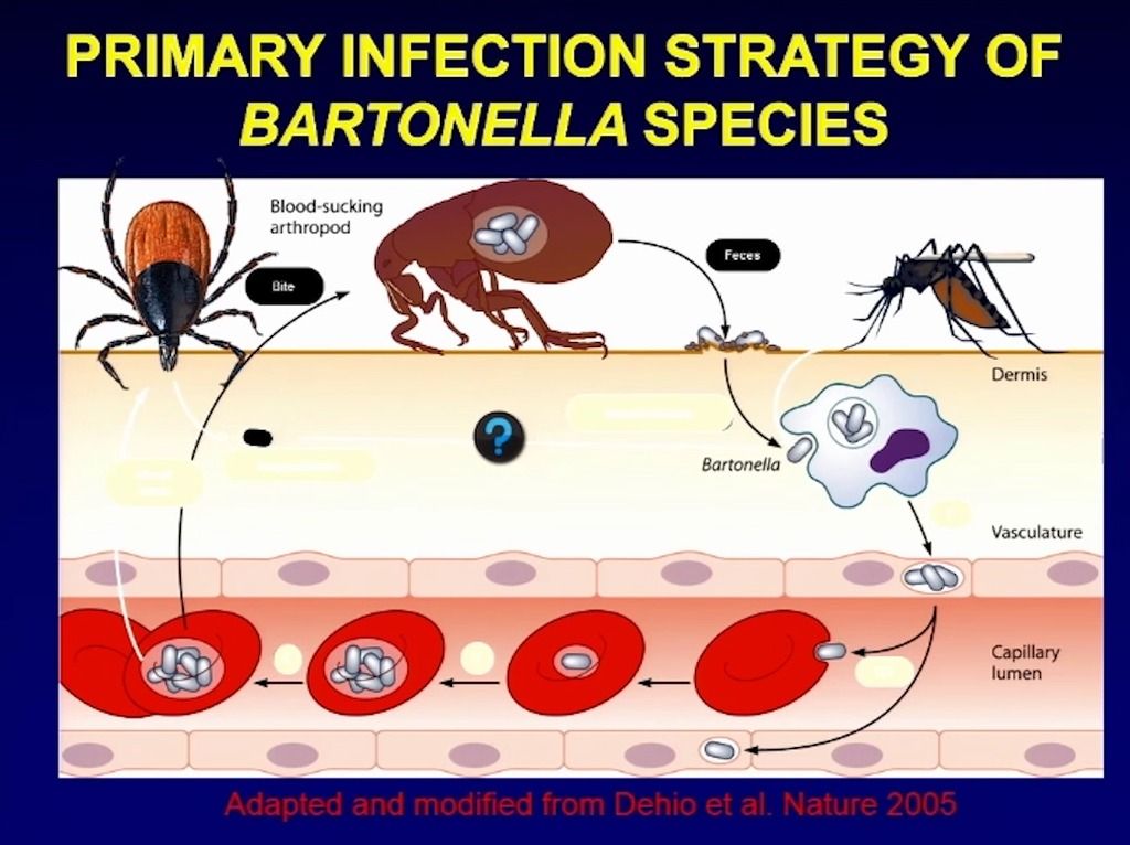
______________________________________ 4) " Common infection strategy of the bartonellae. The drawing illustrates the general concept of reservoir host infections with Bartonella. Following transmission by an arthropod vector (a), the bartonellae colonize the primary niche, which probably involves entry into migratory cells (b) and transport to the vascular endothelium (c), where the bacteria persist intracellularly. From the primary niche, the bacteria are seeded into the bloodstream (d), where they invade erythrocytes and reinfect the primary niche. After limited replication inside the red blood cell (e), they persist in the intraerythrocytic niche (f) competent for transmission by a bloodsucking arthropod (g)."
" FIG 5 Entry of Bartonella into human erythrocytes. The scanning electron micrographs show B. bacilliformis first inducing indentations in the erythrocyte surface (left) and then invading at the resulting pits (right). Note that neither the deformation of erythrocyte membranes nor the actual invasion process appears to involve rupturing of the erythrocyte surface. (Adapted from reference 31 with permission.) "
" The uptake of single bacteria or small conglomerates in the zipper-like mechanism yields Bartonella-containing vacuoles (BCVs) that fail to acidify and to fuse with lysosomes but instead accumulate in the perinuclear space (246). Residence in perinuclear vacuoles was also reported for B. bacilliformis (429) and B. quintana (60, 395), indicating that this mode of intracellular persistence may be a common property of the bartonellae and possibly reflect a cellular niche colonized during infection in vivo." " Intraerythrocytic persistence.It has not been resolved in great detail how erythrocyte infection proceeds after the invasion process, e.g., in what way the bacteria gain access to the nutrients inside the red blood cell. B. bacilliformis was reported to end up in intraerythrocytic vacuoles when the invaginations from membrane deformation bud off during “forced endocytosis.” However, the bacteria also occasionally appeared in the lumen of infected erythrocytes in vitro (31), but it is not clear if the bartonellae are able to actively leave the vacuole under physiological conditions. The examination of rat erythrocytes from in vivo infection with B. tribocorum indicated that the bacteria remain inside a membrane-bound compartment (394). Experiments on the rat model also indicated that red blood cells would be initially infected by not more than one or two bacteria, which divide two or three times, giving rise to eight bacteria per erythrocyte on average, and then persist for the residual life span of the red blood cell, which was apparently not shortened by infection (394)."
Posts: 2094 | From NY | Registered: Oct 2011
| IP: Logged |
Lymedin2010
Frequent Contributor (1K+ posts)
Member # 34322
posted
TNT, thought you might like this.
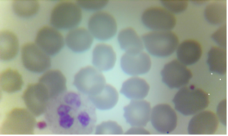
" Blood smear showing a typical Bartonella-like bacteria (office lab)
These images are from my office lab which now performs in house, diagnostic blood smear examinations for our patients. All of these images come from patients with negative serological blood test results.
This a Giemsa stain from my office lab. The tiny dot at the bottom of the slide is typical of Bartonella-like organisms. These are typically very small. The bacteria stains dark blue. Note, the larger purplish splotch indenting a red blood cell comes from platelets. The larger cell is a typical white blood cell called by various names, including: neutrophil, poly, granulocyte. "
TNT
Frequent Contributor (1K+ posts)
Member # 42349
posted
quote:Originally posted by Lymedin2010: TNT, thought you might like this.
" Blood smear showing a typical Bartonella-like bacteria (office lab)
These images are from my office lab which now performs in house, diagnostic blood smear examinations for our patients. All of these images come from patients with negative serological blood test results.
This a Giemsa stain from my office lab. The tiny dot at the bottom of the slide is typical of Bartonella-like organisms. These are typically very small. The bacteria stains dark blue. Note, the larger purplish splotch indenting a red blood cell comes from platelets. The larger cell is a typical white blood cell called by various names, including: neutrophil, poly, granulocyte. "
posted
Possible Babesia in the middle there? It was much more pronounced through the lens; my cell phone doesn't take the most amazing images.
I've done dozens of stains, and this is one of the only halo structures I've seen. I figured if I have Babesia, I would have seen more by now. Maybe I don't have Babesia in my fingers . Starting Mepron this weekend anyway.
I took a shot of this stuff because it stained much more vivid than than anything else I've seen before. Again, not a great quality image, but it was made up of multiple little dots and rods. Definitely not platelets. Check out that inclusion in the RBC to the left. Not sure I've seen anything like that before:
Lastly, I'm still not 100% sure what these dark staining specs are inside of my WBCs:
Hope everyone's having a good weekend!
Posts: 71 | From Canada | Registered: May 2016
| IP: Logged |
posted
Hello everyone, I am here to report that my blood is getting better and better. However, I do want to know what the clusters are in the video. The first cluster consists of black dots. The second one is different from the first one.
posted
One key thing for my feeling better and better is Kelp. I take 6-10 capsules a day. Also every time I ate raw garlic, I herx.
I tried hyperbaric oxygen twice. I tried bee venom twice,but allergic to it.
Hope it helps.
Posts: 41 | From NJ | Registered: Apr 2015
| IP: Logged |
TNT
Frequent Contributor (1K+ posts)
Member # 42349
posted
quote:Originally posted by rainboworiver: Hello everyone, I am here to report that my blood is getting better and better. However, I do want to know what the clusters are in the video. The first cluster consists of black dots. The second one is different from the first one.
I'm glad your blood is looking better and better! What are the elements of your protocol at the present besides kelp and garlic? Too bad you're allergic to bee stings. That's a bummer. Did you have trouble breathing? A reaction is not the same as being allergic. How did you react to hyperbaric oxygen?
The clumps of black dots (at :15 in your video) are clumps I've seen in my blood, too. I have not figured it out, though I think it is probably a collection of cellular debris of some kind or another. I've considered a collection of lipids, too, except lipids should be nearly perfectly-round objects with a different light refraction.
But, the other "clumps" you are referring to (1:00 in your video) are cellular debris and remnants from white blood cells. That I am certain of.
Posts: 1308 | From Eastern USA | Registered: Oct 2013
| IP: Logged |
posted
TNT, to answer your question, I am recapping from my previous posts and now adding new stuff. Here you go.
On Feb 23, I stopped all abx, my blood was loaded with organisms. I started the following herbs: Baical skullcap, 2 dropfuls 3x a day, kelp (2 tablets a day), agrisept 20 drops 3x a day. I did experience herx (not very severe). This is in addition to serrapeptase, Japanese knotweed, nattokinase, protease and probiotics that I have been taking for over one month. Additionally, I have been doing IV EDTA chelation plus vitamin c and b and glutathione once every 1 to 2 weeks for a couple of months. I just finished the last of the 10 EDTA sessions. I believe that EDTA and the enzymes contributed significantly to breaking up the biofilm clusters and red blood cell clustering (the rouleaux) problem. I significantly restricted sugar intake (fruits and startch) for two weeks, but couldn’t last long, so I have relaxed about eating a few of fruits. A few days ago, I added pinella 10 drops twice a day and coptis one dropful twice a day. I also added lauricidin, two teaspoons a day. I experienced herx again (not too severe).
Two weeks ago, I tried HBOT at 2 ATA for 90 minutes. I felt great the first time. I didn’t feel the same drastic effect after the 2nd time. I had to stop to wait to get my doctor’s clearance on brain aneurysm. I tried bee venom therapy, the area blew up to the size of an orange. I stopped after two tries. I may try again in a couple of weeks.
One week ago, I added biotherapy karlovy vary thermal spring salt, it alkalines the blood and adds back the minerals I lost during EDTA chelation. Two weeks ago, I started colonic hydrotherapy, once a week. At the end of the 7th session, they will restore colon flora with probiotics. I have been feeling better after each colonic.
I have been using self-made LED lights (red, yellow and infrared) a couple of times a week for 30 min to 60 mins. I don’t know if this helped with lyme, but didn’t notice any reaction afterwards. It did work magic on the bee sting (orange size) in stopping the unbearable itch within 10 minutes, and calming down the angry redness and reducing sewelling.
I blend lots of garlic in roasted eggplant/pepper, each time I herx from it. I am sticking to this though I probably reek garlic from afar.
Posts: 41 | From NJ | Registered: Apr 2015
| IP: Logged |
Lymedin2010
Frequent Contributor (1K+ posts)
Member # 34322
posted
Clean stains! Both look like it could be babesia. The 2nd one almost looks like a lopsided maltese cross, although it did not stain purple/blue.
Sometimes I do see other babs stains that look slightly pinkish though...you be the judge.
_______________________________ The rbc's stain pink & platelets should stain violet but can also look pinkish if the platelet granules are dispersed and if they are concentrated then slightly more violet or pretty close to purple & wbc's nucleus blue (which may look purplish) & leukocyte chromatin magenta. Bacteria should come out blue & sometimes look purplish as well. All this can also vary upon magnification & your lighting & the camera that picks up the color. Stains are best viewed under 100x oil.
Mustarseed uses this one below & I tried it too...it is great & relatively cheap. You just make the peripheral blood smear just like in this video. You spread & fan out the blood to make a nice corona & then let it air dry in a closed environment (to avoid contamination) for 10-15 min & then stain.
Then you just dip a few times in each container & blot between each container so as to avoid excess drip into the adjacent container (touch the edge of the slide to a tissue to remove excess stain solutions). Mustardseed gave instructions to us a few weeks ago, in 3 or so pages back, as he has managed to perfect the technique.
Great that your blood has less spiros. Another doctor reports that the levels of their blood & their patients blood can go up & down from time to time, as this person has checked for a few years now. So the levels can wax & wane.
Posts: 2094 | From NY | Registered: Oct 2011
| IP: Logged |
Lymedin2010
Frequent Contributor (1K+ posts)
Member # 34322
posted
So tough to say really for sure on the babs stains, but it could be. I am not totally satisfied & don't get that content feeling for either of the images honestly...but they could just as well be.
I would just keep hunting for more evidence.
I have a few what look like babs stains on mine too, but I am not happy with them & dismiss them so far.
Pic 1 issues, no babs organism on the edge & just a plain circular halo.
Pic 2: The white around what almost look like a maltese cross & lack of blue/purple stain. I can forgive the stain, but that white surrounding I wonder & bothers me.
Posts: 2094 | From NY | Registered: Oct 2011
| IP: Logged |
Lymedin2010
Frequent Contributor (1K+ posts)
Member # 34322
posted
I rewrote this for everyone....
CHEAP & ACCESSIBLE Giemsa/Wright type stain: You just make the peripheral blood smear just like in this video. You spread & fan out the blood to make a nice corona & then let it air dry in a closed environment (to avoid contamination) for 10-15 min & then stain.
Here is a lab example of Diff Quick/Dip Quick stain procedure. I would not use tap water as they did here, but RODI or distilled water instead. https://www.youtube.com/watch?v=V9nFq8BvW-Q
1) To stain you just dip in container #1 a few times they say 5. So 5x times up & down to dip. We found that we get better results if we dip 10 times over 5-7 seconds. Then blot dry (touch the edge of the slide to a tissue to remove excess stain solutions)
2) Then don't waste time & go straight to dipping in container #2, it says for 5 dips/seconds. But again 10x dips over 5-7 sec worked better for us. Then blot dry.
3) Quickly into 3rd container, we found best 7 dips over 3-5 sec. Then REALLY QUICKLY rinse it with RODI or distilled water right away. A 5 sec wait between the water rinse & the last dip of #3 might mess it up, so rinse it quickly but GENTLY.
The dipping times are suggested, but there is some free play & you can try and see what works best for you. Some people even try alternative means, for instance instead of air drying the blood sample, they bake it in an oven @100F.
_____________________________________ Will the stain containers have residue after many uses? After many, many uses yes. Most of it will settle to the bottom & so over time try not to shake or disturb the containers as much as possible. If you want an efficient way of managing your stains, then get a Wheaton glass Coplin staining jar with a lid. You can then use this a few times & discard for new solution when you deem necessary.
You can also use these cheap microscope slide holders, but the lid on these is not air/liquid tight & you may have to store them in durable plastic bags & away very safely or just discard the solution after doing multiple slides at one time.
Droppers to use with other staining method, the Diff Quick & Dip Quick (Vet version) do not require the droppers since you just DIP the slides in each jars successively.
***Anyone staining should know that the fumes from these stains is toxic & you should stain in a well vented & open area, although the staining videos that I have seen appear not to have taken those precautions. With Lyme Disease I would not risk exposure to any toxic fumes. _____________________________________ For Giemsa/Wright stain in general:
Structure Colour Erythrocytes Pink/yellowish red Platelets Violet/purple granules Neutrophils Blue nucleus, pink cytoplasm, violet granules Eosinophils Blue nucleus, blue cytoplasm, red granules Basophils Purple/dark blue nucleus, violet granules Monocyte Violet nucleus, light blue cytoplasm Bacteria Blue Spermatozoa Pale blue in the acrosomal region and dark blue in the post-acrosomal region
From the color chart above & you need to view & study the known examples of the various stained pathogens. If you click on the source pictures below sometimes there is description of the stain used & other times you can click on "view page" to see the source page that it came from. You will probably encounter more stained subjects that are artifacts or questionable then to get actual hits that match the known stained pictures. The better you get at staining the easier it becomes, but always expect to discard stained subjects more than you identify.
posted
Lymedin2010, thank you so much for taking the time to write up the how-to guide to staining. I had a mental block about starting, now I feel I can tackle it. I know I have organisms in my RBC. How does one know if it is lyme, babesia or other protozoa in RBC?
I guess before my blood was loaded with so many different organisms, it didn't matter what they were. Now that I have gotten the load way down, I am getting more curious about what else is out there. With the treatment I did in the last month, I have made more progress in one month both in terms of live blood and symptoms than the last 8 years.
Are those pictures from your blood? they are fantastic pictures.
Posts: 41 | From NJ | Registered: Apr 2015
| IP: Logged |
Lymedin2010
Frequent Contributor (1K+ posts)
Member # 34322
posted
rainbow, I answered your question & gave some more important updated info above.
Those are not my samples, but from the links above.
Posts: 2094 | From NY | Registered: Oct 2011
| IP: Logged |
Lymedin2010
Frequent Contributor (1K+ posts)
Member # 34322
posted
"Detection and treatment of babesiosis are important tools to control babesiosis. Microscopy detection methods are still the cheapest and fastest methods used to identify Babesia parasites although their sensitivity and specificity are limited. Newer immunological methods are being developed and they offer faster, more sensitive and more specific options to conventional methods, although the direct immunological diagnoses of parasite antigens in host tissues are still missing. Detection methods based on nucleic acid identification and their amplification are the most sensitive and reliable techniques available today; they are fast, very specific and although most of them relay on sophisticated equipment, new methodologies are being developed without the need of expensive apparatus. Importantly, most of those methodologies were developed before the genomics and bioinformatics era, which leaves ample room for optimization. For years, babesiosis treatment has been based on the use of very few drugs like imidocarb or diminazene aceturate. Recently, several pharmacological compounds were developed and evaluated, offering new options to control the disease. With the complete sequence of the Babesia bovis genome and the B. bigemina genome project in progress [4], the post-genomic era brings a new light on the development of diagnosis methods and new chemotherapy targets."
"Babesia especies in various hosts and tissues. A) Babesia bigemina in bovine erythrocytes. Blood smear stained with Giemsa. B) Babesia bovis in bovine erythrocytes. Blood smear stained with Giemsa. C) Babesia microti in mouse erytrocytes. Blood smear stained with Giemsa. D) Babesia bigemina kinetes in Rhipicephalus (Boophilus) microplus haemolymph. Haemolymph smear stained with Giemsa. E) Babesia bovis in a bovine brain capillar. Histological section of brain tissue stained with Giemsa. F) Detection of antibodies against Babesia bigemina by the Indirect Fluorescent Antibody Test (IFAT). Bovine antibodies were detected by a secondary, donkey IgG anti- bovine IgG bound to Alexa-Fluor 488. Images were obtained with an objective of 100X."
posted
Ya that inclusion in my second picture is really weird. Sort of like some of those pics you posted.
I'm feeling like absolute garbage today, so maybe I'll dive in and try a few more stains.
Posts: 71 | From Canada | Registered: May 2016
| IP: Logged |
Lymedin2010
Frequent Contributor (1K+ posts)
Member # 34322
posted
Staining seemed so easy & straightforward when presented by others, we did not realize there will be this much hunting involved & questioning and second guessing.
Personally, I find microscopy with this horrible disease therapeutic. Rather than just giving into it, we are doing something & trying to figure things out & better than the alternative of doing nothing at all. JMHO.
Posts: 2094 | From NY | Registered: Oct 2011
| IP: Logged |
Lymedin2010
Frequent Contributor (1K+ posts)
Member # 34322
posted
At time 9:24 & onward she says that her doc thinks she has MS. She is on treatment for Lyme Disease.
posted
Hello friends, I've been ill for around 15 years and making steady progress toward recovery over the past three years with detoxification. I had mercury and record-breaking copper toxicity along with mold exposure and chronic inflammatory response syndrome. Now my doctor says I also have Lyme disease so I borrowed a microscope from a friend yesterday. My blood looks awful (massive rouleaux) but I could not see any spirochetes.
I did see a number of red blood cells that were twitching and maybe revolving. They seemed to have some darker curves which would twist and untwist but I'm still unsure as to what I was seeing. I understand that it may take 24 hours or more for spirochetes to emerge, but after 16 hours or so, my slide looks all dried up. Am I doing something wrong? How do I keep it hydrated? My procedure was to place a drop of blood on the slide and then put a slipcover on top.
I should add too that Charles Barker adds pure copper powder to the blood to force the spirochetes right out, but I don't have pure copper. Anyone else experimented with other ways of forcing them out quickly?
Lymedin2010
Frequent Contributor (1K+ posts)
Member # 34322
posted
What magnification are you looking at them?
If you use 100x oil, then add oil immersion to the slip cover & that will seal them & avoid dehydration. If you are not using an oil objective then you can take a Q-tip & pull the tip out to a point & dip it in Vaseline & apply it to the edges of the cover slip only to seal it.
posted
Thank you lymedin2010!! That was very very helpful. With your tip I've been able to keep my blood hydrated with no sign of deterioration going on 24 hours now.
I have not seen any spirochetes, but there are tiny little critters running around that clearly do not belong there. As for size, you could fit a couple hundred of them in a red blood cell. On another YouTube video I saw something similar being described as parasites.
There is also something strange I would love to have your feedback on. I have a number of cells that look to be the same size as red blood cells that twinkle. It's almost as if I had a red blood cell that was full of the little parasite thingies. The twinkling might just be motion of the bright dots.
Unfortunately when I try to capture video, my software crashes. I'm thinking of getting a better camera and maybe microscope also because sure would be nice to zoom in on this stuff...
Lymedin2010
Frequent Contributor (1K+ posts)
Member # 34322
posted
That looks like a white blood cell with granules inside the cytoplasm.
I have tested a few cameras so far & out of Canon 40D, Sony a6300 (HD), Sony FDR-X1000V, & Panasonic G7 (4K), so far the Samsung Galaxy S5 has the best interpretation of lighting (WB) & produces the best video in 4K & the Sony FDR-X1000V comes in 2nd. The Sony is a nightmare in terms of battery life, ease of access. The Samsung phone you can also send video out to a TV via HDMI & or sync with a larger tablet for viewing. If you use the Sync method you can also charge it via USB & have PC access to files instantly while recording & it really works out great. There are time lapse apps on the Google stores that you can use as well.
I show you some of the ways to hookup a phone in this MY MICROSCOPE FaceBook group here:
______________________________________ The ability of the spirochetes to drill forward and instantly backwards is a unique feature of spirochetes. Here is a lab reference B burgdorferi B31 strain that shows this inherent feature at time 26s, but the whole video is worth a watch. Keep this in mind when you watch some of my tick videos & my future tick video that will show spirochetes in a new light.
The Lyme Disease Network is a non-profit organization funded by individual donations. If you would like to support the Network and the LymeNet system of Web services, please send your donations to:
The
Lyme Disease Network of New Jersey 907 Pebble Creek Court,
Pennington,
NJ08534USA http://www.lymenet.org/


 UBBFriend: Email this page to someone!


![[Frown]](frown.gif)


![[Razz]](tongue.gif) . Starting Mepron this weekend anyway.
. Starting Mepron this weekend anyway.


![[Smile]](smile.gif) I know I have organisms in my RBC. How does one know if it is lyme, babesia or other protozoa in RBC?
I know I have organisms in my RBC. How does one know if it is lyme, babesia or other protozoa in RBC? 


 Printer-friendly view of this topic