Never occured to me I could use a DSLR with the microscope? Do you like that better than the purpose built microscope cameras? I do have a G7 here.
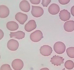
Does everyone have those little white jitterbug's? wondering if those are normal or parasites... I have seen some that are a little larger than others.
That video is really interesting - Those spiros can move!
bluelyme
Frequent Contributor (1K+ posts)
Member # 47170
posted
yes birthday yes, lowdown -i have seen those lil white jitterbugs in a lot of peoples samples some say normal flora other say baby ketes or other pathogens.some one had a name for them do you remember, mustard?
here is a vid from our french counterparts showing a quick encyst on 2nd part , my friend translated and said it says that 2 spirochetes are being birthed from the round form https://www.youtube.com/watch?v=d3dHFBiLac4
-------------------- Blue Posts: 1539 | From southwest | Registered: Dec 2015
| IP: Logged |
TNT
Frequent Contributor (1K+ posts)
Member # 42349
posted
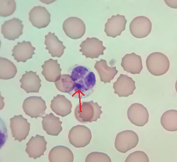
quote:Originally posted by thelowdown: There is also something strange I would love to have your feedback on. I have a number of cells that look to be the same size as red blood cells that twinkle. It's almost as if I had a red blood cell that was full of the little parasite thingies. The twinkling might just be motion of the bright dots.
Lowdown & Blue..... as Lymedin said, those twinkling round cells are white blood cells. I can't quite discern from the picture which white cell it is, but I would lean towards a granulocyte such as basophil or eosinophil. Notice the coarse granules (lysosomes) in the cytoplasm.
Posts: 1308 | From Eastern USA | Registered: Oct 2013
| IP: Logged |
bluelyme
Frequent Contributor (1K+ posts)
Member # 47170
posted
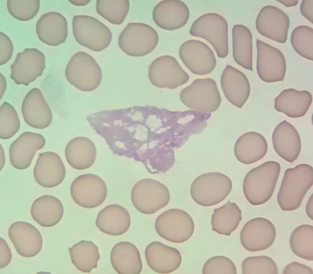
I have seen those disco balls in a few peoples blood now .is it fungal?
-------------------- Blue Posts: 1539 | From southwest | Registered: Dec 2015
| IP: Logged |
TNT
Frequent Contributor (1K+ posts)
Member # 42349
posted
quote:Originally posted by bluelyme: I have seen those disco balls in a few peoples blood now .is it fungal?
They are, without a doubt, white blood cells!
Posts: 1308 | From Eastern USA | Registered: Oct 2013
| IP: Logged |
Lymedin2010
Frequent Contributor (1K+ posts)
Member # 34322
posted
WORLD PREMIER:
For the first time ever (that I know of) I capture time lapse of a SOP (String of Pearls) in squashed ticks, which the pearls then go on to develop into baby spirochete.
Subjects were filmed using various microscopes, objectives, lighting sources, & different camera systems.
**Use the PAUSE button for this video to read long texts, otherwise the video would have been even longer.
I also reveal PCR testing toward the end of the video. This video starts out slow & gets better with more evidence over time...clips of 400x, 630x, & 1000x.
________________________________________________ WHAT WE WILL SEE IN THIS VIDEO:
-What Tick juice looks like without any spirochetes.
-Tick juice with spirochetes.
-Rigid "Needle Form" spirochetes.
-Needle forms wake up to start motility.
-Spirochetes drill/spiral forward AND backwards.
-What stained BSK-H B burgdorferi grown spiros look like.
-Longer & typical spirochetes in ticks.
-Spiros that split into 2, 3, 4...daughter cells.
-Spiros that drill/spiral erratically.
-Spiros that just gyrate and stagnate similarly to Lyme Diseased blood.
-Granular forms (blebs), spiros developing from granules.
-SOP (String of Pearls) & segmented spiros & spiros w/light & dark bands/spotting.
-Time lapse breakup of SOP & blebs/segments grow into baby spiros.
________________________________________________
STAGES IN SPIROCHETE PRESENTATION FROM TICK JUICE:
1) RODI or Distilled water, or other solutions can cause spirochetes to form cysts. Sometimes it can take an hour or more for them to come out of cyst morphology, depending on the solution. Water is just one way to force them to cyst up.
2) Spiros uncyst & can be very rigid and in "Needle Form." This seems to be a shock stage, where they are perhaps still adjusting to osmosis of the added solution.
3) When nutrients are present they can move VERY rapidly & go through rapid splitting once uncysting & adjusting. These are VERY FAST growers & nutrients dictates they produce & split VERY rapidly for these spiros. In this stage many 2 daughter spiros will be seen seperated by a thin bit in between the daughters. Sometimes 3, 4, & more daughters can be seen.
4) When they are left in nutrients for longer periods they tend to grow thicker and longer. Starved nymph ticks may not present with longer or thicker forms of this particular spiro, they tend to remain as smaller units in low nutrients.
5) When nutrients are depleted the spirochetes have less motility or simply gyrate in place in stagnant forms, sometimes very similarly to what we have seen in Lyme Diseased blood. The solution they are in & nutrient availability dictate to some degree how the spirochete evolves and moves.
6) Even in stagnant forms & in forms much smaller than the full blown typical BSK-H grown form, they tend to coalesce, secrete a cloudy/milky substance into the solution, & start forming a slimy biofilm. The cloudiness in the solution over time obscures the visibility of the spiros.
Posts: 2094 | From NY | Registered: Oct 2011
| IP: Logged |
Lymedin2010
Frequent Contributor (1K+ posts)
Member # 34322
posted
40x * Zeiss 15x eyepiece = 600x + cam zoom on a Samsung Galaxy S5 with this microscope adapter. The Zeiss optics are top notch when resolution matters!
Using this light bulb on a standard light fixture right under the microscope condenser: Posts: 2094 | From NY | Registered: Oct 2011
| IP: Logged |
TNT
Frequent Contributor (1K+ posts)
Member # 42349
posted
Lymedin, you are the KING OF SPIROCHETES!!! Great research and a wonderful video production!!! I don't know how a person can improve on that proof. The time-lapse at the end with the string of pearls budding & molting into juvenile spirochetes was just spectacular! And to see them swim away....!!! Incredible!
It would be nice to see similar time-lapse of Bb SOP molting into juvenile spirochetes. Regardless, your documentation of this process is truly remarkable even though they are not Bb spirochetes!
This is cutting-edge research if you ask me! Way to Go!!!
I've got some potential evidence of invasomes that I plan to share before long. Ha Ha, you didn't think you would hear that from me did you? I'm still a bit hesitant to say they are "Bart" invasomes just because there's no way to prove what organisms they are. But, the vacuoles are definitely visible and the organisms inside are unmistakable.
Posts: 1308 | From Eastern USA | Registered: Oct 2013
| IP: Logged |
Lymedin2010
Frequent Contributor (1K+ posts)
Member # 34322
posted
Thanks TNT.
Refinements will produce even better video on the next round.
I am eagerly awaiting your fluoro & bart videos and might even get inspired to get a fluoro scope as well.
Posts: 2094 | From NY | Registered: Oct 2011
| IP: Logged |
posted
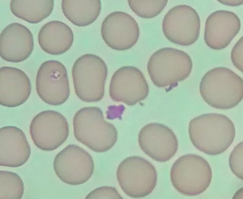
Terrible quality, as per usual with my cell phone camera, but here's a possible Bartonella-like organism. It's hard to tell from this picture, but it's pretty rare something gets colored blue in my stains. Edit: not sure why the image is so small. You'll just have to take my word that there's something there
Anyone see stuff like this next image? I see it quite often. I'm not 100% sure, but I think it's the leftovers of an exploded white blood cell.
Lastly, still getting these dark purple spots inside of my WBC nuclei. This one is particularly big. They never show up outside of the nucleus, and it's possible that it's an artifact. I really don't know.
Posts: 71 | From Canada | Registered: May 2016
| IP: Logged |
TNT
Frequent Contributor (1K+ posts)
Member # 42349
posted
quote:Originally posted by mustardseed2: Terrible quality, as per usual with my cell phone camera, but here's a possible Bartonella-like organism. It's hard to tell from this picture, but it's pretty rare something gets colored blue in my stains. Edit: not sure why the image is so small. You'll just have to take my word that there's something there
Anyone see stuff like this next image? I see it quite often. I'm not 100% sure, but I think it's the leftovers of an exploded white blood cell.
Lastly, still getting these dark purple spots inside of my WBC nuclei. This one is particularly big. They never show up outside of the nucleus, and it's possible that it's an artifact. I really don't know.
Good work! That's a good possibility the first pic could be showing Bart. The two objects could also be other things such as Babesia merozoites, since they look more oblong than most Bartonella on the outside of a RBC. But it's a little hard to tell for sure.
I agree with you on the second pic. It's also referred to as a "smudge cell" in a Giemsa smear. I used to see lots of them too. The past few smears I've done I've seen very few. It seems to have coincided with the addition of Azithromycin (in addition to the other meds). I'm almost certain the smudge cells are a result of the Anaplasma infection, but Azithromycin is not very active against the "spotted" Rickettsias. Then again, the past few smears I've seen very few typical Bartonella on the peripheral of my RBCs. So, maybe the Azithro is hitting Bart which is causing, or at least contributing to, the smudge cells in my case.
As for your 3rd pic, my opinion is that what you are seeing is normal, except that it appears like the Neutrophil is superimposed on the RBCs. It appears ghosted too. This is a bit out of the ordinary. But, the clumpy, non-homogenous nucleus is very typical, and I can't really say that this particular cell looks abnormal (to me),except for what I mentioned.
I've been a bit bummed out lately. I haven't been doing as well and I haven't had the energy or focus to pursue things as I would like. It doesn't help that the past number of AO stains I've done have flopped. I've gotten one good AO dried smear since I've gotten the scope (and one trial wet mount that did ok). I don't know if it's the stain or something seemingly inconsequential I'm doing wrong that is causing my smears to wash (or soak) off. Believe me, I've tried varying my techniques, but no help. I think it's the stain, and will have to talk with the company to see if they can give me any clues as to why I had good success with the first dried smear, but none since. It's SO DISAPPOINTING!!! And, it's at a bad time for me, too. The AO dried smears could really give me some hard evidence concerning a few of these infections. My Giemsa stains are pretty definitive, but that absolute verification would really be helpful right now.
Posts: 1308 | From Eastern USA | Registered: Oct 2013
| IP: Logged |
posted
Smudge cell... awesome! Thanks for putting any doubts I had about those to rest!
These smudge cells could very well be from Anaplasma. I tested positive for anaplasmosis through Igenex and have a moderate neutropenia right now. However, I've looked at hundreds of WBCs and have yet to see any anaplasma inclusions like the great pics you posted a page or two back.
Sorry to hear you haven't been feeling well. I don't know much about AO stains but they look pretty cool when I Google them.
What exactly are you looking for in them?
What infections are you treating right now?
Posts: 71 | From Canada | Registered: May 2016
| IP: Logged |
TNT
Frequent Contributor (1K+ posts)
Member # 42349
posted
quote:Originally posted by mustardseed2: Smudge cell... awesome! Thanks for putting any doubts I had about those to rest!
These smudge cells could very well be from Anaplasma. I tested positive for anaplasmosis through Igenex and have a moderate neutropenia right now. However, I've looked at hundreds of WBCs and have yet to see any anaplasma inclusions like the great pics you posted a page or two back.
Sorry to hear you haven't been feeling well. I don't know much about AO stains but they look pretty cool when I Google them.
What exactly are you looking for in them?
What infections are you treating right now?
Yeah, I would expect Anaplasma to cause the greatest number of smudge cells, but other infections such as Babesia and Bartonella can cause leukopenia as well. Even viruses can. You could try some Azithromycin with a tetracycline to see if that helps rectify your leukopenia. That is what has helped mine. The tetracycline without azithromycin did not do it for me.... which is odd, since effective treatment for Anaplasmosis is typically just Doxycycline. In resistant cases, Rifampin is added, but Azithromycin is usually not used or included.
Yeah, the fluoro stains are mesmerizing. I'm just looking for the things I typically see in my giemsa stains.....just confirmation really. It can be nearly impossible to differentiate between a platelet and a morula in a giemsa stain. Same thing with Babesia ring-forms. I have seen definite presence of both, but just want to see absolute confirmation with AO. The morulas are again the highest load, but just looking for that 100% confirmation.
I have (again) been treating Babs, but have (again) had to stop because of worsening Anaplasma and Lyme symptoms. The recent giemsa smear showed a higher load of morulas. A few months ago, before commencing Babs treatment, I essentially saw none. Interestingly, the last smear also began showing pretty clear evidence of Babs (ringforms). It seems the Malarone is bringing the Babs more to the surface, but allowing the Rickettsia & Bb to grow.
Posts: 1308 | From Eastern USA | Registered: Oct 2013
| IP: Logged |
posted
Wow, super interesting. It seems that even though you stopped seeing the anaplasma, it hid undetected and then started multiplying when you stopped treating it. These infections are difficult.
The problem with Malarone and Mepron is that they don't mix well with Rifampin or Doxycycline, so it's difficult to treat Babesia at the same time as other coinfections.
Like I said, my Igenex came back positive for anaplasma at the same time my LLMD diagnosed me clinically with Babesia. Her idea was to do Zithro, Doxy, and Malarone all together. I definitely herxed on the Malarone so it was still working, despite being mixed with Doxy. Might want to try that if you haven't?
As for the neutropenia, mine was borderline, but stable, on the zithro/doxy combination, but when I added Malarone it rapidly plummeted. Exact same thing happened when I switched from Bartonella medications to Mepron. This article was interesting for me, you may have already seen it:
Best of luck with the new stains, I would love to see the results when you get it working.
Posts: 71 | From Canada | Registered: May 2016
| IP: Logged |
TNT
Frequent Contributor (1K+ posts)
Member # 42349
posted
quote:Originally posted by mustardseed2: Like I said, my Igenex came back positive for anaplasma at the same time my LLMD diagnosed me clinically with Babesia. Her idea was to do Zithro, Doxy, and Malarone all together. I definitely herxed on the Malarone so it was still working, despite being mixed with Doxy. Might want to try that if you haven't?
As for the neutropenia, mine was borderline, but stable, on the zithro/doxy combination, but when I added Malarone it rapidly plummeted. Exact same thing happened when I switched from Bartonella medications to Mepron.
Thanks for the suggestion. I have actually done that combo, and it did seem to have been helping. The problem, though, seems to be when I get to the max dose of the Malarone there is not enough killing power for the Rickettsia or the Lyme. Because, this threshold seems particularly lower -- and coincides with -- the 4-week cycle of the Lyme. In other words, I do ok with full-dose Babs treatment until I hit the anticipated monthly cycle of the Lyme. Then things go south pretty quickly regarding my usual "Bart" (Anaplasma) and Lyme symptoms. Of course, as I said, this is evident in my blood smears, too. I'll probably have to maintain a lower dose of Malarone for longer than normal once I recommence.
That's very interesting that is what happened to you, too....at least according to the neutropenia. It seems unlikely to me that the Babesia would have caused increased neutropenia when you were on the heavy Babs drugs. I tend to think your Anaplasma has not been sufficiently addressed even though you cannot find visual evidence of it in your smears.
Posts: 1308 | From Eastern USA | Registered: Oct 2013
| IP: Logged |
TNT
Frequent Contributor (1K+ posts)
Member # 42349
posted
Fluorescent microscopy will be fascinating once I can delve into it more in depth....and get my smears to turn out.
Here is a pic that Lymedin posted earlier:
And, here is a page on Acridine Orange staining from Counselling Me Microscopy that shows how it is possible to see bacteria (probable borrelia) inside RBCs even when it is impossible to see with darkfield (in live blood):
posted
Wow those bacteria really pop in that stain! Man I hope you can get it working, it looks amazing.
What was your maximum Malarone dose, out of curiosity? LLMDs are prescribing increasingly high amount of atovaquone for Babesia, because the old 750mg (1 tsp Mepron) twice a day just isn't cutting it for people.
For instance, my LLMD wanted to treat with 4500mg per day (3 tsp twice a day). We settled on 3000mg to not freak out the insurance company. 4500mg would be 18 Malarone tablets per day (and remember that the atovaquone tablet is relatively poorly absorbed vs the liquid). I get what you're saying though, you've got to keep on top of the anaplasma too, especially if it's popping up in your stains more frequently.
Of topic slightly, but is this YouTube video from someone in this thread?:
The reason I ask is because it's labeled as Rickettsia. Do we know for sure that's what this is??? Because frig, I see these in pretty much every smear I do. Identical.
Posts: 71 | From Canada | Registered: May 2016
| IP: Logged |
TNT
Frequent Contributor (1K+ posts)
Member # 42349
posted
quote:Originally posted by mustardseed2: Wow those bacteria really pop in that stain! Man I hope you can get it working, it looks amazing.
What was your maximum Malarone dose, out of curiosity? LLMDs are prescribing increasingly high amount of atovaquone for Babesia, because the old 750mg (1 tsp Mepron) twice a day just isn't cutting it for people.
For instance, my LLMD wanted to treat with 4500mg per day (3 tsp twice a day). We settled on 3000mg to not freak out the insurance company. 4500mg would be 18 Malarone tablets per day (and remember that the atovaquone tablet is relatively poorly absorbed vs the liquid). I get what you're saying though, you've got to keep on top of the anaplasma too, especially if it's popping up in your stains more frequently.
Of topic slightly, but is this YouTube video from someone in this thread?:
The reason I ask is because it's labeled as Rickettsia. Do we know for sure that's what this is??? Because frig, I see these in pretty much every smear I do. Identical.
That's an incredible amount of atovaquone your doc wanted you on! I've only ever made it to 4 Malarone tabs daily. I've heard of the 3000mg daily, but not 4500mg! I'm surprised your insurance company would even cover the 3000mg!
Yeah, the morulas are more of an issue for me than Babs ringforms. Only since I started taking Malarone have I seen any probable ringforms in my blood. But, the morulas are definitely prominent.
That video is mine, ha ha. Like you, I've seen plenty of those in my blood! Besides spirochetes, bacilli are the most prominent pathogens in my live blood. I am assuming that these are Rickettsias since that is what I am finding in my stained dry smears. As an example, in the one successful AO dry stain I was able to get, I found 3 very distinct bacilli (rods) that exact size versus only one possible variant ringform.
It sounds to me like Rickettsias are your dominant pathogen as well.....
Posts: 1308 | From Eastern USA | Registered: Oct 2013
| IP: Logged |
posted
Ya it might be. Figured 2 months of Doxy and 6 months Rifampin would have given me some relief.
Tomorrow when I have some time I'm going to start searching for rickettsia blood smears online.
I've seen pictures of morulae and I've seen pictures of thinner, smaller, ehrlichia species, but I haven't ever seen anything like these larger rods mentioned as rickettsial (outside of your video, which is amazing quality btw).
Posts: 71 | From Canada | Registered: May 2016
| IP: Logged |
TNT
Frequent Contributor (1K+ posts)
Member # 42349
posted
Here is a sampling of my fluoro work. Sorry for the large size. I had to change my picture hosting site. Tinypic has become way too slow with too many glitches and possible security issues. I will continue to see if I can minimize the photos from my new site.
A bacillus that is probable Rickettsia:
A closer and crisper pic of the same:
A bacillus on or in a RBC:
Another bacillus:
Typical Bartonella on the peripheral of a RBC:
A possible variant Babesia ringform (Take note of the platelets...they are a very pale green color and do not stain orange because they do not contain a nucleus with DNA or RNA. A Howell-Jolly body in a RBC will stain orange since HJ bodies are left-over nuclear material. But this is no Howell-Jolly body! HJ bodies are perfectly round):
[ 05-10-2017, 11:07 PM: Message edited by: TNT ]
Posts: 1308 | From Eastern USA | Registered: Oct 2013
| IP: Logged |
TNT
Frequent Contributor (1K+ posts)
Member # 42349
posted
Recent finding of Babesia ringforms in family member's blood:
Closer pic of the same field:
Ringform in a neutrophil (at the 2 o'clock position):
Posts: 1308 | From Eastern USA | Registered: Oct 2013
| IP: Logged |
posted
TNT, your first post, the pictures aren't showing up.
Posts: 71 | From Canada | Registered: May 2016
| IP: Logged |
TNT
Frequent Contributor (1K+ posts)
Member # 42349
posted
The Babs pics are, but the fluoro pics aren't??
Posts: 1308 | From Eastern USA | Registered: Oct 2013
| IP: Logged |
TNT
Frequent Contributor (1K+ posts)
Member # 42349
posted
Ok, I just checked with a different device. Yes, the fluoro pics are not showing up. That's odd, because the pictures on both posts are from the same site, my google account. The giemsa pics were smaller format than the fluoro ones, but I don't see how that would make a difference.
Anyone have any ideas?
Posts: 1308 | From Eastern USA | Registered: Oct 2013
| IP: Logged |
posted
Well, I just read this thread from start to finish for the first time. I'm finally caught up. I think the most interesting stuff was the debate about what those rod shaped things in TNT's blood are:
Like I said, I'm finding the same stuff, and I think it was thatdudefromkansas who also found them.
Here's the trouble I'm running into: I can't find any credible information online that rickettsial bacteria are that large. The only thing that we seem to be basing this on is this video from CytoViva:
...as well as the fact that TNT has a persistent HGA infection. However, as originally discussed several pages back, these thing also strongly resemble merozoites:
Notice not only the spirochete changing morphology, but also the two rod like organisms at the 9-10 o'clock position. I'm assuming those are round bodies as well.
All of this is really making my head spin. I'm not saying these rod/round things seen in blood samples is or isn't Rickettsial/Babesia/Borrelia, but what I am saying is it seems likely it could be any of those three.
Anyone know any professional microbiologist we can ask?
Posts: 71 | From Canada | Registered: May 2016
| IP: Logged |
TNT
Frequent Contributor (1K+ posts)
Member # 42349
posted
quote:Originally posted by mustardseed2:
Here's the trouble I'm running into: I can't find any credible information online that rickettsial bacteria are that large. The only thing that we seem to be basing this on is this video from CytoViva:
...as well as the fact that TNT has a persistent HGA infection.
The videos by CytoViva are authoritative, since their videos are with cultured and known organisms.
I struggled with the same questions, but finally had to go with the cumulative evidence (including treatment response), which for me all pointed to Rickettsias.
In the video by Muriel on the conversion to gemma cyst (the last video in your last post), that cyst looks like a rod bacteria only when it's oriented on it's side. Otherwise, you can tell it's a gemma cyst. The organism in my "Rickettsia" darkfield video was rod-shaped no matter what orientation it was.
I never thought about the rod-shaped, partly-dumbell-shaped organism in my phase contrast video resembling two attached merozoites. But, I agree, it could very well be.
Posts: 1308 | From Eastern USA | Registered: Oct 2013
| IP: Logged |
TNT
Frequent Contributor (1K+ posts)
Member # 42349
posted
Ok, I posted those fluoro pics from tinypic, so now you can see them.
Posts: 1308 | From Eastern USA | Registered: Oct 2013
| IP: Logged |
I never thought about the rod-shaped, partly-dumbell-shaped organism in my phase contrast video resembling two attached merozoites. But, I agree, it could very well be.
The organism could also be a bacillus (I'm using the term in it's general sense to describe a rod or coccus) bacteria in the process of binary fission....
Considering the overwhelming evidence I have of an Anaplasma infection, this possibility is more likely.
Posts: 1308 | From Eastern USA | Registered: Oct 2013
| IP: Logged |
It could also be that the rods I see in my blood aren't the same as the rods in your blood, so I'm parlaying my skepticism towards my own sample onto yours. After reading through all that, I'm 99% sure my rods are these "gemma cysts". The color of the rods, the movement, the size and shape variation.
If you're not seeing that stuff, and you obviously see morulae, Rickettsial is probably a good guess.
In the long run, it doesn't really matter. The "best" treatment for HGA is also first line for Borreliosis (not that any of it is guaranteed to work).
Posts: 71 | From Canada | Registered: May 2016
| IP: Logged |
TNT
Frequent Contributor (1K+ posts)
Member # 42349
posted
Yeah, I'm seeing plenty of morulas! Many of them are unmistakable since you can see the individual organisms inside.
Here they are, and I've got some exciting stuff for Lymedin coming tomorrow!!
Posts: 1308 | From Eastern USA | Registered: Oct 2013
| IP: Logged |
TNT
Frequent Contributor (1K+ posts)
Member # 42349
posted
Lymedin, a couple pages back we had this in-depth discussion about Bartonella and invasomes. I felt there was more proof needed to validate what some were calling Bartonella vacuoles, or invasomes, in their live blood. I think that if we are seeing real Bartonella in vacuoles in our wet mounts we should be seeing them in our giemsa stains as well. Up til now I have not seen anything that resembled invasomes, but maybe I was just not noticing, or perhaps I was chalking them up to stain precipitate or artifacts.
I don't know if these are Prokaryotes or Eukaryotes--there is no way for me to tell-- so I'm not going to label them Bartonella. But, the clear presence of intracellular and extracellular vacuoles with multiple organisms inside is unmistakable in these pictures. You can clearly discern and even count the individual organisms within the vacuoles.
Here is what I have found. It is my blood:
Notice that the invasome pictured in these next two pics is still fully encompassed within the red blood cell.
[ 05-12-2017, 07:31 PM: Message edited by: TNT ]
Posts: 1308 | From Eastern USA | Registered: Oct 2013
| IP: Logged |
TNT
Frequent Contributor (1K+ posts)
Member # 42349
posted
A couple more intracellular ones:
This one may not be a good example since it is possible it's a platelet. Though, I'm pretty sure I can see two distinct organisms. It appears as though the organisms just shed the vacuole through the cell membrane.
Posts: 1308 | From Eastern USA | Registered: Oct 2013
| IP: Logged |
TNT
Frequent Contributor (1K+ posts)
Member # 42349
posted
A few extracellular ones:
This first one is a bit unclear on the picture, but there are 4 organisms inside the vacuole. It appears to be on the outside perimeter of the red blood cell. The object beside it on the right I'm pretty sure is a platelet.
And, here is one containing two organisms on the outside of a red blood cell:
Posts: 1308 | From Eastern USA | Registered: Oct 2013
| IP: Logged |
TNT
Frequent Contributor (1K+ posts)
Member # 42349
posted
Wow! Someone just nailed a well-used, albeit decent, laboratory-grade American Optical 110 microscope (brightfield only) for $15.99 plus $35 shipping. A steal of a deal at that!
I love my AO 110 (fluoro) scope! It's a newer version of the 10/20 series. The only thing I caution concerning the 110 series is that accessories such as phase & darkfield condensers are a bit hard to come by and are usually fairly expensive. The 10/20 series scopes and accessories are still plentiful and cheap. I just saw a used oil darkfield condenser for the 110/120 series go for $120 (if I remember exactly), and phase contrast turret condensers are much more expensive and harder to find. As an example, the AO 10 series oil darkfield condensers by themselves (no case or funnelstop) can be had for less than $50.
And, from the limited experience I have (with the AO PLAN achromat phase lenses), the phase objectives and phase contrast condensers are not interchangeable between the 10 and 110 series scopes. The condensers will not mount interchangeably without some customization. The bigger problem, though, is that the annuli in the phase contrast condensers that fit the 10 series will not work with the newer PLAN achromat phase lenses that came with the 110 series. And vice a versa. I had bought a couple PLAN phase objectives (which came new on the 110/120 & 400 series only) and they would not work with the annuli in my 10 series phase contrast turret condenser. So, the annuli must be different in the phase condensers of the 10/20 series versus the 110/120 series scopes.
So, if you are in the market for a used scope and are looking at getting into different microscopic techniques, the AO 10 series scopes and accessories are going to be more plentiful and cheaper than the AO 110/120 & 400 series scopes. Just an FYI.
For brightfield, though, that scope I just linked to, was a steal!
Posts: 1308 | From Eastern USA | Registered: Oct 2013
| IP: Logged |
posted
Great pics TNT! Those vacuoles look very similar to a pic I posted here a few weeks ago, on the left. Great work!
Posts: 71 | From Canada | Registered: May 2016
| IP: Logged |
Lymedin2010
Frequent Contributor (1K+ posts)
Member # 34322
posted
Way to go TNT.....that is some awesome work & findings!!!! Looks like you are the new resident co-infection go to man now.
I am proud of you son!!!
It is great for us to discover & share with one another to make advancements & to learn further...we have all done some amazing stuff & contributed collectively!
My inclination for the clusters & vacuoles is Bart. As you have not found any evidence of Babesia in 2 & 4 pairs yet, with no maltese crosses, we can put Babesia on the back burner. If you look at the science papers & pictures that linked & some of the stains, notice that sometimes the stains show bacillus & other times some odd shape & what look like babesia deformed tear drops shape, or bean shape. I would imagine when the Bartonella is dividing it can present with various morphologies & appear odd and other than the bacillus shape. Just like with the spiro video that I put out, I think there will be various genetic expressions of Bart & some can be smaller & others larger overall as well, besides the mitosis growth stages.
From what I have seen with ricketssia, it is that they can appear as cocci, bacillus, & sometimes even longer as almost string-like. They also tend to grow in larger clusters & I would expect to see a much larger congregation of them in the same cell/spot. They too can form invasomes/phagosomes & can invade endothelial cells. From the evidence that I have seen it is one ricketssia per vacuole though, but I have not looked at all the species & rick's extensively as a whole. Maybe we can collectively spend a week researching rick's & we can post all the evidence and reanalyze. I will come back at some point & make remarks on the pictures individually.
Posts: 2094 | From NY | Registered: Oct 2011
| IP: Logged |
Lymedin2010
Frequent Contributor (1K+ posts)
Member # 34322
posted
I started to write up my thoughts on Mustard's stains, but have to finish that too, but TNT hit it on the nail for the most part anyways.
I too have found similar structures when doing live blood preps, but I was never completely certain & never presented them. Then with the freezing method I was shocked & amazed at the congregation of them, while the wbc's & rbc's disintegrated largely. They also came up in my smears as well, although I wanted to do more stains & perfect them further before I made a true compilation. Now I have gotten them to POP out using fluorescence without even adding a dye yet, as there is a slight pinkish fluorescence they give off when using my Cree LED bulb, but it does not show up when using a halogen bulb though & so the light source has to be really bright & DAYLIGHT (no brown or yellow tinting) for them to be visible it seems. I want to be certain & do the actual fluorescent AO stain & do some stains on normal blood to make sure I am only presenting with these.
But I can put together a small preview video of them, so you see the pop in 4K. It is best when viewed by eye, then downgraded to 4K in terms of what it picks up, & then will probably be downgraded further on YouTube though...I will try to put it up tonight.
Posts: 2094 | From NY | Registered: Oct 2011
| IP: Logged |
TNT
Frequent Contributor (1K+ posts)
Member # 42349
posted
Lymedin, I have found fairly certain evidence of Babs in my blood....pyriforms in a few RBCs to be exact. But no pairs or tetrads. But this was only in the last giemsa smear while I was on Malarone. So, I cannot rule out the possibility that some of what I show in those pictures are apicomplexans. I do think it interesting the difference of morphology between those pictures. They are definitely showing different species, and maybe different lifeforms. It's hard to tell just by looking at them.
Yes, Rickettsias can grow in large clusters, hence the development of morulas in Anaplasma & Erhlichia. Though, I had thought that with A. marginale, they presented as one or two organisms in infected erythrocytes. But, this electron micrograph would indicate that even those little red/purple dots on an erythrocyte is a vacuole containing multiple individual organisms. A. marginale organisms must be extremely small!
Electron micrograph of a bovine erythrocyte infected with Anaplasma marginale (courtesy R.L. Sealock, Animal Parasitology Institute, ARS-USDA)
Just to clarify your comment about them being able to infect endothelial cells. Remember that Rickettsias aren't just capable of infecting endothelial cells, THAT IS THEIR PREFERENTIAL CELL AND TISSUE CHOICE!!! I don't know if they would survive if there was no endothelial --and to a lesser degree-- epithelial cells and tissues to infect. I don't think there would be any persisting infections anyhow. Of course, I don't think we would fare too well without those kinds of cells and tissues, either, ha ha!
So, DAD, when are you gonna show YOUR fluoro stuff? Posts: 1308 | From Eastern USA | Registered: Oct 2013
| IP: Logged |
Lymedin2010
Frequent Contributor (1K+ posts)
Member # 34322
posted
I might be releasing a lessons video, so I will have to hold off. Trying to gather some more evidence.
I definitely have something very interesting that will give us a new twist & perspective on things though. I knew part of it as true, but there is new evidence that is eye opening & serves as a cautionary note to us as well.
TNT, have you tested again? Are you positive for Babs, Bart, Anaplasma, or Rick's...how long ago was your test?
Posts: 2094 | From NY | Registered: Oct 2011
| IP: Logged |
bluelyme
Frequent Contributor (1K+ posts)
Member # 47170
posted
Great work bros ! tnt lymed mustard any one heard from dude? Here is from our french counterparts again .this footage is fascinating he shows a spirochete from canine saliva that is so much faster than any i have ever seen .also at the end it cyst"s and uncyst so quick really is a $hot.
this applies to me because on some of my smears tnt said it appeared to be babesia canis and i remembered a few dog bites back in the day. This kete is scary quik may explain why some euro lyme go so neuro so fast . may make think twice before getting kisses from your dog
-------------------- Blue Posts: 1539 | From southwest | Registered: Dec 2015
| IP: Logged |
TNT
Frequent Contributor (1K+ posts)
Member # 42349
posted
I have not been tested recently for anything. When I was tested, 3 Igenex FISH tests were negative, a Galaxy Bart test was slightly positive (read as a waning infection, ha), and HGE was negative through Igenex. A Brucella PCR was negative as well.
I don't have much faith in the tests. Microscopy and treatment response has been much more reliable.
An original Lyme test through Quest was negative. The original Bb WB through Igenex was CDC positive, the next one only two bands (read negative), and the most recent one was again CDC positive. So, go figure. This illustrates the reason for my lack of faith in tests. This also is the reason I don't believe the old party line that once you've been exposed to Lyme disease, you will test positive for a very long time-- perhaps the rest of your life -- long after the infection is gone. I personally don't think the antibodies last in the body nearly as long as we are told.
Posts: 1308 | From Eastern USA | Registered: Oct 2013
| IP: Logged |
TNT
Frequent Contributor (1K+ posts)
Member # 42349
posted
quote:Originally posted by bluelyme: Great work bros ! tnt lymed mustard any one heard from dude? Here is from our french counterparts again .this footage is fascinating he shows a spirochete from canine saliva that is so much faster than any i have ever seen .also at the end it cyst"s and uncyst so quick really is a $hot.
this applies to me because on some of my smears tnt said it appeared to be babesia canis and i remembered a few dog bites back in the day. This kete is scary quik may explain why some euro lyme go so neuro so fast . may make think twice before getting kisses from your dog
Good find, Blue! And, NO, I won't be getting kisses from any dog!
Last year in the news was a warning from the CDC to wash your hands every time you've held and petted your cat. The concern was about getting Bartonellosis even if one was not scratched! All it took was a lick....in the face or on an open wound!
Here is one article I pulled up real quick from that time. It also talks about a woman dying from sepsis and multiple organ failure all as a result of a lick from her dog. So, Blue, you are right on!
Lymedin2010
Frequent Contributor (1K+ posts)
Member # 34322
posted
TNT, thanks for the testing info.
Yea, I suggested to the FB group they check their dogs/cats tartar a few days ago & someone actually did just did. I had a gut feeling that we will find tons of spiros there. I also believe that one will find tons of spiros in wild animal mouths, including squirrels, deer, & mice.
I got bitten by a squirrel when I was young, as I thought they were cute & wanted to catch one...serves me right though! I also know someone who got bit by a squirrel a few years ago & she now has Parkinson's & is in a lot of pain with many Lyme symptoms. Her non-Igenex test showed negative though.
I just got a report from someone in the hospital that they just died recently of multiple organ failures after a tick bite & Lyme.
Posts: 2094 | From NY | Registered: Oct 2011
| IP: Logged |
Lymedin2010
Frequent Contributor (1K+ posts)
Member # 34322
posted
I hear you about the microscopy, I wish I did not waste my money on the tests, which I could have bought an awesome high-end microscope with all the testing money I spent.
Even if you test positive, can't tell if it was a past infection or current & you cannot keep testing & spending money on tests each 6-12 months.
Posts: 2094 | From NY | Registered: Oct 2011
| IP: Logged |
TNT
Frequent Contributor (1K+ posts)
Member # 42349
posted
quote:Originally posted by Lymedin2010: I hear you about the microscopy, I wish I did not waste my money on the tests, which I could have bought an awesome high-end microscope with all the testing money I spent.
Even if you test positive, can't tell if it was a past infection or current & you cannot keep testing & spending money on tests each 6-12 months.
AMEN! That's right.
Posts: 1308 | From Eastern USA | Registered: Oct 2013
| IP: Logged |
The Lyme Disease Network is a non-profit organization funded by individual donations. If you would like to support the Network and the LymeNet system of Web services, please send your donations to:
The
Lyme Disease Network of New Jersey 907 Pebble Creek Court,
Pennington,
NJ08534USA http://www.lymenet.org/


 UBBFriend: Email this page to someone!


![[Razz]](tongue.gif)


![[Smile]](smile.gif)

![[shake]](graemlins/shake.gif)


 Printer-friendly view of this topic