TNT
Frequent Contributor (1K+ posts)
Member # 42349
posted
Thanks, Lymedin2010. I can't wait to share some of my finds. I'm about as anxious as a schoolboy waiting to show his buddies his pet frog! Though, I'm afraid my videos will be quite humbling compared to some others' on here.
That fluorescent stain video is awesome! The clarity is SUPERB! Peter Kemp is definitely in the big leagues.
Since most of you post on youtube, is that the easiest and safest place to make personal videos public? Is there a size limit?
Posts: 1308 | From Eastern USA | Registered: Oct 2013
| IP: Logged |
Lymedin2010
Frequent Contributor (1K+ posts)
Member # 34322
posted
There are 2 viable options between Vimeo & Youtube, the latter of which has a heck of a lot more monthly viewers.
TNT, you have superpowers & you don't even know it! Besides seeing the world in slow motion because of the chronic exhaustion and stymied pace & being able to sniff out mold & chemicals through chemical sensitivity, you should now realize that you can touch someone with the right force & transfer them what is in your blood.
I don't think anyone, seeing what is in our blood, would want to make direct contact with us. But all kidding aside, if the big guns have not touched the many professionals who are attempting to turn the tides of Lyme, then I think they could care less about you & I.
Make a Youtube account & do not reveal your true name & connect it to a new email account that you should create in conjunction with the youtube account. Then post & feel free with your superpowers Posts: 2094 | From NY | Registered: Oct 2011
| IP: Logged |
Lymedin2010
Frequent Contributor (1K+ posts)
Member # 34322
posted
“Our latest findings indicate that the bacteria can literally outrun our immune cells within the host,” Wooten said. “We figured they would get in the skin and go hide from our immune response. Actually, we are finding that they don’t hide. They continue to move for months or years, and our immune system isn’t clearing them. Why is that? That is what we hope to unravel.”
TNT
Frequent Contributor (1K+ posts)
Member # 42349
posted
That's an interesting article Lymedin. I find the same thing in my blood. The neutrophils readily gather up the candida and other "artifacts," but completely ignore and go right past a kete. It's very depressing if I dwell on it.
So, it's encouraging to see this microbiologist has a found a way to study this phenomenon. Of course, it has to be mentioned in relation to a vaccine!!!
When will the profiteering stop with this disease!!! Posts: 1308 | From Eastern USA | Registered: Oct 2013
| IP: Logged |
TNT
Frequent Contributor (1K+ posts)
Member # 42349
posted
I know you all are going to start getting miffed at me for not sharing these videos (I'm working towards that), but I couldn't wait tell about this find!
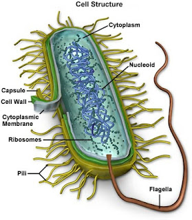
Much to my horror and delight, I finally got video of a bacteria with flagella-3 bacteria in fact! You can CLEARLY see the multiple flagella on the bacteria and see it motoring with them. Two of the bacteria are kind of intertwined but their flagella are distinct. The 3rd one was by itself and very clear to see its whipping "tails" as it moved through the plasma!
INCREDIBLE!!!
What I found interesting is that lysozomes (the little immune particles from the WBCs) were attacking the flagella of the one bacteria.
Posts: 1308 | From Eastern USA | Registered: Oct 2013
| IP: Logged |
TNT
Frequent Contributor (1K+ posts)
Member # 42349
posted
This picture is similar to what the flagella looked like except my bacteria were round, not rod-shaped. And, of course, I don't have near the magnification and resolution as this. Nevertheless, it is unmistakable!
This photo is from the bartonella.org website.
Posts: 1308 | From Eastern USA | Registered: Oct 2013
| IP: Logged |
TNT
Frequent Contributor (1K+ posts)
Member # 42349
posted
Like I said, I am anxious to share my videos, and am working on getting them available. I am also working towards getting equipped with 1000x or more (even 1500x) with my dark phase contrast. I just bought a 100x dark phase objective, and am working on obtaining the other necessities for this.
So, hopefully before too long I (and you all) will benefit from images and videos that will be closer (only closer, sigh) to the pic of that bartonella posted above.
Man, it would have been awesome to have gotten that flagella footage of mine with 1500x dark phase instead of just 400x!
I feel like Galileo wishing for a bigger telescope, lol.
Posts: 1308 | From Eastern USA | Registered: Oct 2013
| IP: Logged |
Lymedin2010
Frequent Contributor (1K+ posts)
Member # 34322
posted
TNT, I too have seen WBC's go right past candida & borrelia. At times though I see WBC's with candida & partly degrades candida vesicles within them, indicating that they must have the surface proteins/antibodies to be able to sweep them up.
We have heard that borrelia suppresses the immune system & perhaps that is why most of the WBC's go past the spiros. Its more clever approach is its retreat into our cells, where our WBC's can't get into.
Lymedin2010
Frequent Contributor (1K+ posts)
Member # 34322
posted
Great find on the bart & can't wait to see your videos!!!
I have never seen a flagella whipping organism in my blood as of yet. I have seen what may be babesia, but it is difficult to say for sure with live RBC's & without staining.
I have seen what lymephotos are calling Horseshoe mites in my blood, at least 6-8 of them when I first started microscopy, but that was early on in my treatment & I have not seen any since.
I have also seen something that resembles an amoeba, but it may have been an underdeveloped phagocyte.
Posts: 2094 | From NY | Registered: Oct 2011
| IP: Logged |
Lymedin2010
Frequent Contributor (1K+ posts)
Member # 34322
posted
Here is a rare time when one of my WBC's picked up a spirochete.
TNT
Frequent Contributor (1K+ posts)
Member # 42349
posted
Lymedin2010, I think you get really good quality brightfield view. Must be the Zeiss optics and the brighter light.
The only thing that salvages my scope is the phase contrast. As the name implies, the technique gives very good visual contrast, and things like ketes really stand out in phase.
The WBC in that video must be a lymphocyte. It's too small and regular shaped to be a neutrophil, although I think I see one come into the field and leave again on the left side.
This answers a question I have had for a while. Apparently some of you find that your lymphocytes are traveling around and "eating" pathogens. I have not seen that yet, only with neutrophils.
Posts: 1308 | From Eastern USA | Registered: Oct 2013
| IP: Logged |
Lymedin2010
Frequent Contributor (1K+ posts)
Member # 34322
posted
TNT, yes it is the objective Zeiss optics that shine on my scope. BUT with the built-in halogen bulbs the image is not as sharp & well lit as it should be. The LED lighting takes it to the next level, just watch out not to burn your eyes out. My USB video camera comes in handy in this respect.
The blebs are the most difficult to spot out of all the morphologies, then comes the cysts & then the full grown spiros.
If you look at the video from my last post, on the WBC that picks up the spiro, there you can see that the WBC is filled with these dark/black & filled dots, but I cannot tell those dark dots from the lysosomes or sometimes lipid droplets & that is why it is hard to know for sure.
One can never really know when it comes to blebs, as you can with the longer & full bodied spiros. One can only gain more suspicion where you see many cut up pieces of borrelia (part of borrelia body + blebs along the body AND many other what may look like blebs or spores in the plasma). Then you can say to yourself, this is MORE LIKELY to be a bleb or spore.
Alternatively, if you really want to prove that it is indeed a bleb. Then you should collect it & grow it in culture media & do some time lapse to see it develop into a full spiro. This is on my list & is what is available from our limited resources. If you had more money & resources, then you could do PCR, DNA probing. or immuno staining, which is always better than a visual to nail down the EXACT type of spirochete we are dealing with.
As I pointed out early when I started this thread I think the growth goes from bleb, to tailed-bleb, to dumbbell shaped, long dummbells & then really long ones. Not all of them get to be long & really long spirochetes, since the internal conditions might force them to become cysts or undergo String of Pearl formation & break apart.
Posts: 2094 | From NY | Registered: Oct 2011
| IP: Logged |
TNT
Frequent Contributor (1K+ posts)
Member # 42349
posted
quote:Originally posted by Lymedin2010: Alternatively, if you really want to prove that it is indeed a bleb. Then you should collect it & grow it in culture media & do some time lapse to see it develop into a full spiro.
I'm afraid I can't.... I don't have tweezers that small.
Sorry, I couldn't resist!
Seriously, though, that seems like it would be quite the undertaking.
Posts: 1308 | From Eastern USA | Registered: Oct 2013
| IP: Logged |
Lymedin2010
Frequent Contributor (1K+ posts)
Member # 34322
posted
No need to get those tweezers dirty with borrelia filth. They make affordable micro-pipettes. Posts: 2094 | From NY | Registered: Oct 2011
| IP: Logged |
Lymedin2010
Frequent Contributor (1K+ posts)
Member # 34322
posted
Fluorescent dyes of bb exiting mouse capillary wall.
I wonder if bb is more aggressive & spiraling when first introduced into humans & becomes less aggressive & spiraling over time? Perhaps it is some component in the blood that inhibits its aggression, which in turn makes it more difficult to treat in humans?
posted
Are you sure its not just a protozoa?
Posts: 387 | From The Netherlands | Registered: Nov 2013
| IP: Logged |
Lymedin2010
Frequent Contributor (1K+ posts)
Member # 34322
posted
Questions: 1) What magnification are you using to view them ?
2) What size do you think the organism is (.5-1 microns)?
3) Do you see the organism right away after live blood prep, or does it come out sometime after?
4) Does it grow, morph, change shape or form in any way over time?
At times at lower magnification even bacilli can look like coccoids, some bacilli start off looking more like cocci early during the life cycle too & then grow more rod like, just take a look at this Vibrio cholerae video. https://www.youtube.com/watch?v=4TWRFF79YsM
Lymedin2010
Frequent Contributor (1K+ posts)
Member # 34322
posted
Without higher magnification we can mistake a rod for a cocci. If one has leaky gut & lowered immune system it can really be any number of organisms. In my experience no professional that I have asked has been able to identify any cocci. Not when I have asked, nor when others who have identified COCCI organisms in their blood & have asked other professionals.
I would buy more clues & perhaps do some time lapse to get closer to the truth. Cocci are the hardest to identify without knowing more.
You mentioned "triangular" shaped in your previous posts. The only thing that I have seen in my blood that is oddly shaped, so not a cocci, rod, spiral, but "imperfectly" round are the following possibilities:
1)Shrunken & ghosted RBC's 2)Borrelia cyst
Some of the borrelia cysts appear more roundish & hollow, while some look more solid & have edges that stick out & can appear triangular for lack of a better term.
Lymedin2010
Frequent Contributor (1K+ posts)
Member # 34322
posted
Your rod shaped with multi-flagella might be Helicobacter Pylori (if magnification is low). This is a common tertiary infection with Lymies & usually in the gut & lives at lowered ph.
With leaky gut & lowered immune it might have traveled from gut into blood stream. How often do you find these in your blood?
It would be really interesting if it was bartonella & you would be the first I have heard of this from live blood microscopy. I have never seen them live in anyones blood, nor those who have made their own videos.
Here is bartonella hensleae, does it look like this?
[ 07-28-2015, 10:38 AM: Message edited by: Lymedin2010 ]
Posts: 2094 | From NY | Registered: Oct 2011
| IP: Logged |
TNT
Frequent Contributor (1K+ posts)
Member # 42349
posted
quote:Originally posted by S13: Are you sure its not just a protozoa?
Could easily be, except with phase contrast, I can usually see nuclei pretty well, and this (these) had no nucleus.
In relation to some of Lymedin's questions, I was viewing under 400x, but zoomed in with my camera to about 4000x. My camera is only a 6mp, so when completely zoomed in, the image quality is not extremely clear. But, when zoomed out or only part way in, the image is fairly crisp, but not very close.
Interestingly, the flagellum are clearer when zoomed out (I guess because of image distortion).
As for size of the organism, let me look again.
I would say .5-1.0um. Definitely no larger than 1um, and almost perfectly round.
The plasma had very slight current at that spot, and the organisms had the flagellum pointed mostly "downstream." In other words, the flagellum were pointed in the direction they were moving.
The shape and amount of flagellum were consistent with this pic. Though, my "cocci" were very bright.
The organisms did not look like these:
The top pic is a cocci bacteria, and the bottom pic (with nuclei) are trichomonas.
I really need to get my videos posted. I will try to work on that.
Posts: 1308 | From Eastern USA | Registered: Oct 2013
| IP: Logged |
posted
Have not had time to read all the posts here, but for those of you interested in microscopy, Dr Alan MacDonald has recently found TWO different Borrelia in the autopsy brain tissue of Alzheimer's Disease victims.
He found both Bb and B. miyamotoi. The Borrelia were labelled with Molecular Beacon DNA Probes specific for each species - the most accurate technology known for identifying bacteria.
These are stunning findings, so important. Dr M is now launching a crowdfunding campaign so that he can test the body fluids and biopsy tissues of LIVING people. Please see my other post here for more info:
I hope you all will support Dr M's fundraiser and circulate the message far and wide - his work could potentially bring hope to millions. Elena
-------------------- Justice will be ours. Posts: 786 | From UK | Registered: Oct 2007
| IP: Logged |
TNT
Frequent Contributor (1K+ posts)
Member # 42349
posted
I'm sorry this is not very good quality published. I lost some video quality in the upload. Did you guys have trouble with your published videos having less resolution than your actual videos? Is there something I'm doing wrong? My videos look much better on my computer before publishing (Of course, actual viewing is even clearer).
Anyways, here is one of the bacteria with the flagellum. Notice also the very long kete.
TNT
Frequent Contributor (1K+ posts)
Member # 42349
posted
S13, I was looking again at some of your videos and am curious how you got that one with candida growing on dead skin cells.
I think that one would have been a difficult one to catch. And how/why were you looking at dead skin cells in the first place?
Maybe I don't want to know, lol....but I'm very curious.
Posts: 1308 | From Eastern USA | Registered: Oct 2013
| IP: Logged |
Lymedin2010
Frequent Contributor (1K+ posts)
Member # 34322
posted
Eight Legs Bad, thanks for the info. His postings have been floating around on Facebook & I have done my part
Thanks for the info & others should help out as well. Dr. Alan MacDonald is one of my new idols post Lyme! His perseverance
Posts: 2094 | From NY | Registered: Oct 2011
| IP: Logged |
Lymedin2010
Frequent Contributor (1K+ posts)
Member # 34322
posted
TNT, great video & it looks as you said it would & that pic serves as a good example. As I said before anything coccoid would be more difficult for anyone to confirm, as I have asked near & far in the past.
But yours has flagella, as you said it does, & it should be easier. I have never seen or heard of such an organisms & most of the babs & bart organisms I have seen are from STAINED RBC slides & never any live ones.
I will do some research on it in the oncoming days & hopefully we can ID this one.
How often do you find this in your blood & about how many can you find on ONE slide? Are they all pretty much the same size or do they come in slightly smaller or larger diameter?
All my past Youtube videos I did not have any conversion & display issues with, except for my last one. I will have to upload another video just to determine if Youtube might have changed any specifications. Perhaps they have moved on & they are expecting most videos nowadays to be in the WIDE screen format (HD) & are no longer 3x4 format friendly?
Posts: 2094 | From NY | Registered: Oct 2011
| IP: Logged |
TNT
Frequent Contributor (1K+ posts)
Member # 42349
posted
quote:Originally posted by Lymedin2010: How often do you find this in your blood & about how many can you find on ONE slide? Are they all pretty much the same size or do they come in slightly smaller or larger diameter?
This is the first time I have seen cocci with flagellum. There may have been one or two cocci in previous samples, but it was very rare and never before with flagellum.
The bart-like bacteria I HAVE seen before... maybe 3 other times/samples (one organism per sample those other times). The one that was pummeling the kete was identical to this one. Actually, all the previous bart-like ones were identical to this one.
Would anyone be interested in the video where the bart-like bacteria was attacking the kete? It may have resolution issues, too, since I zoom in most of the time, and, since I may lose some clarity in the upload.(I will be more careful with the zoom from now on-at least until I get a better camera).
Thanks for your help Lymedin!
Posts: 1308 | From Eastern USA | Registered: Oct 2013
| IP: Logged |
Lymedin2010
Frequent Contributor (1K+ posts)
Member # 34322
posted
Was the bart like organism a rod shape in any form, or strictly round?
How large is original video, as it may not pass the max file size requirement?
Posts: 2094 | From NY | Registered: Oct 2011
| IP: Logged |
TNT
Frequent Contributor (1K+ posts)
Member # 42349
posted
The bart-like organism that attacks the kete is identical to the one in the second video I posted. It is rod-shaped. The bart-like organisms I refer to are all rod-shaped.
The kete-attacker video is a few minutes long, depending on which one I select.
Posts: 1308 | From Eastern USA | Registered: Oct 2013
| IP: Logged |
TNT
Frequent Contributor (1K+ posts)
Member # 42349
posted
The bart-like organism (rod-shaped one) I am learning could easily be E. coli bacteria. According to Wiki, cells are typically rod-shaped, and are about 2.0 micrometers (μm) long and 0.25–1.0 μm in diameter, with a cell volume of 0.6–0.7 μm. That would fit the bill for the "bart-like" (rod-shaped) organisms.
Here are some pics of E. coli from Dennis Kunkel Microscopy Inc.:
Here is an interesting one in which it looks like there could be a cocci form, but I think it is just on end:
Some E. coli strains do possess flagella, but my rods did not. Only the cocci looking organisms had flagella.
So, this only gives me a possible answer for the rods, but not for the round flagellate organisms. And a quick search did not reveal a "cocci" morphology for E. coli. So, I don't think the round flagellates were E. coli.
So, I'm still looking for suggestions on what the round flagellates could be.
Posts: 1308 | From Eastern USA | Registered: Oct 2013
| IP: Logged |
Lymedin2010
Frequent Contributor (1K+ posts)
Member # 34322
posted
Here is a great video of E. Coli & it shows both rod & rounded shaped forms, I would imagine it being dependent on where in its growth cycle it is at. Also, the rounded ones appear to have flagella & is more noticeable when one pauses the video & reviews it frame by frame.
Another one of my colleagues suggest that it might be a platelet (aka thrombocyte), which are 2-3µm in diameter. As you make more microscopic observations see if any appear without flagella. Or do they ALL appear with flagella all the time? This is a Giemsa-stained peripheral blood smear. https://upload.wikimedia.org/wikipedia/commons/thumb/5/51/Platelets2.JPG/1280px-Platelets2.JPG
[ 08-01-2015, 08:15 PM: Message edited by: Lymedin2010 ]
Posts: 2094 | From NY | Registered: Oct 2011
| IP: Logged |
TNT
Frequent Contributor (1K+ posts)
Member # 42349
posted
Hey, thanks for the video of the E. coli! I, too, think it appears as though the round ones have flagella. The rod shaped ones, too. Though, I wish the video was of a higher magnification to tell for certain.
I don't think those round flagellate organisms in my blood are platelets.... I can see platelets very well, and are fairly numerous. The round and rod-shaped "bacteria" are very rare, and are quite different from the platelets. To name just one major difference, platelets look dark in my dark phase view. When I upload more videos, I can try to get ones with some platelets in the field.
I would tend to think my organisms are E. coli, especially if there is round-form stages with flagella. It seems to me that it would be very easy for a predominant organism (E. coli bacteria) of the GI tract to get into the blood stream in few numbers, especially if the mucosal membrane is not completely healthy and there is an imbalance of gut flora.
The scary thought is that the gut could seed other parts of the body via the bloodstream in a situation as that...or just as scary, lead to sepsis.
Posts: 1308 | From Eastern USA | Registered: Oct 2013
| IP: Logged |
Lymedin2010
Frequent Contributor (1K+ posts)
Member # 34322
posted
One thing that I have learned, especially from watching the Animal Planet "Monsters Inside Me" series, is that we are susceptible to many different varieties of tertiary infections with the immuno suppression of Lyme.
If you watch the series you will become all too familiar with the wording "most people can fight off the infection, however the young, elderly, or immuno compromised individuals are at risk."
Posts: 2094 | From NY | Registered: Oct 2011
| IP: Logged |
quote:Originally posted by TNT: I would tend to think my organisms are E. coli, especially if there is round-form stages with flagella. It seems to me that it would be very easy for a predominant organism (E. coli bacteria) of the GI tract to get into the blood stream in few numbers, especially if the mucosal membrane is not completely healthy and there is an imbalance of gut flora.
The scary thought is that the gut could seed other parts of the body via the bloodstream in a situation as that...or just as scary, lead to sepsis.
I think you are very right here! The wild life i have encountered when watching growth in the blood is staggering and the diversity just doesnt seem to match with a single borrelia infection, or even one with a couple of known co infections.
So the idea that gut flora is constantly seeding the body with a wide variety of bacteria is a very good possibility. The growing candida hyphal forms in my blood sort of proves its possible. And the mechanism is leaky gut.
One of the clear observations i have made is that this wild life growth under the microscope is much more abundant when you press the slide with a small object, basically rupturing some of the RBC's and WBC's. So i suspect most of this wildlife is contained in the WBC's. At least an indication that the WBC's are mopping up this gut overflow.
And even without rupturing the WBC's i have pictures showing this wild life just growing from inside whole (but probably dead) WBC's :
Its a bit fuzzy, but it shows 2 WBCs with unknown bacteria and or fungi growing out of them.
So gut flora overspill is probably a very important factor for some of the lyme patients. Continuous overstimulation of the Th1 branch of the immunesystem causes chronic inflammation. Lyme itself with its LPS also adds to this problem of course. Continuous chronic inflammation of the gut causes more bacteria to leak out and possibly causes more SIBO by damaging the ileocecal valve for example. So its a vicious cycle.
quote:Originally posted by Lymedin2010: One thing that I have learned, especially from watching the Animal Planet "Monsters Inside Me" series, is that we are susceptible to many different varieties of tertiary infections with the immuno suppression of Lyme.
So are we immune compromised? Or is it just continuous overstimulation of the immune system that is causing trouble?
Posts: 387 | From The Netherlands | Registered: Nov 2013
| IP: Logged |
Lymedin2010
Frequent Contributor (1K+ posts)
Member # 34322
posted
I have seen a few "normal" & "healthy" blood samples & THEY ALL HAVE FOREIGN ORGANISMS!!!!
The difference is that our Lyme blood is overloaded & overstimulated from many different angles & sources and hence our blood shows the very large loads.
Posts: 2094 | From NY | Registered: Oct 2011
| IP: Logged |
Lymedin2010
Frequent Contributor (1K+ posts)
Member # 34322
posted
Did you guys see Peter Kemp's new centrifuge work with Lyme blood? I almost bought a centrifuge during the first year I bought my scope, since I thought I could sequester & concentrate the spirochetes in a blood layer & have visible masses of spirochetes for time lapse. After my disease spread the spirochetes became more plentiful & I lost the need for a centrifuge.
So in a nutshell this is basically how Advanced Labs & Dr. Burrascano would do a Borreliosis blood culture to prove Lyme Disease in our blood. It is not the exact recipe & but the gist is here. Add a culture medium, add your blood & store in a temp controlled environment for a few days & then check via microscopy.
The only difference here is that Peter was interested in determining which blood fractionation layer would host mot of the spirochetes. I always suspected that the blebs or cysts would be very buoyant & would "float" to the top plasma layer.
Posts: 2094 | From NY | Registered: Oct 2011
| IP: Logged |
TNT
Frequent Contributor (1K+ posts)
Member # 42349
posted
OMG! I watched Peter Kemps video. I thought the buffy coat layer was bad! But, when the video got to the lower RBC layer, I about fell out of my chair! That's BAD! Those RBCs are basically colonies of spirochetes! I do wonder sometimes how we are still alive.
I viewed the blood of a very healthy friend of mine a couple months ago. I was very surprised, yet not surprised, by what I saw. He had one definite spirochete, perhaps 7-8 um long, and two probable shorter ones.
I am certain he has been exposed to lyme one way or another, and probably has been tick-bitten before. So, it was not a complete surprise to me that he did harbor a very small presence of the germ. But, he is as healthy and strong as an ox.
Though, if something would severely stress his immune system, I am sure there is a chance he could "come down" with Lyme disease.... the same way many people "come down" with it (not necessarily right after exposure, but after a STRESSOR on the immune system).
Oh, Lymedin. Do you think there is anyway you could shorten the URL address of that picture of the giemsa stained blood smear to get this page back to normal size?
[ 08-01-2015, 09:10 PM: Message edited by: TNT ]
Posts: 1308 | From Eastern USA | Registered: Oct 2013
| IP: Logged |
Lymedin2010
Frequent Contributor (1K+ posts)
Member # 34322
posted
TNT, the blood sample was CULTURED to get that many spirochetes.
.3ml blood from various layers after the centrifuge + 1ml BSK medium, which was then incubated at 35C for 4 days.
So the few spirochetes in your blood became many by the time he looked at them on the 4th day.
Posts: 2094 | From NY | Registered: Oct 2011
| IP: Logged |
TNT
Frequent Contributor (1K+ posts)
Member # 42349
posted
Phew! That's a relief! I didn't see that it was cultured. NOW that I read the video description, I see that it SAYS it was cultured. Thanks, Lymedin.
If there would have been a snake in that description I could have been bitten.
Thanks also for fixing the page. It's so much easier on my eyes.
Posts: 1308 | From Eastern USA | Registered: Oct 2013
| IP: Logged |
Lymedin2010
Frequent Contributor (1K+ posts)
Member # 34322
posted
TNT, I know a lady who has Lyme so bad & so much circulation issues that she cannot go to bed with her arms & legs crossed , because it hurts/burns so much from the Lyme.
I am willing to bet anything that her blood will look close to that.
There must be some limiting factors that prevent our blood from totally building up with spirochetes though & it must be the utilization of an essential growth component of bb, maybe something like availability of manganese or something along these lines.
Posts: 2094 | From NY | Registered: Oct 2011
| IP: Logged |
Lymedin2010
Frequent Contributor (1K+ posts)
Member # 34322
"In this report we present a method to cultivate Borrelia spirochetes from human serum samples with high efficiency. This method incorporates improved sample collection, optimization of culture media and use of matrix protein. The method was first optimized utilizing Borrelia laboratory strains, and later by demonstrating growth of Borrelia from sera from fifty seropositive Lyme disease patients followed by another cohort of 72 Lyme disease patients, all of whom satisfied the strict CDC surveillance case definition for Lyme disease.
The procedure resulted in positive cultures in 47% at 6 days and 94% at week 16. Negative controls included 48 cases. The positive identification of Borrelia was performed by immunostaining, PCR, and direct DNA sequencing."
Posts: 2094 | From NY | Registered: Oct 2011
| IP: Logged |
Lymedin2010
Frequent Contributor (1K+ posts)
Member # 34322
posted
Just as they mentioned in Under Our Skin 2: Emergence, that they closed down the microscopy work of Prof. Morten Lanne because of what was revealed.
Microscopy visualization of Borreliosis in Chronic Lyme is HUGE.
"Prof. Morten Laane held the presentation: "Easy Detection of Bacteria and Parasites in Infected Human Blood by Microscopy. Some Simple, Low-cost Methods" at the NorVect conference 2014.
Because his and Prof Mysterud's study on Lyme patients were stopped and all results were destroyed by the Norwegian Board of Health Supervision, Prof Laane was not allowed to talk about this particular study. He showed some of his work using two films. One is seen here in this excerpt. His presentation can be watched in full length at http://www.norvect.no
dbpei
Frequent Contributor (1K+ posts)
Member # 33574
posted
I recently came across this thread and saw some pictures that resembled what I have been seeing with my eye floaters for the past few years.
Photos posted by Lymedin2010 7/28/15 at 9:55 a.m. - both pictures look very similar to what I see daily! I could not tell if these photos posted were from H Pylori or Bartonella.
I also have some small cyst shaped floaters, but the ones I see the most, I describe as looking like earwigs with a bunch of long, thin hairs on one end. Do any of you see these same floaters? After seeing these photos, I wonder if it could be bartonella or H Pylori that is keeping me sick.
Lab testing shows that I am making lots of antibodies for brucellosis. Is there a place I could find out what that bacteria looks like under microscope?
I don't own a microscope but am seeing my eye doc this morning. I wonder what she will say when I tell her what my floaters look like. I hope she won't add me to her nutty patient list.
Posts: 2387 | From New England | Registered: Aug 2011
| IP: Logged |
Lymedin2010
Frequent Contributor (1K+ posts)
Member # 34322
posted
Many Lymies have strings as floaters, as I have had for years & continue to have as well, which I suspect are the Borrelia spirochetes.
The pics I posted on that day are of H. Pylori, but your floaters could be Brucella because of your antibodies clue. If you describe them as you do, "like earwigs with a bunch of long, thin hairs on one end," then they could just as easily be H. Pylori escaping from your gut, bart, or another organisms we have not thought of yet.
H. Pylori: Posts: 2094 | From NY | Registered: Oct 2011
| IP: Logged |
dbpei
Frequent Contributor (1K+ posts)
Member # 33574
posted
Thanks so much! It definitely resembles H Pylori missing the bands I see at the end near the hairs, but actually looks more like an earwig than the rod I see here. I saw my eye doc today for my annual exam and told her about them.
She examined my eyes really well and found one of the floaters I described. She told me they are vitrious detachments and not to worry. I hope she is right.
I really appreciate the help in identifying these critters! I will have to follow this thread. It is very interesting.
Posts: 2387 | From New England | Registered: Aug 2011
| IP: Logged |
Lymedin2010
Frequent Contributor (1K+ posts)
Member # 34322
posted
Eye floaters are really hard to prove in either direction, but it is interesting how many Lymies report them.
If anyone is interested in a break down of which cameras are best for pics, video & time lapse, here is a breakdown with focus specifically on microscopy.
Thankfully, it turns out that I did not lose much resolution in the upload.
Posts: 1308 | From Eastern USA | Registered: Oct 2013
| IP: Logged |
Lymedin2010
Frequent Contributor (1K+ posts)
Member # 34322
posted
TNT, the big one sure looks like borrelia!!!! Awesome video & thanks for sharing!
It has the bulbous tips on both ends & it spirals/undulates in that very distinctive way with a bit more aggression than just a stagnant string or anything that may be of human origin & an actual artifact.
BUT, the one you are calling Bart like I also think is Borrelia, just a pre-mature one. It looks like a dumbbell, right?
From the beginning of my thread: "The progression might be DOT--> DOT WITH TAIL-->STUBBY DUMBBELL-->MEDIUM DUMBBELL-->LONG DUMBBELL (STRING)."
At that time I thought it was spirochaeta gallinarum, because it was the only spirochete that I could find where someone discusses the blebs or spore formations. Now I know it is borrelia for sure & the blebs (dots) grow to be tailed blebs at first & then look like very small dumbbells, which are basically growing borrelia organisms.
One of my future wishes is to catch the blebs grow into tailed blebs & then the small spirochetes (small dumbbells).
Posts: 2094 | From NY | Registered: Oct 2011
| IP: Logged |
TNT
Frequent Contributor (1K+ posts)
Member # 42349
posted
Yes, the longer one is definitely borrelia.
Why is the shorter one acting the way it is? It sure appears to me to be attacking the longer one. And the longer one appears to be losing energy.
The shorter one could possibly be borrelia, but I'm not sure. I don't have enough information to go on. I will pay attention to your videos with dumbbell organisms that size.
If you find any dumbbell footage from Sapi or MacDonald, please let me know.
Posts: 1308 | From Eastern USA | Registered: Oct 2013
| IP: Logged |
The Lyme Disease Network is a non-profit organization funded by individual donations. If you would like to support the Network and the LymeNet system of Web services, please send your donations to:
The
Lyme Disease Network of New Jersey 907 Pebble Creek Court,
Pennington,
NJ08534USA http://www.lymenet.org/


 UBBFriend: Email this page to someone!


![[Smile]](smile.gif)
![[Roll Eyes]](rolleyes.gif)
![[Mad]](mad.gif)

![[dizzy]](graemlins/dizzy.gif)
![[Wink]](wink.gif)
![[Big Grin]](biggrin.gif)
![[lol]](graemlins/lol.gif)


![[bonk]](graemlins/bonk.gif)
![[Frown]](frown.gif)


 Printer-friendly view of this topic