TNT
Frequent Contributor (1K+ posts)
Member # 42349
posted
Keep up the good work. We would definitely be interested in seeing your time-lapse if you get that working.
quote:and it might only be oil immersion, but you can buy condensers for this purpose.
Now that you mention it, I think I have seen info on oil condensers for darkfield.
quote:I draw blood into vacationers.
I assume you mean you draw blood into vacuum containers, and not vacationers? Wow, a phlebotomist and a microscopist! Do you refrigerate the blood until you view it? Why do you draw the blood that far in advance?
Lymedin2010 has a blood slide making tutorial in which he shows how to seal off the coverslip with immersion oil to "preserve" the blood for longer viewing. That would help with getting a good time-lapse capture.
My brain isn't working very well today, so I can't go into much detail, but you definitely are positive for borreliosis: Band 39 is specific for Lyme, and your microscopy confirms it.
Bands 31 and 41 are not always specific for lyme and can cross-react with other infections. Those bands are also specific for Brucella, so they could possibly indicate a concurrent undiagnosed infection with that. (Very) possibly (I feel).
In fact, I found a description page about a test for Brucella that actually suggests that the Multiple Sclerosis epidemic could be connected to Brucellosis. So, it's not just because of Lyme. Read this description about Brucellosis:
Notice the last sentence in the first paragraph:
"...it is possible that there is a link between an infection with Brucella and the outbreak of multiple sclerosis."
And, here are PubMed articles showing the specificity of Osp (outer surface proteins) or Omp (outer membrane proteins) -- (referred to as "Bands" on the Western Blot) -- 31 and 41 for Brucella bacteria!!
I personally feel this is an unseen epidemic even in the U.S. But, I won't take the time to go into that.
So, if you ever want to broaden your microscopy techniques, we have plenty of opportunity to delve into things like staining, fluorescence, DNA probing, culturing, etc.
Some of which we can possibly do ourselves with a little learning and practice (AND money).
These advanced techniques have the ability to help us better understand what each of us is dealing with!
Of course, simple microscopy, time-lapse, and staining is well within each of our abilities and means. We just need to keep at it and bring in more recruits!! Posts: 1308 | From Eastern USA | Registered: Oct 2013
| IP: Logged |
posted
I don't refrigerate it. I keep it at room temperature.
The blood I view over this longer period is blood I have drawn from others. It's just easier this way then saying hey, Come over to my house so I can draw blood real quick.
On top of that, I am trying to compare what I see in these longer samples than a straight draw.
I'd imagine the change in environment has an effect (98.6 degrees in body versus 70 degrees room temp), stuff like that.
Posts: 163 | From USA | Registered: Oct 2015
| IP: Logged |
Wow-- kansas dude----amazingly excellent video!! I can clearly see at least two spirochetes hanging off that red blood cell-- trying to suck the life out of you.
What microscope , magnification and stain did you use? I really like the resolution on your microscope. ( I want to buy a microscope but have been gun shy because I want to get a good one for spirochetes with dark field--- without having to spend $1,000s of dollars. Anyway-- amazing work -- you deserve a "Spiro" award....(It looks like the oscar but is squiggly) LOL..
Posts: 696 | From New York | Registered: Aug 2006
| IP: Logged |
I have the T490B-DK. 400x. (40x objective and 10x eyepiece).
I believe it was 375 dollars, off amazon. Dark field useful up to 400x, which is what those videos are. I filmed with my DSLR, however, through the eyepiece. I am waiting for an attachment to see if I can get better images with the DSLR in the trinocular port. You can get excellent microscopes with dark field for cheap. This is obviously not lab/research quality, but things are still quite visible.
No stains, just dark field. I have to use and LED headlamp, as the bulb in the scope is not strong enough. 214 lumen headlamp for 10 dollars. (I only look in the microscope until i find something, then record. LED is bad for the retina, can be damaging).
This is a good breakdown: T490B-DK (375 on amazon) 400x Dark field Slide and slip covers (cheap, like 12 dollars for 75 or so, and you just clean them with alcohol to reuse them all) Samples tested up to 7 days after drawing (vacutainers), stored at room temp. Some samples tested on blood slide over the course of 12 hours or more, as long as sample has not dried (start seeing the most around 2-4 hours after placing on slide)
Posts: 163 | From USA | Registered: Oct 2015
| IP: Logged |
posted
I am looking for info on a research quality scope if anyone has advice?
I have a very specific reason that I need something good enough to capture pristine, sharp, clear images up to 1000X or higher. I am looking at around 1500 dollars to 2000 dollars.
This microscope is adequate for finding, viewing, and recording, but I need something much greater in quality. Not sure if just updating the 40x and 100x objectives with a higher quality objective lens would do the trick or not.
Posts: 163 | From USA | Registered: Oct 2015
| IP: Logged |
TNT
Frequent Contributor (1K+ posts)
Member # 42349
posted
It's kind of hard to know what to advise without knowing exactly what you need it for.
But unless you want something special like an inverted scope (which are actually some of the cheaper research grade scopes from what I have seen), I would recommend an Olympus BH series. The BH series have a little age, but are definitely research grade.
Olympus microscopes are excellent! I don't think they make a cheap line like even Nikon has. Of course, I don't speak from actual experience, just from looking and watching them sell.
They can run well over $1000.00 (used). But, I saw a nice BH series with a case sell thru Ebay from a thrift store go for a little over $200.00. So, if you look and wait, you can sometimes get a really good deal.
Whatever brand you decide on, you can get a VERY nice (used) scope for the amount of money you have available.
I personally have fallen in love with (dark) phase contrast. They tend to be considerably more money, but you should still be able to get a pretty nice dark phase Olympus with that kind of money. A dark phase Olympus with a turret in excellent shape runs $1500.00-$2500.00.
Your idea about just swapping lens is an idea to consider if you would rather stash that cash away for something else (depending on how many lens you buy). The Objectives and oculars are really the main components on a scope, so if you would buy some high-end lens, you would in effect have the same grade of scope for some less. Buying lens individually can get expensive, though.
I think all objective lens nowadays (especially high-end ones) are infinity-corrected, so it wouldn't matter what scope you installed them on. Regardless of lens brand, I wouldn't consider any that are not "Plan Achromat."
Posts: 1308 | From Eastern USA | Registered: Oct 2013
| IP: Logged |
I have a few more videos, but it has been hard to get videos of them actual motile, moving freely in the serum and not attached to/coming out of RBC's.
Posts: 163 | From USA | Registered: Oct 2015
| IP: Logged |
Thanks for the info on the scopes-- I will be buying one soon and am anxious to see whats in my own blood. I want to take vis of before and after herbal regimens.
I saw the spirochetes in the video of your blood above-- very good film!
I hope your red blood cells in the video above were old and drying-- because if not, the sharp spiky points on all your red blood cells might indicate that you have toxicity in your body, including heavy metals (aluminum or mercury) and/or pesticide exposure. How long does it take before round rbc's begin to look spiky like that?
Posts: 696 | From New York | Registered: Aug 2006
| IP: Logged |
posted
Yea, the crenation is from the blood simply drying/being exposed to the environment outside of the tubes/body.
It varies on when I start seeing more crenated RBC's. I havne't paid much attention to that.
Posts: 163 | From USA | Registered: Oct 2015
| IP: Logged |
TNT
Frequent Contributor (1K+ posts)
Member # 42349
posted
Here are a couple pics of biofilm in my blood:
An earlier pic with brightfield:
A recent pic with dark phase contrast:
Closer (notice a couple red blood cells at the top, and some platelets at the bottom, partially entrapped in the film):
Really close. You can easily see the partially entrapped RBCs at the top. I don't know what the object in the middle is...perhaps a uric acid crystal or a piece of contaminant on the slide:
Posts: 1308 | From Eastern USA | Registered: Oct 2013
| IP: Logged |
TNT
Frequent Contributor (1K+ posts)
Member # 42349
posted
A Fry smear showing biofilm
Posts: 1308 | From Eastern USA | Registered: Oct 2013
| IP: Logged |
Can you see any of that on live blood smears? Or is it only visible with staining?
Posts: 163 | From USA | Registered: Oct 2015
| IP: Logged |
TNT
Frequent Contributor (1K+ posts)
Member # 42349
posted
All 4 pics of my blood (the brightfield and the dark phase) are live blood samples. There is no stain. The above (Fry) smear...originating from Fry Labratories, is stained.
It appears like the Fry smear might be a (modified?) Giemsa stain.
Posts: 1308 | From Eastern USA | Registered: Oct 2013
| IP: Logged |
Any idea what this is? I've seen reference to a Medusa's head?
They also appear segmented, similar to the string of pearls that I have seen photos of. My microscope video is better and seeing the segmentation, but that's on another computer.
Any ideas?
Posts: 163 | From USA | Registered: Oct 2015
| IP: Logged |
TNT
Frequent Contributor (1K+ posts)
Member # 42349
posted
quote:Originally posted by thatdudefromkansas: https://www.youtube.com/watch?v=-WDmCzJNnUo&feature=youtu.be
Any idea what this is? I've seen reference to a Medusa's head?
I'm sorry dude, I'm not sure. Lymedin2010 or S13 will have to weigh in on that one. It looks similar to what I think could be l-forms in my own blood. If you could get a little more magnification on it, it may help narrow down the possibilities.
I just published the video of that biofilm in my blood. The video shows slightly more detail and proves it is live blood.
posted
Wow-- I just viewed this person's blood (channel name jasmine) on youtube-- this has got to be a very sick person, or perhaps its cotton tissue left on the slide--- the blood is riddled with long tendrils. Do you guys think that some of these are spirochetes?
TNT
Frequent Contributor (1K+ posts)
Member # 42349
posted
quote:Originally posted by WakeUp: Wow-- I just viewed this person's blood (channel name jasmine) on youtube-- this has got to be a very sick person, or perhaps its cotton tissue left on the slide--- the blood is riddled with long tendrils. Do you guys think that some of these are spirochetes?
Some of the blood looks pretty good. The blood has plenty of immune components bouncing around (lysosomes), so it appears like their immune system is not too suppressed. I think they were viewing at the edge of the sample where the condition is often worse. The junk usually accumulates there.
That being said, I have never seen any parts of my samples look like that. I can't say what those "strings" are, but my OPINION is that they COULD be l-form (cell-wall-deficient) spirochetes, though they don't look like what I normally refer to as l-form ketes (the thicker, fainter-looking ketes with darker/brighter ends). I see lots of round forms among those strings that could POSSIBLY be round form (cyst) spirochetes. These are just guesses.
Without seeing active undulating (thinner, shorter) spirochetes with bulbous tips in this person's blood, I would be a bit hesitant to say they are definitely infected with borrelia.
We need more research about some of these "artifacts."
Posts: 1308 | From Eastern USA | Registered: Oct 2013
| IP: Logged |
posted
The most informative live blood spirochete microscopy video I have seen to date--- I think the narrator is a microbiologist.
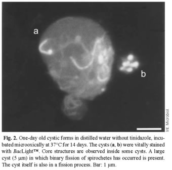
She covers identification of the many morphologies of spirochetes(including IDs of cell wall deficient forms versus cysts and blebs, string of pearls, etc), as well as IDing different types of lyme biofilms. One astounding piece of information she divulged is that cysts undergo reproduction inside the cyst wall, and when they burst open they contain 8-12 living spirochetes!!
This does not bode well for doxycycline treatment alone, which creates a large increase in cysts, while killing the living spirochetes. I got a screen shot of the chart of the morphologies for future reference..
quote:Originally posted by TNT: All 4 pics of my blood (the brightfield and the dark phase) are live blood samples. There is no stain. The above (Fry) smear...originating from Fry Labratories, is stained.
It appears like the Fry smear might be a (modified?) Giemsa stain.
Hi TNT-
Your live blood biofilm pics are excellent-- I think I can detect what Dr. MacDonald referred to as "water channels" inside your very own personal biofilm sludge! It would be very interesting to count biofilm blobs in an average 1/2 hour live blood session, and then begin to start a diet high in biofilm busting substances such as pomegranate, rosemary, cloves, mango, cinnamon, sarsaparilla, curry, etc..... or just take Eva Sapi's Samento/Banderol combination--- and then see if the count of biofilm blobs progressively goes down over time.
I think that liveblood microbiollogist (in the video I posted above) had stated that by the time she sees less than 5 live spirochetes in a 1 hour live blood session, the subject is usually symptom free. Of course our goal is ZERO spirochetes/cysts/lforms/blebs. LOL....
Posts: 696 | From New York | Registered: Aug 2006
| IP: Logged |
posted
Excellent photograph of a borrelia cyst, by the amazing Brorsons research team in Norway --- revealing multiple spirochetes inside-- Posts: 696 | From New York | Registered: Aug 2006
| IP: Logged |
posted
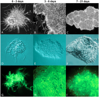
Recognizing the stages of borrelia biofilm (Sapi 2012)-- Dr. MacDonald's famous "water channels" seem to be clearly visible in the older biofilm colony on the right of the image-- also the older biofilm has no spirochetes hanging off the edges-- Im assuming they are all sheltered inside "the structure." Does anybody know if the structure is composed of alginate or dead organisms-- or metals?
Posts: 696 | From New York | Registered: Aug 2006
| IP: Logged |
TNT
Frequent Contributor (1K+ posts)
Member # 42349
posted
quote:Originally posted by WakeUp: Your live blood biofilm pics are excellent
Thanks WakeUp! And, thanks for your great contribution here on the microscopy thread, and the Spirocheticidal & Antibiofilm Compounds Thread!
As for breaking up the biofilm, my experience has been that I get more debilitated if I address biofilms and my antimicrobials are not strong enough to address what's being released out of the colonies' protective niche.
If you have sufficient firepower there when the bugs are flushed out (anti-biofilm agents are used), you will improve your health. If not, you will make yourself MUCH sicker. That was confirmed by my LLMD who is big into biofilm treatment.
I, too, think the number of organisms in the blood is a good indication of how well you are. Just don't forget we can't always easily see (or identify) the different morphologies. So, just because there are few ketes, doesn't mean there are few cysts or l-forms.
Also, there are unseen colonies in various body tissues that can seed the blood and rest of the body. So, NO ketes seen over an extended time period with multiple viewings is a safer gauge.
Posts: 1308 | From Eastern USA | Registered: Oct 2013
| IP: Logged |
TNT
Frequent Contributor (1K+ posts)
Member # 42349
posted
quote:Originally posted by WakeUp: Excellent photograph of a borrelia cyst, by the amazing Brorsons research team in Norway --- revealing multiple spirochetes inside--
Whoa, the cyst in that picture is HUGE!
I didn't realize they got that big. 5 microns? I wonder what the average size would be? Perhaps some of "candida-like" stuff I have seen could be cysts. They are as big as that many times.
Usually the "candida-like" objects are more uniform in roundness without the slightly ragged edge seen in this pic.
I'll try to look more carefully the next times.
Does anyone know the average size of cysts?
Posts: 1308 | From Eastern USA | Registered: Oct 2013
| IP: Logged |
You will clearly see Bartonella AND gut bacteria in my blood.
I am working on other videos, but I'll post in the appropriate topic areas later.
-------------------- My biofilm film: www.whyamistillsick.com 2004 Mycoplasma Pneumonia 2006 Positive after 2 years of hell 2006-08 Marshall Protocol. Killed many bug species 2009 - Beating candida, doing better Lahey Clinic in Mass: what a racquet! Posts: 830 | From Mass. | Registered: Aug 2006
| IP: Logged |
TNT
Frequent Contributor (1K+ posts)
Member # 42349
posted
Thanks for joining the thread, Cold Feet!
Your video from Tijuana is intriguing. I am fairly new to this "hobby" so I hate to question what Dr. Francisco says, but what he points out as Bartonella at 5:00 (the first video) looks to me to be the end of the spike of a crenated RBC.
He claims that the Bartonella will hide around the side of the cell when it feels like it is identified. (???) I don't think it knows it's being viewed, nor can it go to the back side of a cell and escape the immune system (only inside the cell to escape) because the immune system is all around the cells.
I would agree that the black dot on the bottom of that RBC (6 o'clock position) resembles Bartonella, but not the one he points to at the 2 o'clock position on the same RBC (when he says they will go around the side of the cells to hide). As I said, I think that one looks like the end of the spike and only looks like a dot because of how the microscope is focused.
I could very well be wrong.
Please don't take my comments personally. This is the first I have viewed that video and this was just one thing that I couldn't quite see eye to eye with him.
But, I have watched some of your other videos before, and am very glad you have this wonderful library on your channel. I particularly like the ones with Dr. Alan McDonald and the one in Sapi's lab. EXTREMELY helpful!
Thank You!
Do you personally have a scope and look at your own blood?
Posts: 1308 | From Eastern USA | Registered: Oct 2013
| IP: Logged |
You will clearly see Bartonella AND gut bacteria in my blood.
I am working on other videos, but I'll post in the appropriate topic areas later.
Hi Coldfeet-- Wow-- International Biocare's microscope setup is amazing-- interesting that he could immediately spot the intracellular Bartonella. Thanks for sharing this valuable information and video. Next time I go to California, I might make a trip down to Tijuana to have them analyze my blood too. In the last half of the video was he analyzing dried blood that had been centrifuged? I was confused. Also what did he recommend for the Bartonella?
Posts: 696 | From New York | Registered: Aug 2006
| IP: Logged |
Lymedin2010
Frequent Contributor (1K+ posts)
Member # 34322
posted
I too love the MacDonald videos & I am very grateful for them. However, the video from IBH, there are a few holes we can punch in & they appended an extra zero on the magnification reporting as well.
Interesting read on Alan's continuing work & microscopy.
"“…A hunch led me to stain peripheral blood from a heavily infected Chronic Lyme patient with dementia with Congo red Stain for Amyloid.;;
The biofilms in blood were completely covered with Amyloid, and the Amyloid stain demonstrated the textbook partly red/partly green color – [ i.e.; Birefringence under white polarized light illumination]
Water channel spaces are clearly seen inside of the Congo Red stained amyloid covered Biofilms.
Summary point: Borrelia biofilms in Alzheimer’s brain exist and are always wedded to amyloid plaques which completely cover the Borrelia biofilm.
Biofilms of Borrelia in circulating blood exist in some dementia patients and are …wedded to Amyloid covering the biofilms in blood …”
Please click on the link below for details and microscopic images of amyloid-coated Borrelia biofilm in the blood of a victim of Alzheimer’s Disease.
Amyloid-coated Borrelia Biofilms in Blood of Alzheimer’s Patient"
Posts: 2094 | From NY | Registered: Oct 2011
| IP: Logged |
TNT
Frequent Contributor (1K+ posts)
Member # 42349
posted
In some of my videos I have shown possible merozoites. The organisms could very possibly be bartonella (or BLO). Their shape suggest apicomplexans, but their size would suggest BLO.
What I am about to post resemble released apicomplexans from apparent ruptured red blood cells. In these pictures, the shape AND size both resemble merozoites, not to mention the proximity and position of the ruptured RBCs. The only anomaly is that these objects did not take up the Giemsa stain.
It is impossible to determine what these objects are. The only clues I have are my positive lab tests and my correlating symptoms.
That all said, these objects could be platelets beside ruptured or maybe even lopsided RBCs. But the scenario and placement (and shape/size of the objects) would suggest my earlier-stated possibility as well.
So, basically two possibilities.
I'll let you be the judge.
These pictures were taken from my Giemsa slide. I simply toggled the neutral density filter below my condenser to get more contrast on the specimen.
1.
2.
3.
4.
5.
6.
7. Posts: 1308 | From Eastern USA | Registered: Oct 2013
| IP: Logged |
TNT
Frequent Contributor (1K+ posts)
Member # 42349
posted
Typical platelets:
*The larger objects (in clumps) are what I'm referring to. They are approximately 2-4 microns in diameter. (Red blood cells are 6-8 microns in diameter). I don't think the smaller objects (in clumps) are platelets. But, they could be.
[ 10-22-2015, 06:04 PM: Message edited by: TNT ]
Posts: 1308 | From Eastern USA | Registered: Oct 2013
| IP: Logged |
posted
Hey TNT, nice to see you are trying stuff with giemsa staining.
However you need to change your staining technique a bit. Your stains lack a lot of color and that makes it difficult to recognize bacteria from platelets, inclusions, stippling etc.
Take a look at some of the giemsas ive made:
Perhaps your solution is too diluted, or you are not allowing sufficient time for staining. Also the yellow bulb in your microscope is not allowing you to see the blue spectrum of the light very well, and blue is a very important part of the giemsa stain.
I suspect a lot of the humps you posted in your last post are in fact platelets. In my stains the platelets are clearly visible as pink, not blue.
Also perhaps you need to try different types of water you use to make the giemsa solution. Different types of water with different pH values. The pH of the solution has a significant impact on the coloring of the sample.
I hope this message gets through (im using a proxy server now to post here since my internet provider gave me another IP address, one which has been banned apparently from lymenet flash).
Posts: 387 | From The Netherlands | Registered: Nov 2013
| IP: Logged |
TNT
Frequent Contributor (1K+ posts)
Member # 42349
posted
S13, I love your lymphocyte! What are the round white objects in the cytoplasm? Also, the bacteria on your one RBC is pretty cool, too (NOT)!! That RBC is very parasitized! Oh my!
One thing I notice about your platelets is that they are pretty uniform in roundness. That is a difference I see in the objects I pointed out (which are more pointed). But, I admit, it is hard to see this in my pics. In your last pic, the platelets have more similarities to the smaller (pointed) objects I pointed out in my sample, so you are probably right about my smaller objects being atypical platelets. What do you think about how they appear to be bursting from ruptured red blood cells?
I appreciate your suggestions. I definitely need more practice. I'm so glad I have you and Lymedin2010 to help me out!
The Giemsa solution I purchased is "ready to use" and no mixing needed. I suspect the main issue is the fixative I'm using. A friend of mine had methanol available, but it is dyed (very light blue). Since it was free, I tried it anyhow and thought it wasn't much of a problem since the white blood cells and some bacteria stained fine. Though, I did notice that my lymphocytes didn't stain with both hues and my color overall was not typical.
I let it stain at least 30 minutes, and it probably was closer to an hour.
Also, I don't know if it matters, but I'm using Reverse Osmosis water to rinse the stain off the sample.
I guess my next step is to get methanol without any dye in it.
I might have to try Lymedin's suggestion on the light to see if I can get better views.
Thanks for sharing those pics.
How have you been doing, lately? Are you still doing mHBOT? Is your diet still helping? Keep us posted (when you can get through). It sure is good to have you check in from time to time, especially here on this thread.
Also, I don't think I'm missing any steps in my staining technique, but it might help me to have a more experienced microscopist post step by step instructions from start to finish. If you get time, I think we all would benefit. Even if it is just a link to the instructions that perhaps helped you get it under your belt.
(The videos that Lymedin linked me to earlier were great, but I still think it would be helpful to have steps written out here on the thread).
Posts: 1308 | From Eastern USA | Registered: Oct 2013
| IP: Logged |
posted
The white objects are some kind of inclusions, not sure what. It may have something to do with ehrlichia, thats why i took a picture of it.
The blue bacteria on the RBC are not necessarily inside the RBC. You can see the focus plane of the microscope is not on the RBCs (they appear a bit out of focus) but a little bit above. So my guessing is the bacteria are just sticking on top of the RBCs.
Im not sure about your bursting RBCs. I know they burst all the time, thats just from the smearing procedure. So the guts can just spill out, and that may be what you see on your photos. I think if you get the staining a bit optimized you will be able to tell if it is inner RBC guts, platelets or some kind of bacteria.
If your methanol is not 100% pure you can always try without and see if that improves color. Its only used for fixation anyway. If you are careful when applying the giemsa solution and the rinsing procedure afterwards you probably can get good results without fixation. For rinsing you can use any type of water, so reverse osmosis is fine.
I think the giemsa solution you have is a bit too diluted. I usually do 45 minutes and that already overstains some of the objects (especially wbcs). Perhaps you need to try 2-3hours, and gently move the sample around a bit a couple of times so the solution gets redistributed during the proces.
I used this pdf for staining information that also talks about the pH value: www.tropeduweb.ch/parasitology_methods_pdf/2_blood_giemsa.pdf But i dont use chemicals to create a certain pH value, i just use different kinds of spring water. They all seem to have slightly different pH values which is ideal to create the solution with. I guess since you have a prediluted giemsa solution, you cannot easily change the pH value without diluting it even further. So lets hope your pH is not too bad. Making the smear is not difficult at all. Just remember to let the sample dry sufficient between each step and dont forget to use oil when viewing under the microscope (at 1000x), that seems to work best.
Im still doing the mhbot yes. And the GAPS diet is still really helpful. But im stuck on the introduction diet. Somehow my dysbiosis will not resolve, and its causing a lot of symptoms. So im still looking for solutions to solve that. Im probably gonna do some more experiments with candida herbs or sibo herbs.
For example the candida coursing through my blood: Here shown in its yeast form (budding), but i also have dark field time lapse showing true hyphal forms growing in my blood.
Posts: 387 | From The Netherlands | Registered: Nov 2013
| IP: Logged |
TNT
Frequent Contributor (1K+ posts)
Member # 42349
posted
S13,
I see that ethanol works as good as methanol for fixing. Only, it takes longer.
Denatured alcohol is supposedly the same thing as ethanol and can be obtained from my home improvement store locally. My question is then, can I use denatured alcohol as my fixative?
You reminded me to use oil when viewing at 1000x. That is a given. But do you place oil directly on the sample, or do you use a cover-slip and place the oil as usual? So far, I have not been able to see anything by using oil directly on the sample. I have had to use a coverslip.
That's scary to see candida budding in your blood. You said you will have to try more candida and SIBO herbs. Have you tried Thorne's product "SF722?" It is undecylenic acid and supposedly potent for killing candida. I have been using it along with Nystatin and it seems to be effective. According to Thorne, it is 6x more potent than caprylic acid.
TNT
Frequent Contributor (1K+ posts)
Member # 42349
posted
I just don't understand what I'm doing wrong!
So, I did a thick smear and let it dry. I used no fixative. Then I put it in the "ready to use" Giemsa for 2.5 hours. I rinsed it very carefully (very little of the blood rinsed off). Then, I let it dry for another 2 hours.
I can see no RBCs and the WBCs are not clear. There is more color this time, but I can distinguish very little.
WHAT?
According to the seller's description of this Giemsa, it is suited for seeing malaria parasites.
posted
Very weird. Its supposed to be diluted (1:50) stock giemsa solution.
Normal giemsa solutions are diluted to about 1:20 or 1:15, so they are a bit stronger. But still yours should still color the sample, though it would take a bit longer. So if you dont get coloration after 2 hours, something is wrong. Perhaps the pH is completely off? I suppose you wouldnt be able to measure it?
Perhaps you should have gotten the 26154-03 instead of the 26154-01 solution: https://www.emsdiasum.com/microscopy/technical/datasheet/26154.aspx I think that is the "pure" stock giemsa solution (undiluted). Then by diluting you can play with the pH level yourself. Ive noticed huge differences with RBC colors and the pH value of the solution.
Posts: 387 | From The Netherlands | Registered: Nov 2013
| IP: Logged |
TNT
Frequent Contributor (1K+ posts)
Member # 42349
posted
I don't know what happened. I see on the distributer's data sheet (the link you posted) that the instructions are to allow the sample to air dry 18-24 hours BEFORE staining. Maybe that's what the issue was.
The first stains I did I used the dyed methanol and fixed them. This time I did not fix, but I did not allow it to "set up" for longer than it took the blood to look totally dry.
I will keep trying. Practice makes perfect.
Thanks for posting your own Giemsa pics. It's very helpful to compare the finished "product" since I am so new to staining.
If my next couple tries do not turn out, I may have to buy the undiluted Giemsa. I'd rather not have to do that because mixing the solution looks complicated to my Lyme brain.
Thanks again for your help.
Posts: 1308 | From Eastern USA | Registered: Oct 2013
| IP: Logged |
TNT
Frequent Contributor (1K+ posts)
Member # 42349
posted
Hey thatdudefromkansas,
Here is a nice Olympus scope with phase contrast. All it needs is a 100x Olympus phase objective.
TNT
Frequent Contributor (1K+ posts)
Member # 42349
posted
This one is even better. Phase contrast (with a turret) as well, and looks like it may be the 100x objective that is phase. A trinocular at that, but needs 220V current. Nevertheless, a dandy!
TNT
Frequent Contributor (1K+ posts)
Member # 42349
posted
1. What's nice about the CH scope (the first link) is that it has phase objectives for 20x and 40x already. You would only need the 100x. AND, the turret is equipped with all the annuli!
You can use phase objectives as regular lens if you only want to do regular brightfield viewing. So, you don't need other lenses with this scope (besides the missing 100x phase objective).
This is a nice scope!
2. The second scope is an extremely NICE trinocular. The BH series is a better grade (and a newer line I think) than the CH series. A dandy scope!
You would have to message the seller to find out which annuli the turret is equipped with. I would assume it has the 100x annulus since the 100x objective appears to be the phase lens, but you never know for sure without asking.
This scope may be more suited for your needs if you need a trinocular port.
Posts: 1308 | From Eastern USA | Registered: Oct 2013
| IP: Logged |
I'll post one soon once it is uploaded to youtube.
I may look into getting a new microscope soon. The only downside to the one I currently have is poor resolution at anything over 400x. The 100x oil immersion lens on my current scope just isn't that good. As well, I don't have a dark field lens that can be used with the 100x anyways. I have 20x eye pieces, but the resolution still lacks a bit. If I could find a better way to film, I would. The resolution at 400x is crystal clear, but the limited ability to film is what hurts it in the videos.
It's good enough now for my current purposes.
Posts: 163 | From USA | Registered: Oct 2015
| IP: Logged |
At 2:30, 2:45, 3:30, and 6:30 are some good ones.
Posts: 163 | From USA | Registered: Oct 2015
| IP: Logged |
TNT
Frequent Contributor (1K+ posts)
Member # 42349
posted
You definitely are loaded, dude! Are you on any antimicrobials?
I just put my 100x dark phase lens in service. I thought lyme blood was scary at 450x! It is absolutely incredibly bad-looking at 1000x!!!
There are ketes that aren't even visible at 450x that I can see at 1000x. Quite a few of them, actually. I was looking at an area with my 45x and it looked pretty clean. Then I flipped to the 100x and couldn't believe my eyes. Many tiny baby ones. Hard to believe, I know, but true.
Dude, I hope that if you have that much money for a scope that you don't settle for just a high-quality brightfield, but get one that is equipped with a phase contrast turret and the corresponding phase lens. With that, you are able to use it as brightfield. Even if said scope is not equipped with darkfield, you can add a darkfield condenser pretty cheaply very easily. Then, viola, you have a wonderful high-grade scope you can do phase contrast, brightfield, and darkfield.
But, don't settle for the cheap package deals for phase (like Omax or Amscope) seen all over Ebay. Get something good like an Olympus. Like the ones I showed you.
Posts: 1308 | From Eastern USA | Registered: Oct 2013
| IP: Logged |
I know there are some that make scopes with both Phase Contrast and Darkfield (and obviously brightfield).
For my intents and purposes, bright field would be useless, other than for any stains I may do.
I am not on antibiotics of any sort currently. I am currently, recently diagnosed with MS. None of the docs will listen, but I am trying to get them to look into Neuroborreliosis. To no avail, at this point.
Also, for anyone curious, my approach right now is fairly simple.
I store blood samples in EDTA Lavender top tubes, and I store them in an incubator (Egg incubator off of amazon) at roughly 98.6F. I am gonna do various samples incubated at different temps out of curiosity.
Posts: 163 | From USA | Registered: Oct 2015
| IP: Logged |
TNT
Frequent Contributor (1K+ posts)
Member # 42349
posted
If you are not on any antibiotics, you are the perfect candidate for WakeUp's "trials." (See page 4 on the microscopy thread... WakeUp's post on 8-11-15).
There are many different natural substances that are potent antimicrobials. You could try some of them and see if they make a difference (you might herx first, though).
I prefer Stephen Buhner's protocols, as his choices seem to be pretty effective for many people. I don't care for Cowden. Way overpriced and many people find his products ineffective.
WakeUp has an active thread right now about these type of substances. It's a pretty good list. You can be his first guinea pig on the microscopy thread.
That's an awesome idea about the incubator for culturing!
Posts: 1308 | From Eastern USA | Registered: Oct 2013
| IP: Logged |
However, with the potential for this to be a neurological infection, it's not something I may have the time to mess around with.
I'll take a look at that, though.
I needed an incubator, but normal lab incubators are expensive. I just bought a cheap, still air incubator and it does what it needs to do.
Posts: 163 | From USA | Registered: Oct 2015
| IP: Logged |
TNT
Frequent Contributor (1K+ posts)
Member # 42349
posted
Honestly, if there was only one thing I could take, and nothing else, it would be CBD oil. It has the most promise to be a cure than any other compound. Check out WakeUp's post on 10-17-15 on the Spirocheticidal & Antibiofilm Compounds Thread.
It's worth a try, at least for the time-being, if you cannot get a doc on board to issue antibiotics.
Hemp CBD oil is legal in every state. And, it's exactly the same thing as CBD from Marijuana.
If you look into it, CBD oil is quoted as illegal except in Medical Marijuana legal states. But, that's not true.
The confusion surrounds CBD oil derived from Marijuana (the "Charlotte's Web" strain of CBD oil). THAT IS ILLEGAL in non-legal states. But CBD oil from hemp is not.
That's my understanding of it.
Posts: 1308 | From Eastern USA | Registered: Oct 2013
| IP: Logged |
The Lyme Disease Network is a non-profit organization funded by individual donations. If you would like to support the Network and the LymeNet system of Web services, please send your donations to:
The
Lyme Disease Network of New Jersey 907 Pebble Creek Court,
Pennington,
NJ08534USA http://www.lymenet.org/


 UBBFriend: Email this page to someone!


![[Smile]](smile.gif)


![[Wink]](wink.gif)


![[Big Grin]](biggrin.gif)


 Printer-friendly view of this topic