Lymedin2010
Frequent Contributor (1K+ posts)
Member # 34322
posted
This could be it!
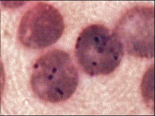
B. henselae
More bart. Posts: 2094 | From NY | Registered: Oct 2011
| IP: Logged |
TNT
Frequent Contributor (1K+ posts)
Member # 42349
posted
quote:Originally posted by Lymedin2010:
It says that arrows are pointing to the coccobacilli. The Fry photos I have seen do show actual arrows, but this photo has none, only boxes with numbers with a line attached to the organism (except on #3). I know I'm being technical, but Fry normally has a couple pictures, one of which does normally have actual "arrows."
Could there have been another pic with these results that could be posted that would give us more info? Is the description actually referring to THIS pic? I know I'm being technical....since it does look like coccobacilli.
Posts: 1308 | From Eastern USA | Registered: Oct 2013
| IP: Logged |
Lymedin2010
Frequent Contributor (1K+ posts)
Member # 34322
posted
Actually, to the left of the square box surrounding the number IS a pointer (arrow) & it is very faint. I guess they consider that the "arrow" or pointer to the bacteria.
All of the arrows seem to be properly aligned to the dots (bart coccobacilli) on the peripheral of the rbc extracelluarly, except for #3 which I think is just not aligned properly.
You can right click the image & save as, then zoom in to see it better.
Posts: 2094 | From NY | Registered: Oct 2011
| IP: Logged |
TNT
Frequent Contributor (1K+ posts)
Member # 42349
posted
I made my first Giemsa stain! The bottle of stain came from a local supplier thru Amazon, so it came much quicker than I expected it to.
I know I should know this, but, do Bart-like organisms stain with Giemsa? Or, is it only parasites with a nucleus?
My brain is not working well enough right now to try find the answer myself.
Posts: 1308 | From Eastern USA | Registered: Oct 2013
| IP: Logged |
Lymedin2010
Frequent Contributor (1K+ posts)
Member # 34322
posted
Warthin-Starry staining is used to identify it histologicaly or another type of SILVER stain.
Posts: 2094 | From NY | Registered: Oct 2011
| IP: Logged |
TNT
Frequent Contributor (1K+ posts)
Member # 42349
posted
S13, what do you use for bacteria or bart-like organisms?
Posts: 1308 | From Eastern USA | Registered: Oct 2013
| IP: Logged |
TNT
Frequent Contributor (1K+ posts)
Member # 42349
posted
S13, what I meant was what stain do you use for bacteria or bart-like organisms.
Has anyone seen these new videos yet? They are pretty good for what appears to be a home setup. The proposed cyst video has over 60 views already!
posted
For bacteria you can use the giemsa stain yes. Though if you want more differentiation you would need a Gram stain (so you know if its gram negative or gram positive bacteria).
For BLO i dont know. Nobody knows, since we dont know what BLO is. Is it a bacteria? Does it even exists? Perhaps its just a collection of other known bacteria?
Anyway, take a look at some of the posts over in this blog: http://lymemd.blogspot.nl/ He has posted several examples of giemsa smears showing babs and what he thinks is bart or BLO. Though even he regards some of the bacteria just as "mystery bugs".
Posts: 387 | From The Netherlands | Registered: Nov 2013
| IP: Logged |
Lymedin2010
Frequent Contributor (1K+ posts)
Member # 34322
posted
The last video sure looks like Borrelia & fits the 3 criteria I mentioned previously.
I don't think Giemsa is reliable for many Bartonella species, as most DO NOT stain well. That is why when you see many of these studies they use Warthin-Starry staining, Steiner silver stain, or other type of SILVER stains, which are BEST used for Bartonella.
I think I read somewhere that Fry Labs has used a MODIFIED Giemsa stain with success, but they did not mention any details as to the exact modification process.
One could also culture Bart on chocolate blood agar or soy agar, but it takes DAYS (9-40) for cultures to appear.
Posts: 2094 | From NY | Registered: Oct 2011
| IP: Logged |
Lymedin2010
Frequent Contributor (1K+ posts)
Member # 34322
posted
Dr. Willy Burgdorfer crushed ticks & viewed them under a microscope.
Are you guys willing to try this? Now I can't wait to find the next tick.
Posts: 2094 | From NY | Registered: Oct 2011
| IP: Logged |
TNT
Frequent Contributor (1K+ posts)
Member # 42349
posted
I hate the nasty, filthy creatures! Who knows what each one may be carrying. There's no way I am going to be tampering with a tick. They go down the toilet unless they have been attached. Though, I have been known to burn and torture them.
Posts: 1308 | From Eastern USA | Registered: Oct 2013
| IP: Logged |
TNT
Frequent Contributor (1K+ posts)
Member # 42349
posted
Ok, I tried posting a picture from my Google account (then deleted it) and now I know it will work.
To those on the forum that are more computer savvy than me, will this be a potential security hazard for my Google account or my identity?
I love sharing about my microscopy, but I can't afford to invite a breach into my personal info.
The main thing I'm concerned about is the URL address of the picture being visible to the whole world, but it didn't appear like any personal info was in the URL, and even if it did link my profile, I don't really have any personal info visible on the profile (just in the account).
So, I feel pretty safe, but would like some input.
Thanks
Posts: 1308 | From Eastern USA | Registered: Oct 2013
| IP: Logged |
Lymedin2010
Frequent Contributor (1K+ posts)
Member # 34322
posted
You can email it to me & I can place it on photobucket, or you can make your own photobucket account.
Posts: 2094 | From NY | Registered: Oct 2011
| IP: Logged |
TNT
Frequent Contributor (1K+ posts)
Member # 42349
posted
Lymedin, why do you use photobucket instead of your google account?
Are you saying there is a security threat with posting from one's google account?
The fewer accounts that I open the better!
Posts: 1308 | From Eastern USA | Registered: Oct 2013
| IP: Logged |
Lymedin2010
Frequent Contributor (1K+ posts)
Member # 34322
posted
I would not give out my main email account to the public no matter what I was doing.
Also, you can keep your hobbies & interests private from your friends & families if that is what you desire also.
Posts: 2094 | From NY | Registered: Oct 2011
| IP: Logged |
Lymedin2010
Frequent Contributor (1K+ posts)
Member # 34322
posted
At 25:38 Dr. Alan MacDonald presents pictures of Alzheimer's Diseased dead brain tissue that he was able to culture aggressive adult spirochetes from.
Notice how in the brain tissue they appear squiggly & worm like, just like we see in our blood. Whilst the cultured versions appear more aggressive & textbook style types.
Wow-- these are amazing videos. Luckily for her, her live blood doesn't look too bad in terms of spirochete infestation-- but classic spirochetes are clearly visible and actively trying to suck the life out of her red blood cells! I wish I still had my youtube channel so that I could comment and give her a thumbs up and heap praise on her for her videography work.. If anyone here has a youtube channel, please give her our congratulations and praise. Thanks for sharing these. All people out there doing Lyme videography deserve academy awards.
Posts: 696 | From New York | Registered: Aug 2006
| IP: Logged |
Are you guys willing to try this? Now I can't wait to find the next tick.
This is a great vid, too lymed2010... In this specific video of the crushed tick's blood, it looks like there are more borrelia cysts than actual live spirochetes( I only saw a couple of live spirochetes) which is a useful piece of scientific info. I was hoping to see filaria also, but I don't know exactly what they look like. I also think that youtube may be suppressing the view count on these types of videos--- if you watch that video 5 times, the view count stays static... LOL...
Posts: 696 | From New York | Registered: Aug 2006
| IP: Logged |
Lymedin2010
Frequent Contributor (1K+ posts)
Member # 34322
posted
Hi Wakeup, filtered water was added to the crushed ticks & I think water forces them into cysts & blebs as they prepare for adverse conditions. I think more are inclined to the relative quick cyst conversion, as opposed to the time consuming SoP formation & hence more cysts are seen.
The water level concentrations are not the same for every point of the mixture & at one point it balances out as tick cells lyse & change the overall balance, so a few spiros might never encounter high enough water concentrations to induce morphological change & as a result appear atypical.
What is surprising is that they are atypical in a tick as well, just like we see in our blood & not the text-book style aggressors (for at least this case).
Posts: 2094 | From NY | Registered: Oct 2011
| IP: Logged |
posted
Hello. I wanted to post that I will start doing some research on my own too and do what I can to contribute.
I have a Nikon laboratory microscope. I work as a chemist but this microscope is MINE. I am ordering some slides and I am also going to order some gram staining kits and other kits too.
I am missing my oil immersion lens .
However I will need to get one of those.
I find some of these videos fascinating. I work with two microbiologist but one of the guys I work with is very intelligent.
He worked in Cali producing Botulinum Toxin from C. Botulinum. I have worked in microbiology for two years prior to becoming a chemist however there is still a great deal to learn.
My background: I spent two years working in food and pharmaceutical microbiology and going on 3 years as a chemist with 1 year in food chemistry and 2 years in pharmaceuticals. Was completing biochemistry degree but all this hit me and I was too ill to finish.
I am still making an attempt but to be honest, almost everything you need to know is available online and can be self learned.
I hope I can be of some help as I was just diagnosed yesterday and having this for ~4 years.
Talk soon, ANeg
-------------------- Life is short, live it without regrets. Posts: 33 | From DFW | Registered: Sep 2015
| IP: Logged |
posted
Hello. I am more of a chemist than anything and my knowledge set is not strongest in microscopy however I noticed something strange with my blood stains when they dry.
Now I noticed when this was fresh last night there were what looked like thousands of tiny proteins that I could see and some still seem to be moving among the smear.
There is an artifact in the photos that looks long and rod shaped, that is from the camera and I was unable to get that off. Not sure what it is.
However I have no idea what the network looking strings are. I was thinking they could be potentially the plasma crystallising while drying. What are your thoughts.
This is at 1000X. Sorry for poor lighting, the camera was not the best.
-------------------- Life is short, live it without regrets. Posts: 33 | From DFW | Registered: Sep 2015
| IP: Logged |
TNT
Frequent Contributor (1K+ posts)
Member # 42349
posted
Whoa, ANeg, you got an oil lens pretty quickly!
Those "network-looking strings" may be fibrin spicules.
Do you think the "proteins" you are seeing could be lysozomes (they would be the same tiny particles you see flowing around in the WBCs, but can also be seen bouncing around in the plasma from brownian motion)? What I notice is that they are the most abundant in the plasma when first viewing the slide, but diminish as the sample deteriorates.
Thanks for your contribution! I don't know if you have any other hobbies, but, be forewarned, microscopy may become hobby #1 for you now, LOL!
Posts: 1308 | From Eastern USA | Registered: Oct 2013
| IP: Logged |
TNT
Frequent Contributor (1K+ posts)
Member # 42349
posted
[ 10-09-2015, 11:02 PM: Message edited by: TNT ]
Posts: 1308 | From Eastern USA | Registered: Oct 2013
| IP: Logged |
posted
They could be. I borrowed the lens from the microlab at work. I am going to order a Nikon brand as for this one was Olympus. not really a big deal but my microscope is a Nikon. Old school alphaphot YS but it works well and I can see things that I could not ever be able to using college microscope.
I do enjoy it, I cannot look too long under the scope or I will get dizzy. I was looking at getting a camera but I have yet to find anything good. Any ideas?
-------------------- Life is short, live it without regrets. Posts: 33 | From DFW | Registered: Sep 2015
| IP: Logged |
TNT
Frequent Contributor (1K+ posts)
Member # 42349
posted
The pic I posted above is from my first Giemsa-stained slide. It shows a Basophil among erythrocytes.
I plan on posting various pics of my Giemsa stain.
Aneg, a Nikon is a nice scope to have. As for a good camera, I use an old Canon A540 and that works pretty well just shooting through the eyepiece. But, you cannot do time-lapse with that; I hope to get an eyepiece camera sometime for that purpose. I'll probably get an Amscope eyepiece camera unless I find a better one for a better deal.
The problem I'm hearing with time-lapse is that between shots the scope can get out of focus. S13 has overcome that problem, and I wish I knew more about how he did it.
If you get a chance S13, do share the details of how you accomplished that (like what program you used and how you integrated it practically). Hopefully it's not too challenging for those like me to understand.
Posts: 1308 | From Eastern USA | Registered: Oct 2013
| IP: Logged |
posted
Yes the camera will get out of focus with timelapse recording. I still think it has to do with the heating from the light bulb that slowly warps the frame of the microscope. So the solution i use is to switch off the bulb between pictures. This works really well.
I do use a complicated solution, but thats just because i used to be an electronics engineer before lyme and i had some old projects laying around that i could use.
Basically i use a standard microscope camera with PC software. Ive written a small software program that commands the camera PC software to take a picture with a programmable interval. It also communicates with an FPGA board via USB. By toggling a relay on the FPGA board it can enable and disable the microscope light bulb.
So every 2 minutes or so, the program will enable the light, wait a couple of seconds, command the camera software to take a picture, wait another couple of seconds, and disable the light.
So you end up with hundreds of pictures that can be converted to a video by a video editing tool (i use powerdirector). And then you get something like this for example: https://youtu.be/l4t7UKi4ma8 (24h candida growing time lapse)
Time lapses can be very fun to see things grow Posts: 387 | From The Netherlands | Registered: Nov 2013
| IP: Logged |
posted
Can someone take a look at this and tell me what I am seeing? I followed it for over an hour. It appears to be an RBC (portion of) with a spirochete or some other mechanism providing motility, as the cell matter traveled around to some degree. It's not easy to see in the video, but when viewing through the microscope, you could see the thin portion protruding from the cell matter moving and whipping around. HERE IS THE VIDEO (filmed on my cell phone) https://www.youtube.com/edit?o=U&video_id=7wOwSebreVcPosts: 163 | From USA | Registered: Oct 2015
| IP: Logged |
TNT
Frequent Contributor (1K+ posts)
Member # 42349
posted
Hey dude,
Welcome to the forum, and to this thread!
It looks like your link is a dud. It appears as though you didn't "publish" your video when you were done uploading it.
Try again, and we may be able to give you some possibilities of what you may be seeing.
Care to share some of your story??
Posts: 1308 | From Eastern USA | Registered: Oct 2013
| IP: Logged |
I've actually been diagnosed with MS, but was also reactive on a few bands on western blot IgM and IgG. So I've been using my microscope to do some of my own stuff, out of curiosity. I have some formal training in this kind of stuff, but it's been so long, and I am not a professional.
The video is from my cell phone, as it was the only way to capture it at the time.
This video is also roughly two hours after I started viewing it, so the motility is much lower, likely due to the blood drying.
Posts: 163 | From USA | Registered: Oct 2015
| IP: Logged |
tbh i cant make much of this video. Perhaps use a steady digital camera with some optical zoom?
Posts: 387 | From The Netherlands | Registered: Nov 2013
| IP: Logged |
TNT
Frequent Contributor (1K+ posts)
Member # 42349
posted
There it is! Thanks S13. Before my last post I even searched youtube for the last 10-15 characters of the link dude gave us, and still couldn't find it.
Yeah, I couldn't make anything out of the video either. I would 2nd S13's suggestions.
A smaller digital camera works great if you hold it up to the eyepiece. A smaller camera works better than a larger one. My Canon A540 works great, whereas my Canon SX160is is very unsuitable even though it is a newer and better version (the diameter of the lens is too wide to practically hold against the eyepiece since the 160 has a wide angle lens).
Posts: 1308 | From Eastern USA | Registered: Oct 2013
| IP: Logged |
posted
Yea, unfortunately this was 2 hours after I had started viewing it. It's the hemolyzed RBC right below the arrow. The only one that is not a full RBC.
It's hard to see, but I did follow it and had others look and confirm that I wasn't just seeing things.
The small portion extending out of the RBC on the right side was moving, and providing motility. I'll see if I have any other videos.
Posts: 163 | From USA | Registered: Oct 2015
| IP: Logged |
I uploaded another video I had pointing at what I was looking at.
Unfortunately, you have to look closely to see it moving. The portion extending out of it WAS moving, and providing motility. I had numerous other people also view it to tell me what they saw.
It moved through a small portion of the slide before the blood had started to dry, which is why it is not moving around like it was. However, the RBC was being moved, as you could see that small portion wiggling/flicking.
Posts: 163 | From USA | Registered: Oct 2015
| IP: Logged |
TNT
Frequent Contributor (1K+ posts)
Member # 42349
posted
I'm sorry, dude, but I couldn't see any more than with the first video. They make cradles that you can attach your phone to a microscope with if you don't have access to a point and shoot camera. It will keep your phone still and "may" help your phone get a better capture.
But, even with a point and shoot camera, you may still have similar trouble keeping it steady. Then again, there are adapters for that, too.
Posts: 1308 | From Eastern USA | Registered: Oct 2013
| IP: Logged |
TNT
Frequent Contributor (1K+ posts)
Member # 42349
Lymedin2010
Frequent Contributor (1K+ posts)
Member # 34322
posted
Will post more in future, for now just a quick answer to a question.
I have found that running a ceiling fan & keeping room temp constant AFTER bulb has been on a while, to help drastically with focus creep issue. I would imagine that a dedicated fan properly placed should diffuse the heat. The whole trick is to keep a constant temp, no matter what that temp is. Expansion & contraction due from the heat is what causes the focus creep.
Posts: 2094 | From NY | Registered: Oct 2011
| IP: Logged |
Best photos I could get for the moment. I have a video uploading currently if anyone wants to look. It appeared to be a spirochete to me, linking to RBC's. The free RBC that is crenated was actually pulled closer, across the open plain of plasma, so it is about twice as close as it started.
4:30 is a good point to see what I am talking about. Focusing on it and getting it to be clear in the video is hard right now.
[ 10-10-2015, 02:44 PM: Message edited by: thatdudefromkansas ]
Posts: 163 | From USA | Registered: Oct 2015
| IP: Logged |
Lymedin2010
Frequent Contributor (1K+ posts)
Member # 34322
posted
It is really hard to tell what that is. Since there are string like objects in human blood, I would disregard anything that does not fit the criteria I mention in the past (with bulbs on both tips...etc).
There will be spiros without bulbous tips, but in order to avoid false identification it is best to disregard anything without those criteria. You have actually posted a beautiful video from another Youtuber that met the criteria.
To make instant improvements to your video & image, I would use any DAYLIGHT 5000K CFL bulb, such as the Philips CFL with TuffGuard Protection that I showed you video of.
Basically, place the bulb in a standard lamp & then directly under your condenser & shift the bulb with your hand until you witness best lighting & shading via the binocular port. Keep in mind the heat will cause more focus creep though & the LED daylight bulbs will produce less heat but can be damaging to your eyes. So if you use an LED bulb, then it is best to only use your camera to view the images & not damage your eyes with prolonged LED exposure.
Posts: 2094 | From NY | Registered: Oct 2011
| IP: Logged |
posted
Will do. Unfortunately, the other one I did see would more closely match that criteria. Another one protruding out of an RBC, with a bulbous end and very clearly moving.
I need to get a shorter lens on my camera. It'll clear it up more.
Posts: 163 | From USA | Registered: Oct 2015
| IP: Logged |
TNT
Frequent Contributor (1K+ posts)
Member # 42349
posted
Here are some pics from my first Giemsa stain:
1. Two possible Bart-like organisms that took up the stain:
2. A zoomed in pic of the same organisms:
3. Another possible Bart-like organism:
4. A zoomed in pic of the same organism:
5. 3 WBCs and a possible kete:
6. A possible kete among RBCs and a few platelets:
7. A zoomed in pic of the same organism:
If the above organism is a kete, it's noteworthy that it did not take up the Giemsa stain.
[ 10-11-2015, 03:37 PM: Message edited by: TNT ]
Posts: 1308 | From Eastern USA | Registered: Oct 2013
| IP: Logged |
TNT
Frequent Contributor (1K+ posts)
Member # 42349
posted
Have I ever told you guys how HAPPY microscopy makes me?
This HAPPY:
It's too bad a smiley-faced-shaped WBC is not the sign of a happy immune system!!
Posts: 1308 | From Eastern USA | Registered: Oct 2013
| IP: Logged |
Lymedin2010
Frequent Contributor (1K+ posts)
Member # 34322
posted
Hey that last pic looks like me.
I feel the same way about microscopy. Rather than being in the dark, it gives us power.
"6. A possible kete among RBCs and a few platelets:" That one looks like it could be a spiro & undergoing blebbing.
TNT, I am surprised you are not seeing as many in your blood as I am in mine, which might be a good thing actually. I find mine everywhere & the only thing that has been able to reduce them was Cowden Protocol so far, but it has wore off.
As I said before the only other thing that MUST have cleared my blood of spiros, but sent them to all my joints, organs & nerves, was IV Rocephin. Since I could not find any spiros in my blood at the tail end of that drug.
FYI, check my other post out about Dr. Eva Sapi's work on Stevia & Bee Venom, which both showed anti-Borrelia activity. CBD oil was also shown to be effective on ALL FORMS of bb.
Posts: 2094 | From NY | Registered: Oct 2011
| IP: Logged |
TNT
Frequent Contributor (1K+ posts)
Member # 42349
posted
quote:Originally posted by Lymedin2010: TNT, I am surprised you are not seeing as many in your blood as I am in mine, which might be a good thing actually.
That may be a good thing, unless they are in L-form, as I am suspecting. The last wet mount that I did about a week ago showed a higher load of what I have been suspecting are L-forms (the forms I pointed out in a couple of my videos).
(As for that last pic resembling you), I imagined you with bigger ears, lol!
Ha Ha, I'm just kidding!
I have recently been giving Bee Venom treatment a little more consideration. It has done wonderful things for some people.
Posts: 1308 | From Eastern USA | Registered: Oct 2013
| IP: Logged |
TNT
Frequent Contributor (1K+ posts)
Member # 42349
posted
quote:Originally posted by Lymedin2010: As I said before the only other thing that MUST have cleared my blood of spiros, but sent them to all my joints, organs & nerves, was IV Rocephin. Since I could not find any spiros in my blood at the tail end of that drug.
As I pointed out in a previous post, Penicillins and Cephalosporins CAUSE cell-wall deficient bacterial forms!!!!!
This could be why you saw no ketes in your blood. It could be the ketes sought safe haven in your joints and nerves, OR, it could be that the Rocephin caused them to convert to L-forms and the L-forms caused the damage at these places. Cell-wall deficient forms cause the most damage, and are the hardest to eradicate! Thus, when you discontinued the Rocephin, you eventually got much worse than you previously were. Because, not only did the ketes come out to play once you discontinued the Rocephin, you now had l-form ketes to deal with.
I personally feel that LLMDs should not treat with Cephalosporins or Penicillins UNLESS concurrent treatment with a macrolide or tetracycline is given!
[ 10-13-2015, 01:39 PM: Message edited by: TNT ]
Posts: 1308 | From Eastern USA | Registered: Oct 2013
| IP: Logged |
Lymedin2010
Frequent Contributor (1K+ posts)
Member # 34322
posted
I feel the same way & I thought to myself what type of backward thinking is this.
It is useless to treat mid to late stage without a cyst buster, simply moronic & I have had that stance since the first month of starting treatment.
Posts: 2094 | From NY | Registered: Oct 2011
| IP: Logged |
TNT
Frequent Contributor (1K+ posts)
Member # 42349
posted
quote:Originally posted by Lymedin2010: I feel the same way & I thought to myself what type of backward thinking is this.
It is useless to treat mid to late stage without a cyst buster, simply moronic & I have had that stance since the first month of starting treatment.
Even if one does treatment with a penicillin or cephalosporin AND a cyst-buster, you will STILL CAUSE CONVERSION TO L-FORMS and perhaps be worse off.
That is why it is important to hit all three forms at the same time for effective treatment.
Though, I am afraid of cyst-busters because I have had horrible reactions with them. Did they cause conversion to L-forms? Is that one reason (among others) for why I had bad experiences with them? Quite possibly.
I find it interesting that there is so much hype about the overuse of antibiotics causing antibiotic-resistant super-bugs and so little research into the fact that most bugs are pleo-morphic.
This really could be a huge component of why the drugs aren't working as well anymore. I mean, when someone has strep throat and the normal course of Amoxicillin doesn't clear it up, the patient is then given Azithromycin. That clears it up.
ABX-resistance?
That's what they say (and it probably is).
Pleomorphism?
Very possibly, too, in many cases.
It's interesting that the drug of choice for eliminating persister cells is Daptomycin. If you look Daptomycin up you'll find that it is one of the most effective drugs against L-form bacteria. It ruptures the cell MEMBRANE of cell-wall-deficient bacteria.
So, are the real persister cells (and the reason many don't get well with ABX treatment even with anti-cyst drugs) L-form and not cysts?
Posts: 1308 | From Eastern USA | Registered: Oct 2013
| IP: Logged |
This is dark field, but filmed with my DSLR instead of microscope cam. Where the indicator is pointing. Watch in 1080 HD, full screen. It's on 400x in this video.
Easier to see now?
I have more videos I can upload as I go.
Posts: 163 | From USA | Registered: Oct 2015
| IP: Logged |
Watch in HD. Where the indicator is pointing.
Posts: 163 | From USA | Registered: Oct 2015
| IP: Logged |
TNT
Frequent Contributor (1K+ posts)
Member # 42349
posted
Very good, dude! Now that's what we are after! It appears like that RBC on the second video may be a "colony" as I see multiple ketes swinging out of it.
That's a nice crisp view of things! Your DSLR does a great capture!
What kind of scope do you have? Did it come with a darkfield condenser when you bought it, or did you just get the darkfield?
Isn't microscopy SO exciting!? I'm glad you joined the thread! We need more recruits.
Is it possible to do darkfield at 1000x? I think I remember S13 mentioning that it's not possible at 1000x. That's too bad if not, because you could have REALLY been able to see that cell at 1000x if it would be possible.
How long after drawing you blood did you make the videos? It appears like you are not heavily infected, thankfully.
Some of the other members are doing time lapse. Can you do time lapse? I sure would like to but I need to get an eyepiece camera.
What bands showed on your Western Blot if I may ask? Some bands can cross react with other infections.
Posts: 1308 | From Eastern USA | Registered: Oct 2013
| IP: Logged |
posted
I have a microscope camera I am gonna try as well.
I bought an Amscope T490-DK. Was only about 375 dollars, including dark field, useful up to the 40x.
It is possible to do dark field on 1000x. However, you need a dry or dark field lense with the aperture large enough. I could be wrong, and it might only be oil immersion, but you can buy condensers for this purpose.
I draw blood into vacationers. Generally, I have been viewing samples anywhere between 1 and 3 days after drawing the sample, and anywhere between 1 and 6 hours after applying to a slide.
Time lapse on my microscope camera won't seem to work, but I'll keep trying.
My history: I have been diagnosed with MS. IgG: 31, 41 IgM: 39, 41 That's it. I am also testing the blood of a co-worker, also diagnosed with MS. I find the same in his blood.
Posts: 163 | From USA | Registered: Oct 2015
| IP: Logged |
The Lyme Disease Network is a non-profit organization funded by individual donations. If you would like to support the Network and the LymeNet system of Web services, please send your donations to:
The
Lyme Disease Network of New Jersey 907 Pebble Creek Court,
Pennington,
NJ08534USA http://www.lymenet.org/


 UBBFriend: Email this page to someone!


![[Frown]](frown.gif) .
.


![[Smile]](smile.gif)


 Printer-friendly view of this topic