Lymedin2010
Frequent Contributor (1K+ posts)
Member # 34322
posted
Your symptoms because of this might be: 1) CFS 2) Exercise or high activity intolerance. 3) Circulatory issues. 4) Night sweats. 5) Brain fog...not enough O2 to the brain.
???
Ever positive for bart test?
Posts: 2094 | From NY | Registered: Oct 2011
| IP: Logged |
TNT
Frequent Contributor (1K+ posts)
Member # 42349
posted
"Ever positive for bart test?"
Slight positive with Galaxy culture (read as a waning infection, ha). But I have not been tested for Anaplasma that I can remember. HGE, but not Anaplasma. Not sure if they would cross over on antibody tests or not, though. I just got tired of spending money on tests that were never conclusive. Especially Medical Diagnostic Labratory's tests, and the local labs antibodies tests (not for Anaplasma).
Microscopy has been more insightful, enlightening, educational, and even entertaining. Not to mention more conclusive. Posts: 1308 | From Eastern USA | Registered: Oct 2013
| IP: Logged |
Lymedin2010
Frequent Contributor (1K+ posts)
Member # 34322
posted
Most definitely, most of the ones who have done microscopy can tell there is something terribly wrong with their blood & it is telling of a multi-system disease that involves the blood as a super highway.
In case this ever comes up & just to get this out of the way, I don't think these are Howell-jolly Bodies. Your objects appear to be consistently on the outer edge of the rbc's as if they are exiting.
Lymedin2010
Frequent Contributor (1K+ posts)
Member # 34322
posted
BorreJaakko, below is a sample pic from my 50d at the time with the Amscope adapter I linked you. I sold the 50d before the prices had dropped any further & I ended getting a 40d for $100.
Here is the video from 40d, BUT it is much better to watch the original file on my PC as Youtube downgrades the quality. In another video it actually even flipped the video to the vertical mode...dunno why. : https://www.youtube.com/watch?v=iSTDQN-3Cb0
Here is a pic from a 50d:
[ 01-14-2017, 03:43 PM: Message edited by: Lymedin2010 ]
Posts: 2094 | From NY | Registered: Oct 2011
| IP: Logged |
I also purchased a 8 megapixel camera board for rasperry pi, and just got it today. I am just now playing with it.
This camera will give me all the flexibility to control the camera exactly how I want. The software will not be a problem for me as I work with software professionally, so I can develop my own utility.
Rasperry pi camera is quite cheap, so I can leave it to record time lapse or video extended periods.
It can do 2592×1944 at 15fps.
Quality seems reasonably good.
Posts: 31 | From Finland | Registered: Sep 2016
| IP: Logged |
The adapters are VERY sturdy & metallic. Good quality & it does what it is supposed to. The custom ones are very similar & I don't think too far away. The only difference would be the eyepiece type optical & Amscope is good enough to produce quality images for that.
My TL delays are normally 10s & 15s & all depends on the details I want to capture. I can always speed it up, which mostly I do in the software processing. If I want capture lets say a rbc or wbc degrading, I would do 1-5 minutes & again speed up in software, and as much as 15 min intervals. So it depends on what your capturing & what details you are looking for. If you miss something in between & it was hard to capture, you will kick yourself & I tend to go lower with plenty of hard drive space.
posted
I got my Rasperry PI camera. It seems pretty decent, although processing power seems a little limited, which means taking full HD video with high frame rate is not possible.
It seems I did not find anything, until today I cleaned a slide with water and paper and then put a drop of fresh blood on it, and things started happening.
Some white tiny things seem to be attacking the blood cells quite viciously, and the cells become spikey. It seems like some of RBCs are killed and eaten by dozens of them -- alive.
It seems like the video is moving with extra speed, I guess I need to set up the recording frame rate.
This raises a question: do you, and if yes, how do you clean your slides?
Posts: 31 | From Finland | Registered: Sep 2016
| IP: Logged |
TNT
Frequent Contributor (1K+ posts)
Member # 42349
posted
quote:Originally posted by BorreJaakko:
Some white tiny things seem to be attacking the blood cells quite viciously, and the cells become spikey. It seems like some of RBCs are killed and eaten by dozens of them -- alive.
This raises a question: do you, and if yes, how do you clean your slides?
The little "white" things in your video are immune components called lysozomes. Basically they are the same as the little granules of the white blood cells except they are free in the plasma. They attack pathogens. I've seen many spirochetes being attacked by them. I assume they release enzymes that dissolve pathogens just like the ones inside the WBCs do. I doubt they were attacking your blood cells since they don't attack "self." I've never seen that anyways.
Your video is good. The clarity with that 60x objective is notable. Now all you need to do is seal off your cover-slip with immersion oil and wait to see some ketes. They will surely come out if you are infected. But, you may have to wait and look a little if you are not heavily infected.
I buy pre-cleaned slides. But, they are usually cloudy so I wash them with soap and running water and let them air dry in a drainer. That removes any bacteria that may be on them out of the box. It's important to have clean slides especially when one is staining blood. Otherwise, you think you see bacteria in the blood when it's just contamination.
Great job!
Posts: 1308 | From Eastern USA | Registered: Oct 2013
| IP: Logged |
quote:The little "white" things in your video are immune components called lysozomes. Basically they are the same as the little granules of the white blood cells except they are free in the plasma. They attack pathogens. I've seen many spirochetes being attacked by them. I assume they release enzymes that dissolve pathogens just like the ones inside the WBCs do. I doubt they were attacking your blood cells since they don't attack "self." I've never seen that anyways.
Interesting. I thought I saw that happening live, and that it would be visible from the video too.
For example at 0:20 you see those lysozomes "ride" the blood cells. At 1:08 you see a big formation of them, which I figured would be a cell eaten by losozomes. And at 1:30 you see some cells that are completely covered with those lysozomes. Also, I find it interesting that some of the cells have spikes, whereas some of them are nice and round....
quote: Your video is good. The clarity with that 60x objective is notable. Now all you need to do is seal off your cover-slip with immersion oil and wait to see some ketes. They will surely come out if you are infected. But, you may have to wait and look a little if you are not heavily infected.
Thanks! My problem with sealing off right now is that I bought wrong kind of cover slips... twice! First ones were too long and wide, and it is not feasible to seal them off because the slip is almost as wide as slide, and there is no space for the oil on the side... and the next ones are nice and round, but only 5mm in diameter so they are way too small. So I need to find a place that sells proper size cover slips.
quote: I buy pre-cleaned slides. But, they are usually cloudy so I wash them with soap and running water and let them air dry in a drainer. That removes any bacteria that may be on them out of the box. It's important to have clean slides especially when one is staining blood. Otherwise, you think you see bacteria in the blood when it's just contamination.
Yeah, I have pre-cleaned ones too, I just thought that maybe it is easiest to just reuse the slides as I don't need the old dried up blood for anything..anyway I'll use fresh ones from now on, thanks for the tip.
Posts: 31 | From Finland | Registered: Sep 2016
| IP: Logged |
posted
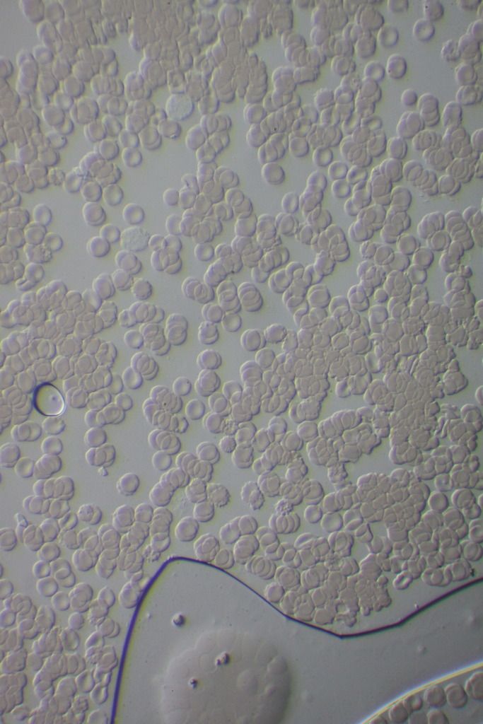
Hey guys, hopefully you can help me out again (and this might be relevant to some of the earlier posts from TNT).
Any idea what these round inclusions in the RBC's could be?
They seem too big to be Bartonella.
At the same time, I don't see any typical Babesia forms anywhere in my blood (halo's, crosses, etc).
It leaves me scratching my head as to what these large, round circles are.
Any input would be appreciated. Thanks!
Posts: 71 | From Canada | Registered: May 2016
| IP: Logged |
TNT
Frequent Contributor (1K+ posts)
Member # 42349
posted
quote:Originally posted by mustardseed2: Hey guys, hopefully you can help me out again (and this might be relevant to some of the earlier posts from TNT).
Any idea what these round inclusions in the RBC's could be?
They seem too big to be Bartonella.
At the same time, I don't see any typical Babesia forms anywhere in my blood (halo's, crosses, etc).
It leaves me scratching my head as to what these large, round circles are.
Any input would be appreciated. Thanks!
Those are Heinz bodies. They can be caused by hemolytic anemia due to unstable hemoglobins, exposure to oxidizing drugs, chemical poisoning, G-6PD deficiency.
Good job on the staining. Blood staining can be extremely helpful in giving us insights into our bodies and clues about what may be going on. That's good you are not seeing any signs of Babesia. Are you seeing any dots on the outside perimeter of the RBCs typical of Bartonella? Any Anaplasma morulas? Any other morphologies?
Posts: 1308 | From Eastern USA | Registered: Oct 2013
| IP: Logged |
posted
Thanks TNT! That a great page you posted there, I bookmarked it.
When I started staining, I was convinced I had a Babesia problem. I have a 24/7 headache, chills, night sweats, weakness, tingling, and ringing in the ears. On top of that, I've herxed on Malarone, Artemisinin, Alinia, and Beyond Balance MC-BAB-2.
But alas, haven't seen anything Babesia-like in my blood. A little perplexed by it to be honest.
As for Bartonella, the only dots I see on the perimeter of RBC's are big ones like I posted. I didn't herx at all on high doses of Levaquin for 3 months and Rifampin for 6 months. So I don't think I have Bart.
I haven't seen any anaplasma inclusions in WBC's, but I have seen some rod-like bacteria, similar to the pictures you posted as rickettsia. Too small to be sure though.
I did test postitive for anaplasma through IGENEX. I took several months of Doxycycline, followed by 6 months of Rifampin, so I think I would have hit anaplasma pretty well.
Posts: 71 | From Canada | Registered: May 2016
| IP: Logged |
TNT
Frequent Contributor (1K+ posts)
Member # 42349
posted
quote:Originally posted by mustardseed2: Thanks TNT! That a great page you posted there, I bookmarked it.
When I started staining, I was convinced I had a Babesia problem. I have a 24/7 headache, chills, night sweats, weakness, tingling, and ringing in the ears. On top of that, I've herxed on Malarone, Artemisinin, Alinia, and Beyond Balance MC-BAB-2.
But alas, haven't seen anything Babesia-like in my blood. A little perplexed by it to be honest.
As for Bartonella, the only dots I see on the perimeter of RBC's are big ones like I posted. I didn't herx at all on high doses of Levaquin for 3 months and Rifampin for 6 months. So I don't think I have Bart.
I haven't seen any anaplasma inclusions in WBC's, but I have seen some rod-like bacteria, similar to the pictures you posted as rickettsia. Too small to be sure though.
I did test postitive for anaplasma through IGENEX. I took several months of Doxycycline, followed by 6 months of Rifampin, so I think I would have hit anaplasma pretty well.
Glad I could be of some help! What treatment are you currently on? Are you still on the anti-protozoan meds that you herx on?
That's interesting you are seeing minute rod-shaped objects. Definitely describes Rickettsias. Where are you seeing them? Inside WBCs or in the plasma? Could you post some pictures of them??? It does seem unlikely you could still have a Rickettsia with months of Doxy followed by Rifampin. Perhaps it's still possible if you were not on them together. In Burrascano's guidelines he says Doxy plus Rifampin may be needed.
"However, there are reports of treatment failure even when higher doses and long duration treatment with doxycycline is given. In such cases, consideration may be given for adding rifampin, 600 mg daily, to the regimen."
How long are you canvassing a stained slide? It has taken me quite a while at times before I've found some of the morulas.
Your herxing pattern suggests a protozoan, unless the "herx" was a flare (a worsening) on account of hitting a symbiotic organism. That has definitely happened to me.
Posts: 1308 | From Eastern USA | Registered: Oct 2013
| IP: Logged |
Lymedin2010
Frequent Contributor (1K+ posts)
Member # 34322
posted
BJ:
1) Great video. What can make it even better is if you increase the contrast & details will begin to pop even further. There might even be sharpness setting on there as well & maybe they have modes such as "vivid" to further enhance the quality. I believe that is a 5MP camera? I think the new Raspberry Pi 2 cameras are 8MP.
2) At time 1:09 it looks like you have an Eosinophil there & sometimes Lysosomes can escape from the wbc & they can be what those white dots dancing around in Brownian motion are. The peroxisome from the wbc escape too & are bigger than the lysosomes. But they can also be platelets or micelles (dissolved fat droplet particles). If you eat a very fatty diet & check your blood 1 hr later, you will see TONS of the micelles in your blood in the plasma.
Your video recording is too jerky & it almost seems like those dancing dots can be pathogens & it will be important for you to fix that in order sometimes to pickup on slight cues as to whether something is normal vs foreign in your blood.
3) I buy new boxes of slides & cover slips & never have to clean them, but if you did then I would wash them off, autoclave them in an oven at high temperatures & then maybe clean them off again with alcohol & cotton. I don't like to do that, as for me it is not worth mistaking an artifact for something interesting in blood, which is precious info for all of us.
You can use one slide & make 3 different samples on it if you like (left, middle, & right) and you can save this way. A lot of times I take a sample & do a simple experiment on the same slide using a 2nd cover & so make a 2 slip-covered slide.
4) I talk about crenation & the spiky rbc's here, as that is a normal occurrence. Sickly blood may have ammonia or other pathogen toxins that can make the rbc's degrade quicker though, but all blood can degrade like this & sometimes super quick based on air exposure on the slide.
Lymedin2010
Frequent Contributor (1K+ posts)
Member # 34322
posted
mustardseed:
WOW, really nice staining work!!!
They look like Howell-jolly Bodies, which are basophilic nuclear material that never got expelled from the cell by the late erythroblast precursors.
Have you ever been scanned, as the spleen normally gets rid of these in normal blood. Do you have Splenomegaly, enlargement of the spleen? I did when they last scanned me a few years ago & I could feel the tenderness on my left side, under my ribs sometimes. Especially when I am taking a heavy combo/dose of abx.
I wonder sometimes if they can also be platelets, just stuck on the outside or on top of the rbc. Your first pic the 1 micron object is nice & pink with stain & I wonder if it can be a platelet on top of the rbc.
Why is your last pic outside the rbc & not within, sometimes the rbc's lyse & release the Howell-jolly Bodies. This one looks more darkly stained & may not be a platelet. Are the rest of your platelets nice & pink in the plasma?
TNT
Frequent Contributor (1K+ posts)
Member # 42349
posted
They could be Howell-jolly bodies, and I originally thought that. But, the fact that he is seeing them on the outside of the RBCs would lend to the stronger possibility that they are Heinz bodies. But, it is possible they could be HJ bodies.
Posts: 1308 | From Eastern USA | Registered: Oct 2013
| IP: Logged |
Lymedin2010
Frequent Contributor (1K+ posts)
Member # 34322
posted
I think Heinz bodies are more irregular in shape & his are nice and more rounded, almost like a perfect circle. Perfect circle because it used to be a nice round nucleus that did not expel. And they are on the slightly larger side too.
At least that is what I think, but you guys be the judge. I think the spleen is overworked in us Lymies, because of all the pathogens and when it suffers our pathogen load gets higher & we get sicker. That has been my own personal account & witness with microscopy & my pains/tenderness in the spleen area & scan.
Sometimes rbc's can also lyse & release them and they can appear external and can be confused with platelets.
"This is a darkfield microscopy of my father's blood sample, revealing presence of spirochete like structures. my father has been officially diagnosed with ALS." https://www.youtube.com/watch?v=pkvwRS5vqYI
Lyme test was negative though & this is what she had to say....."No, my father has been diagnosed only with ALS. We did testing for Lyme, and all tests came back negative, except the microscopy we performed in Romania and they managed to film spirochetes in my father's blood specimen. He does have some of the symptoms that you listed in your post, so he has have a lot of sweating, skin itching, fasciculations, unusual dermatological issues on his face, GI irregularities, constant conjuctivitis (like he has have a sand in his eyes he tells me), bladder disfunction-frequent urination, constipation, bloating. at this very moment he is bed ridden most of the time, he developed a very serious muscle atrophy, his hands are completely paralysed, a lot of atrophy on his legs, but he can still make a step or two on his own. Lately his breathing became bad.... I don't know could I connect all of this with ALS alone."
This sounds like an infection of some sort for sure, as it is not just an atrophy of the main nerve bundles & maybe multiple infected.
Posts: 2094 | From NY | Registered: Oct 2011
| IP: Logged |
Since I wasn't getting anywhere on antibiotics or anti-protozoal medications, I've switched gears completely for a few months.
I'm currently doing one month of Diflucan + Ketoconazole nasal spray. This will hopefully hit any candida or other fungus, as I have some consistently nasty oral thrush.
This will be followed by a six week parasite treatment where I'll go through Praziquantel, Ivermectin, Pyrantel pamoate, Albendazole, and Alinia.
My eosinophil count sky rocketed when I tried Alinia, plus I have pictures a few pages back of some stuff in my blood I thought were parasites. It's possible that my herxing on anti-babesia meds was really parasites reacting to those medications (I know Alinia and Artemisinin are anti-helminth, not sure about Malarone)
I usually canvas a slide for about half an hour. I've done probably over a dozen stains at this point, so I'd say I'd be surprised if I found anything new now. Or maybe I don't have Babesia in my finger tips where I'm taking blood from
Either way the herxing is weird. I hadn't thought that it could be hitting a symbiotic organism... I'm not even sure what organism would do that when killed.
Lymein2010,
I've never had my spleen tested. I don't have any pain or tenderness. I do blood work every month, but I'm not sure there's anything on there that relates to spleen hmmm.
I'm not sure these 3 are platelets, simply because I find the platelets to be a slightly more irregular, and slightly more diffuse in color.
You are correct to question it though, because I do regularly find them showing up on top of RBC's. It's hard to really show the difference with these pictures, as they're taken with my crappy cell phone camera through the objective.
I'm leaning towards HJ bodies for my pics.
Anyway thank you both! I love this thread!
Posts: 71 | From Canada | Registered: May 2016
| IP: Logged |
TNT
Frequent Contributor (1K+ posts)
Member # 42349
posted
quote:Originally posted by mustardseed2:
My eosinophil count sky rocketed when I tried Alinia, plus I have pictures a few pages back of some stuff in my blood I thought were parasites. It's possible that my herxing on anti-babesia meds was really parasites reacting to those medications (I know Alinia and Artemisinin are anti-helminth, not sure about Malarone)
I usually canvas a slide for about half an hour. I've done probably over a dozen stains at this point, so I'd say I'd be surprised if I found anything new now. Or maybe I don't have Babesia in my finger tips where I'm taking blood from
Either way the herxing is weird. I hadn't thought that it could be hitting a symbiotic organism... I'm not even sure what organism would do that when killed.
That's very interesting that your Eosinophil count increased when on Alinia. That definitely says something. Especially since Eos count raises in response to worm/helminth/parasite infection.
Depending on the presentation of your herx, though, I think it's possible that you could be causing a flare of some other symbiotic organism by using the anti-helminth/anti-protozoal meds. What I mean by that is this. Many times these microbes live together symbiotically, or similarly, in such a way that keeps the others in check. When you kill one part of that "community," it allows the proliferation of other competing or perhaps other symbiotic organisms. Take Heartworm infection in dogs as a good example. Veterinarians know that if you treat Heartworm in heavily-infected dogs, it can actually kill the dog. NOT because the die-off (herx) of Heartworm is so great, but because of the MASSIVE release of symbiotic Wolbachia (a Rickettsia) bacteria into the system of the dog. The resulting MASSIVE immune response to the Wolbachia antigen release causes so much inflammation that it kills the dog!
Not the Heartworm, not the die-off, but the immune response to the sudden release of antigenic material kills the dog via a huge immune response. And, if the immune response doesn't kill the dog, I imagine that the unchecked proliferation of the Rickettsia eventually would.
That's why vets use Doxy to treat heartworm in heavily-infected dogs. There is no sudden release of Wolbachia because it kills them first.
So, it's very possible that the antihelminth medication was causing a release of bacteria into your system causing a flare of symptoms relating to a bacterial infection, and not necessarily related to a parasite die-off.
It's the Russian doll effect. That's why order of treatment really does matter. And, a good example of why microscopy can be helpful.
Posts: 1308 | From Eastern USA | Registered: Oct 2013
| IP: Logged |
My question would be would this still happen in the presence of antibiotics as well?
For instance, while I was herxing on Malarone, I was also taking zithromax and doxycycline.
When I herxed on Artemisinin, I was also taking minocycline and ceftin.
When I herxed on Alinia, I was taking biaxin and tindamax.
I suppose the herx I was feeling could be from these secondary medications attacking bacteria that was being released?
Posts: 71 | From Canada | Registered: May 2016
| IP: Logged |
TNT
Frequent Contributor (1K+ posts)
Member # 42349
posted
quote:Originally posted by mustardseed2:
I suppose the herx I was feeling could be from these secondary medications attacking bacteria that was being released?
That would be my guess. That, or a true parasite die-off herx. I'm not sure exactly how a true parasite herx presents.
But, like I said, your "herx" symptom picture may give some clues. My experience is that true DIE-OFF herxes make me sleepy and flu-like. Flares (worsening of normal symptoms), or especially the onset of new and strange symptoms, although touted by many people (and LLMDs) as true herxes, I feel are really the worsening of some other concurrent infection. Just like the Heartworm model.
Posts: 1308 | From Eastern USA | Registered: Oct 2013
| IP: Logged |
posted
I wanted to post one more picture of the rod-like bacteria I mentioned before.
The picture is not the best quality because it's just from my cell phone. Also the RBC's got a little wonky/spiky when I put the cover slip on, so ignore that.
In reality, the thing in question is actually less angular than it appears in the picture. It's definitely an oval, not a circle.
It was moving around in brownian motion, moving through my blood plasma.
Not quite sure what it is.
Posts: 71 | From Canada | Registered: May 2016
| IP: Logged |
TNT
Frequent Contributor (1K+ posts)
Member # 42349
posted
I see the exact same things in my wet mounts even now. Rod bacteria the same shape and size. I was seeing them in my earliest samples and all along.
Interestingly, I did not see morulas UNTIL I was on Doxy and that followed a few months of Levaquin. I was on both those ABX for a month at the end and AFTER THAT I saw the morulas. I'm still not sure why that was or what it means. I believe I've been infected with Anaplasma from the beginning, though.
Posts: 1308 | From Eastern USA | Registered: Oct 2013
| IP: Logged |
TNT
Frequent Contributor (1K+ posts)
Member # 42349
posted
One thing I've noticed that gives me a clue that I've been infected with Anaplasma all along is that in my wet mounts most of my WBCs are anesthetized and motionless, whereas in most other people's blood I notice their WBCs are active.
Also, in my stains I see many disintegrated WBCs throughout the sample.
This points to a WBC infection. Most people who have Borrelia still have active WBCs in their videos I've noticed. This would suggest my WBC infection is from a different WBC pathogen than just Borrelia.
Posts: 1308 | From Eastern USA | Registered: Oct 2013
| IP: Logged |
posted
Also, wow, I've never seen my WBCs in any sort of motion. I also have neutropenia.
Posts: 71 | From Canada | Registered: May 2016
| IP: Logged |
TNT
Frequent Contributor (1K+ posts)
Member # 42349
posted
quote:Originally posted by mustardseed2: Interesting. Did you have any improvement in the meds?
Yes, the meds definitely helped. In fact, I did a wet mount a few weeks after starting Levaquin and it was one of only a couple samples that my WBCs were very active. And, I definitely made clinical gains on those meds. Very noticeable too. I've also been doing BVT & herbs along with the meds.
I suspect it makes a difference in WBC movement & appearance with how much fluid is in our samples, just like it does in the appearance of our RBCs with how much crenation they show. But, I've done many samples and I don't see as much correlation concerning the WBCs as I do with the RBCs. Besides, the dried stains should not be affected by that (at least not the issue of my WBCs). And, even in the stains, the condition of my WBCs are pretty consistent.
Posts: 1308 | From Eastern USA | Registered: Oct 2013
| IP: Logged |
quote: 1) Great video. What can make it even better is if you increase the contrast & details will begin to pop even further. There might even be sharpness setting on there as well & maybe they have modes such as "vivid" to further enhance the quality. I believe that is a 5MP camera? I think the new Raspberry Pi 2 cameras are 8MP.
Thanks for the tip! I used Rasperry Pi 2.1 camera, so it is capable of 8MP. However, the Rasperry itself is old, so it was not able to process the encode the data fast enough. I was streaming it over WLAN to my laptop. This is my backup setup.
quote: 2) At time 1:09 it looks like you have an Eosinophil there & sometimes Lysosomes can escape from the wbc & they can be what those white dots dancing around in Brownian motion are. The peroxisome from the wbc escape too & are bigger than the lysosomes. But they can also be platelets or micelles (dissolved fat droplet particles). If you eat a very fatty diet & check your blood 1 hr later, you will see TONS of the micelles in your blood in the plasma.
Hmm, from what I read, eosinophils are not dangerous..
quote: Your video recording is too jerky & it almost seems like those dancing dots can be pathogens & it will be important for you to fix that in order sometimes to pickup on slight cues as to whether something is normal vs foreign in your blood.
The jerkiness comes from two things. One is that recording speed is 5 fps, but some program in the middle seemed to speed it up. I guess I can fix that with Youtube video editor.
Other even bigger problem is that the magnification of the camera is so big that it is impossible to move around the specimen without jerkiness happening. Even slight movement causes it to move around quite a lot.
quote: 3) I buy new boxes of slides & cover slips & never have to clean them, but if you did then I would wash them off, autoclave them in an oven at high temperatures & then maybe clean them off again with alcohol & cotton. I don't like to do that, as for me it is not worth mistaking an artifact for something interesting in blood, which is precious info for all of us.
Great tip. I'll avoid washing them from now on.
quote: You can use one slide & make 3 different samples on it if you like (left, middle, & right) and you can save this way. A lot of times I take a sample & do a simple experiment on the same slide using a 2nd cover & so make a 2 slip-covered slide.
Sounds sensible.
quote: 4) I talk about crenation & the spiky rbc's here, as that is a normal occurrence. Sickly blood may have ammonia or other pathogen toxins that can make the rbc's degrade quicker though, but all blood can degrade like this & sometimes super quick based on air exposure on the slide.
Yeah, I see now that these spiky rbc's are the crenation. All this terminology is new to me...
Thanks for the answers!
Posts: 31 | From Finland | Registered: Sep 2016
| IP: Logged |
However, I am still having problems sealing off cover slides. I saw a tip of using vaseline somewhere, and I'll try that from now on. I used a cotton swab to put vaseline around the blood, and then put cover slip on top.
Anyway, here is a video of specimen that is one day old. On this one, I did put a cover slip on top, but did not seal the specimen in any way.
Very weird stuff showing in the video, I hope you can comment on this.
This long thing, starting from 0:07, ending in a big greeny thing at 0:33, is that fungus? Candida?
The huge ugly looking thing on the right corner around 0:59... what on earth is that?
How about the black dots at 1:12?
Of course it is possible that these things come from the air... but scary looking stuff anyway.
I also watched my partner's blood. She is healthy, and wow her blood looked good. Just round rbc's, floating separately by themlselves.
Posts: 31 | From Finland | Registered: Sep 2016
| IP: Logged |
Lymedin2010
Frequent Contributor (1K+ posts)
Member # 34322
posted
1) That is some really good & crisp video with the 7d & at only 1920x1080 video resolution. Imagine now what a 4K video would look like with a Sony a6300. Your camera can take pics at 5184 x 3456 & time lapse should be even better and breath taking with your camera.
2) I don't think your blood can look that dirty, especially with how cleaner it was for your last video. Most likely you have a dirty slide. Is this one of the slides you previously used & if so, this proves a point that it is not worth to reuse them as then it becomes hard to tell what is what.
3) The black dots are contamination. The long branches are probably fungi or just the result of manufacturing, as I have seen them before but very rarely. Sometimes with the clean slides we get some dirtier ones, but with my set it does not happen too often. I keep mine enclosed in a box with minimal air exposure to prevent dust particles and contaminants from settling on them.
4) The big thing at 0:59 might be an air pocket with some morphed rbc's within them.
5) The rbc's look 2d & squashed. Sometimes if you do not add enough blood or if you spread it too thin or if you push down on the cover slip the rbc's get squashed. Sometimes I like to do it intentionally & sometimes I like to leave the slip cover a little loose to see the rbc's in 3D & to see the wbc's move more freely. This all depends on you & what you want to do. Generally I see & get more out of a slide by not squashing it, but the field of view becomes larger & so there is a trade off. When you squash rbc's it is harder for spiros to come out of the top & bottom of the slide, but they can come from the sides...just a FYI.
Great job on the video overall!!!
For Vaseline: -Seal the slide with cover slip.
-Take a Q-tip & stretch out the cotton into a point & then dip in Vaseline in a twisting motion.
-With one hand hold down the corner of the cover slip so it does not move. With the other hand do 1 side of the cover slip, then the adjacent other side (2 sides).
-Then release your hold on the corner & it becomes easier to add Vaseline to the other 2 sides & you may need to dip the Q-tip in Vaseline another time.
The prepared slide should last you for days & sometimes weeks.
Posts: 2094 | From NY | Registered: Oct 2011
| IP: Logged |
Lymedin2010
Frequent Contributor (1K+ posts)
Member # 34322
posted
Watch these 2 videos, as it is important to study normal blood so as to know what to look for in Lyme infested blood.
About the wbc's, I find sluggishness in my wbc's too & I need to do time lapse to watch them move, but every now & then I got some actively moving ones without time lapse. If you look at the 2nd video above you will see how active they are in that person's blood.
Posts: 2094 | From NY | Registered: Oct 2011
| IP: Logged |
quote:1) That is some really good & crisp video with the 7d & at only 1920x1080 video resolution. Imagine now what a 4K video would look like with a Sony a6300. Your camera can take pics at 5184 x 3456 & time lapse should be even better and breath taking with your camera.
Thanks! I really liked the quality too. I think beyond 1920x1080 the bottle neck will be the hard disk space and uploading speeds. Even this few minutes of video took 500GB and uploading took 1,5 hours. (There is something wrong with our internet connection, it seems.)
quote: 2) I don't think your blood can look that dirty, especially with how cleaner it was for your last video. Most likely you have a dirty slide. Is this one of the slides you previously used & if so, this proves a point that it is not worth to reuse them as then it becomes hard to tell what is what.
Yes, something probably went wrong and there was contamination. I need to learn how to avoid that. However, these were not reused slides.
quote: 3) The black dots are contamination. The long branches are probably fungi or just the result of manufacturing, as I have seen them before but very rarely. Sometimes with the clean slides we get some dirtier ones, but with my set it does not happen too often. I keep mine enclosed in a box with minimal air exposure to prevent dust particles and contaminants from settling on them.
I've seen this kind of fungi before too a few times, so that is why I was thinking that it is also possible that it is coming from my blood. But I do not see it always.
I am also assuming some kind of fungi in me is quite strong still. Maybe candida. I am herxing quite badly on anti-fungals. I can only take one drop of Nutramedix Avea (which is basically turmeric), and cannot eat turmeric almost at all without getting brain fog. Also, taking 1/4 tea spoon of coconut oil gives me headache. It is getting better, though. I was on a strict diet for almost 6 months which seems to have helped a lot already. I no longer get extremely tired from just small amount of sugar. I was also able to get up to 2x 3 drops of oregano oil per day.
But, probably it is contamination. Though I did see it today on another one-day-old slide, which I had completely sealed with vaseline. However, I believe it may have been on the top of the the cover slip...because contrary this video, I needed to focus on a different spot to see it.
I am still learning how to use the vaseline. Before reading your instructions, I was putting vaseline between the slide and the cover slip. When it dried little bit, the distance between the slide and the cover slip became too much, and I could not focus the microscope on the cells anymore on 40x and 60x.
Anyway, I am trying to keep the clean slides, cover slips, and the prepared slides protected from air. But maybe somewhere there is contamination in my preparation process.
quote: 4) The big thing at 0:59 might be an air pocket with some morphed rbc's within them.
Hmm, OK.
quote: 5) The rbc's look 2d & squashed. Sometimes if you do not add enough blood or if you spread it too thin or if you push down on the cover slip the rbc's get squashed. Sometimes I like to do it intentionally & sometimes I like to leave the slip cover a little loose to see the rbc's in 3D & to see the wbc's move more freely. This all depends on you & what you want to do. Generally I see & get more out of a slide by not squashing it, but the field of view becomes larger & so there is a trade off. When you squash rbc's it is harder for spiros to come out of the top & bottom of the slide, but they can come from the sides...just a FYI.
Great tips! Thank you. What I am trying to see right now is just if there are spiros or not. And see if I could somehow trace the progress in eradicating the fungus/candida. As well as to see if there are hints about other co-infections besides candida.
With the positive lab results for borreliosis there was also positive results for chlamydia pneumonia.
Based on the symptoms I am suspecting either bartonella or babesia, maybe both. My body temperature is very low, I am feeling very cold in hands. I am having pain/pressure behind my eye, in my neck, in the shoulder, in the hip, and on my foot and sole.
quote: Great job on the video overall!!!
Thank you!
quote: For Vaseline: -Seal the slide with cover slip.
-Take a Q-tip & stretch out the cotton into a point & then dip in Vaseline in a twisting motion.
-With one hand hold down the corner of the cover slip so it does not move. With the other hand do 1 side of the cover slip, then the adjacent other side (2 sides).
-Then release your hold on the corner & it becomes easier to add Vaseline to the other 2 sides & you may need to dip the Q-tip in Vaseline another time.
The prepared slide should last you for days & sometimes weeks.
Thanks for the tips. The only cover slips that I have been able to order are of size 60x24mm. This means that putting vaseline on the long side makes the slides very messy, messing up my microscope. I don't want to do that. And putting vaseline on the short side, I have trouble keeping the cover slide in place. But I have already ordered better cover slips...
One more question I have about mail ordering stuff. Are there any limitations in ordering staining things by mail? It seems that they contain materials that are prohibited to be delivered by regular mail at least it seems.
Posts: 31 | From Finland | Registered: Sep 2016
| IP: Logged |
Lymedin2010
Frequent Contributor (1K+ posts)
Member # 34322
posted
Yes, they can deliver it as that is how the labs order it. They put a special label on the box when shipped that contains warning info.
Posts: 2094 | From NY | Registered: Oct 2011
| IP: Logged |
TNT
Frequent Contributor (1K+ posts)
Member # 42349
posted
Hey Dude, if you're still in the market for a nice scope, I found one that might fit the bill for you. It has fluorescence, phase contrast, and darkfield. It has research grade objectives. It says local pick-up, but if you read the description, they will ship inside the U.S. for extra cost.
posted
I am, but it is schedule dependent. Thanks for the link.
It is almost a better deal to spend some money on a new microscope then the get a kit for the one I have. A bit more money, but I get a whole new system to use.
We'll see. I am sticking with what I have until I get time in my schedule, which is very busy now. That's why I haven't been doing or uploading anything recently.
Posts: 163 | From USA | Registered: Oct 2015
| IP: Logged |
Lymedin2010
Frequent Contributor (1K+ posts)
Member # 34322
posted
"Abstract : The following medium was found satisfactory for the culture in vitro of Borrelia [Spirochaeta] gallinarum: - A. Basic salt solution 1, 000 cc. ; proteose Difco peptone 4 gm. ; dextrose 1 gm. ; thioglycollic acid 0.1 gm. B. Before inoculation add to 10 cc. of A, 1 cc. of fresh rabbit serum and 0.6 cc. of chicken red cells resuspended in an equal volume of basic salt solution.
Instead of adding the chicken red cells they may be haemolysed with 3 volumes of water and the supernatant fluid, after centrifugation, used instead of the red cells. The basic salt solution has the following composition: NaCl 5, Na2HPO4 2.5, KH2 PO4 0.25, MgCl2 0.3, FeSO4 0.0005, MnSO4 0.0005, and H20 1000. "
At 56:34 using high powered microscopy at Fry Labs to visually see her infection. And a minute later they mention FL1953 https://youtu.be/So2K68r8pOY?t=3394
TNT
Frequent Contributor (1K+ posts)
Member # 42349
posted
It is amazing that with all her treatment, the only way Christa got well was with the help of microscopy. Microscopy is indispensable.
She had the typical "Fry smear." It shows the typical Bartonella-like bacteria, protozoans (particularly Fry's "novel" protozoan- Protomyxzoa Rheumatica), and biofilm in a blood smear with a proprietary stain that Fry himself developed (I think it's a modified Giemsa) at a magnification of 400x.
That's hardly high-powered microscopy, but I think he uses computer enhanced magnification that allows him to see pretty small detail.
I find it slightly amusing and irking that blood sent to this lab did not find Borrelia, but only his novel organism and biofilm (two of his pet interests). I believe it's extremely unlikely that we ever eradicate Bb from our bodies once we have it.
On the other hand, this is a good example of the other belief I have that it's the co-infections that set us up to become sick with borrelia in the first place, complicate our illness, and keep us sick in spite of adequate treatment.
Posts: 1308 | From Eastern USA | Registered: Oct 2013
| IP: Logged |
Lymedin2010
Frequent Contributor (1K+ posts)
Member # 34322
posted
I could have sworn a saw an atypical form video clip when I first saw this documentary a few years back. I did not find it now, but did not go through the whole thing.
Yup, co-infections can make or break the LD for sure. I wonder too if one can get Bb & then later on get Bart or other pathogens from other sources & then LD rears its torture chamber?
Posts: 2094 | From NY | Registered: Oct 2011
| IP: Logged |
Lymedin2010
Frequent Contributor (1K+ posts)
Member # 34322
posted
This one uses silver nitrate and stains spirochetes, including Treponema pallidum, Leishman-Donovan bodies, tuberculosis mycobacterium, & Bartonella henselae.
Dr. Eva Sapi's lab uses silver stain as one of the Borrelia proofs in their research & they used it in the STD study.
" 3. Dieterle silver staining Dieterle silver staining was performed using two fixation methods. In the standard method, formalin-fixed, paraffin-embedded pellets were sectioned and stained with Dieterle silver stain as previously described (Aberer & Duray, 1991; Middelveen et al., 2013a). In the newer method, culture fluid was spread and dried on a SuperFrost™ Plus microscope slide (Fisher Scientific) and fixed by incubating the slide in acetone for 10 minutes at -20°C, as previously described (Sapi et al., 2013). Dieterle silver staining was performed on the acetone-fixed slide."
" Indeed in plaques, amyloid is regularly represented by the ‘‘congophilic core’’ structure which is so named because the waxy amyloid material binds the congo red stain and is congophilic. "
I have tried the modification of the Steiner microwave technique introduced by Bosma in 1984*, and later by Elias and Bosma in 1987. These methods employ the use of 'alpha-amylase' to predigest the sections. My preference is the Steiner microwave modification by Garvey W. et al: Modified Steiner for the demonstration of spirochetes. J. Histotechnology 8: 15-17, 1985. I have also stained spirochetes with the classic and modified microwave methods of Dieterle, Steiner and Steiner and Warthin-Starry. The choice of methods appears to be a personal one. All the methods differ slightly with the use of uranyl nitrate as a sensitizer, pH, reproducibility and turn around time.
I have all of the above mentioned methods and would be happy to fax you a copy.
*Boon ME, Kok LP (1988) Microwave Cookbook of Pathology: The Art of Microscopic Visualization. Leiden: Coulomb Press Leyden.
Lymedin2010
Frequent Contributor (1K+ posts)
Member # 34322
posted
Has anyone seen any stained spirochetes in their blood? I think I might have, but they were far from typical & hard to tell for sure & I was just too tired to take pictures.
From Dr. Burgdorferi below. Just don't give him grief about not being able to see them via standard light microscopy as sometimes, depending on the setup of the scope & lighting, you may not see them. Even I can change the settings & change the light so as not to see them with my scope & a quick change again can reveal them.
" The cells are gram negative and stain well with Giemsa and Warthin-Starry stains. Unstained cells are not visible by bright-field microscopy but are visible by dark-field or phase-contrast microscopy. "
posted
Any difference in image quality between A and B?
Posts: 71 | From Canada | Registered: May 2016
| IP: Logged |
TNT
Frequent Contributor (1K+ posts)
Member # 42349
posted
I don't remember there being any quality difference, optically-wise.
Posts: 1308 | From Eastern USA | Registered: Oct 2013
| IP: Logged |
Lymedin2010
Frequent Contributor (1K+ posts)
Member # 34322
posted
I did not notice any optical difference either, but preferred the thickness of B's viscosity.
Posts: 2094 | From NY | Registered: Oct 2011
| IP: Logged |
The Lyme Disease Network is a non-profit organization funded by individual donations. If you would like to support the Network and the LymeNet system of Web services, please send your donations to:
The
Lyme Disease Network of New Jersey 907 Pebble Creek Court,
Pennington,
NJ08534USA http://www.lymenet.org/


 UBBFriend: Email this page to someone!


![[Wink]](wink.gif)


![[Razz]](tongue.gif)


 Printer-friendly view of this topic