quote:Originally posted by mustardseed2: I started doing some staining. This showed up fairly bright. Any idea what this could be? OR just an artifact from a poor stain?
I seems to have a lot of random stuff like this kicking around:
I agree with Lymedin, I see no particular morphologies in those pics. As a tip: When you put stain on top of your dried smear, make sure you sufficiently flood the top of the slide. That way it will not begin to dry before you rinse. This will minimize the possibility of artifacts.
Yep, make sure you flood the top of the slide. I always flood the top of the slide just to the point of the stain's surface tension breaking and flowing over the edges, if that makes sense. You can work with it however you desire, especially if you want to conserve resources, but I always completely fill the top of the slide.
Posts: 163 | From USA | Registered: Oct 2015
| IP: Logged |
TNT
Frequent Contributor (1K+ posts)
Member # 42349
posted
Has anyone seen this video and do any of you know anything more about this website (lyme-n.com)? It appears that you have to join the membership to view the website.
I have sent off an email to them. We'll see what they say.
Is that a collagen coated slide or something? It appears those spirochetes are maybe on a different plane than what you can see in the background (you can see lots of movement out of focus).
Posts: 163 | From USA | Registered: Oct 2015
| IP: Logged |
It appears to be a website for a study. However, it appears as though the website came down. Perhaps because of outside pressure from medical community, etc.
posted
It's a nebulizer treatment based off of organic and inorganic compounds.
60 day treatment is 3,800 dollars. Yikes.
My instincts tell me it is likely a moneymaking scheme, as reviews I have read online by people who have gone through the treatment report no changes.
Allegedly, it is a nebulizer treatment with a compound that allows penetration of the blood/brain barrier. I'd be interested to see what the ingredients are. Seems like a nebulized solution of something that is nebulizer simply as a gimmick to make it appear more legitimate or something. Of course I could be wrong, but that is what I am getting from the stuff I have been reading online in the short time I have been reviewing it.
On a side not, I need to get a fresh blood sample under the microscope. I have noticed that the BSK creates a noticeable change on light refraction in samples. So samples in the BSK do not appear as bright/clear as samples of whole blood...which makes sense.
Posts: 163 | From USA | Registered: Oct 2015
| IP: Logged |
TNT
Frequent Contributor (1K+ posts)
Member # 42349
posted
In this video it doesn't appear that the spirochete is a Bb spirochete. It's too BIG. Notice the comparison bar. It's an incredibly long and thick Bb spirochete if it is one.
It wasn't clear. It says Lyme....but are they using that as an all-encompassing term?
Also........would it's appearance in those videos suggest that it is a laboratory specimen?
How often, even in video produced by reputed laboratories/pathologists/scientists, do you see BB take a form like that? You'll still see videos of BB in the spiral form, but even then, the wavelength is generally much longer and less rigid.
These videos give the appearance of something much closer to treponemes and not borrelia.
Posts: 163 | From USA | Registered: Oct 2015
| IP: Logged |
posted
You could also give benefit of the doubt to the micrometer bar in the video being an estimate, and begin WAY off. It'd be an easier estimate next to an RBC. However, that could be generated by software/camera that is fairly accurate.
Posts: 163 | From USA | Registered: Oct 2015
| IP: Logged |
quote:Originally posted by mustardseed2: I started doing some staining. This showed up fairly bright. Any idea what this could be? OR just an artifact from a poor stain?
I seems to have a lot of random stuff like this kicking around:
I agree with Lymedin, I see no particular morphologies in those pics. As a tip: When you put stain on top of your dried smear, make sure you sufficiently flood the top of the slide. That way it will not begin to dry before you rinse. This will minimize the possibility of artifacts.
Yep, make sure you flood the top of the slide. I always flood the top of the slide just to the point of the stain's surface tension breaking and flowing over the edges, if that makes sense. You can work with it however you desire, especially if you want to conserve resources, but I always completely fill the top of the slide.
I'm using a veterinary quick dip, so I'm not flooding the slide, just dipping for 5 seconds in the fixative, stain, and counter stain. I'll have to keep trying to get rid of the artifacts I guess.
Posts: 71 | From Canada | Registered: May 2016
| IP: Logged |
In fairness to the board, I would ask that you be honest.
I have two pieces of equipment I can get rid of:
I have a DRY darkfield condenser....useful with any objective 40X and below, or a oil 100x as long as it has an iris that will adjust the numerical aperture below 0.90.
I also have an OIL 100x 1.25 objective.
Is there anyone in need of a 100x objective that is adjustable, and is there anyone in need of a DRY darkfield condenser?
If you have a condenser, but want to upgrade to a capable objective at 100x, email me.
If you don't have a darkfield, and want to experiment/use that, email me.
They are both Amscope brands. The Objective will fit any standard microscope annulus. (Outside of Nikon, and I believe Leica, but I could be wrong on the second one, just ask).
The D/F may, MAY, only fit amscope brand microscopes. If you research the Amscope T-490, and the condenser it uses will fit your microscope, let me know. I am not gonna research it myself.
However, these two items may come as being excess for me, and I will not need them.
I also don't intend to sell them, but will share them.
If you have a need, perhaps I can provide it to you and you won't have to spare money that you need for other things in this "hobby" , or in life itself.
I was gonna post earlier, but got busy. Now I'm a bit drunk from enjoying a night of movies and wine!
Posts: 163 | From USA | Registered: Oct 2015
| IP: Logged |
Seems like it is also tailored towards CBC differentials, and is not a gram stain kit or a Giemsa/Wright Stain. It gives coloration "similar to", but is that coloration of the RBC's and WBC's similar to a Geimsa/Wright, or also bacterial staining?
If not, DM me directly please!
Posts: 163 | From USA | Registered: Oct 2015
| IP: Logged |
Lymedin2010
Frequent Contributor (1K+ posts)
Member # 34322
Nice video with new APO objective. A better test might be on a stained & fixed slide and then you can compare & contrast the same image & get a better appreciation for your purchase, as APO's are definitely better. If you doing 100x, with a greater depth of field, it will be harder to notice outright, but on a 40x the clarity is more apparent.
Spirochetes can be very long sometimes 30,40,50 & 60 microns long. Dr. MacDonald & Eva Sapi have seen them this long as well & they attest to it. I have seen them that long in my blood, but never that long in a tick. So I think it requires some excess nutrient input to stimulate longer growth. The longest I have seen in a tick is about 15-20 microns.
I put out 4 dental videos from my mouth. I brush with baking soda every so often & so probably end up killing a majority of the spiros. At first the baking soda burned my mouth & it felt like a local mouth herx & weird. After some time those feelings went away, as the spiros dies. The 4th video is most revealing with some plaque/biofilm & many rods. https://www.youtube.com/watch?v=eeRsUR-TWD0Posts: 2094 | From NY | Registered: Oct 2011
| IP: Logged |
Lymedin2010
Frequent Contributor (1K+ posts)
Member # 34322
posted
Also, guys I have evidence that the SOP may be RBC cell wall shedding & can shed what look like spiros, so they are false-spiros too. I will have to put a video together eventually, as I have been meaning to do it for quite some time now & it is time consuming to go through all my video to get the perfect representation & proof.
The SOP is definitely another morphology in Borrelia & the big dogs admit to finding them in lab grown & BSK-H cultures. I have seen segmented ones in ticks, so I knew they breakup, but I have never seen a SOP in ticks yet.
For instance I think thatdude's video there is a true spiro, as it moves with some aggression from spiraling motion.
Posts: 2094 | From NY | Registered: Oct 2011
| IP: Logged |
I see the same SOP stuff in areas of a slide with lots of cellular degradation.
I have written that stuff off for the moment, and don't consider/pursue that as spirochetal. Would need to see a transition to some other form for that to be a thing for me.
It doesn't mean that doesn't occur with Borrelia, or any other species of spirochete, as many prominent (AND smarter) trained pathologists and microbiologists have noted this form. They would know what they are seeing, and would know what the different components of blood appear as during different stages of cellular degradation. So it may be cell wall debris.
I stick to the long, undulant forms for viewing, as well as the cyst/L -forms present.
Especially after that video I recorded showing the transition point of the spirochete into that form.
It'll be interesting to see your video to see if it is the same thing I have seen. I might need to prep a slide and let it take its course without sealing the edges of the cover slips and take a look at that again.
Also, you're right on the comparison of objectives. The difference in depth of field is also noticeable, even though the objective NA is only .05 higher than my original one (1.25 original vs. the 1.30 new). The resolution is much better simply due to the greater degree of correction and higher quality glass, etc. I don't like that the new one does not have a spring, though. So until the specimens settle on the slight, I see a lot of movement of the entire plane.
Posts: 163 | From USA | Registered: Oct 2015
| IP: Logged |
posted
I've looked up different culture methods as well.
I have reviewed Ichelson's culture media from the 1950's. There is also a US patent that describes the entire culture method utilizing it.
Seems readily recreatable from components you can easily purchase. Nothing that would require working for a lab, etc.
It requires animal serum, but that MIGHT be substitutable with human serum if you have a centrifuge. Centrifuge your own blood in a vacutainer, or allow gravity precipitation in a syringe to separate it, and use that. So it wouldn't be exact, but it is doable.
I also want to try something with either gelatin or collagen, or both, in a culture medium. There are conflicting results with gelatin in studies, but collagen is affective. I've thought about acquiring a skin punch to use my own biopsied skin tissue for that purpose. Probably need a prescription for it, though.
Posts: 163 | From USA | Registered: Oct 2015
| IP: Logged |
posted
With the spirochetes we film....I can see the wave like motion and twisting. You can see that, perhaps, there are still flagella present in the cell. Perhaps they have simply shed the component that gives them rigidity (as a method of eluding detection). But you CAN still sill that movement. As those the flagella are flexing, but lack the ability to provide movement.
Posts: 163 | From USA | Registered: Oct 2015
| IP: Logged |
Have you thought about trying a brighter light source?
I use a 1000 lumen headlamp instead of the built in light source.
And I have a 5000 lumen one on the way.
you can achieve higher resolution with brighter light, and see more, especially in brightfield.
Posts: 163 | From USA | Registered: Oct 2015
| IP: Logged |
bluelyme
Frequent Contributor (1K+ posts)
Member # 47170
posted
Dude skin punch ...hardcore ! Nice new vids
Mustard keep the stains coming good work
-------------------- Blue Posts: 1539 | From southwest | Registered: Dec 2015
| IP: Logged |
Lymedin2010
Frequent Contributor (1K+ posts)
Member # 34322
posted
An alternative to BSK that is homegrown is welcomed. I have some collagen & my own frozen blood on hand. I have tried some other combos before, but no cigar.
The collagen itself can be mistaken as spiros themselves, that was the problem with the Advanced Lab Culture test and denied approval & on phase 2 they had to degrade the collagen.
I have a really strong feeling that my thin skin will produce some Borrelia in culture. I've been meaning to check it forever & waiting on some good culture medium.
Check out my 63x no oil lens, but there is a caveat to this video as the video was taken without a slip cover & with slip cover it is blurrier (closer to 100x oil). I may have to fine-tune it some & play with the adjustment collar.
I'll have to look up the information for their culture and see what they did and, allegedly, did wrong.
I saw the Dr. MacDonald came out in support of the culture/testing method, and that there was no contamination contrary to what the CDC stated.
Posts: 163 | From USA | Registered: Oct 2015
| IP: Logged |
Lymedin2010
Frequent Contributor (1K+ posts)
Member # 34322
posted
I've been following Advanced Labs from other posts.
Contamination claims & then collagen that looks like spiros too.
I need a new slip cover sealant...any ideas?
1) Vaseline: The Vaseline tends to succumb to ambient room temp over time.
2) Elmer's Glue: Works great temporarily, but after it dries it becomes porous. I've even tried applying clear nail polish as atop coating on the Elmer's once the glue dries out, but ultimately the sample tends to dry & we get loss of Brownian motion in samples.
3) Nail Polish: Direct application is too harsh & seeps into the sample from what I have witnessed in live blood preps.
4) Immersion oil: Avoiding that with 40x,but haven not tried JUST doing slip cover edges as of yet.
Any other ideas? Candle wax may be too hot & hot glue gun may impact sample. I need some oil or paste that is robust at room temp AND non-reactive with samples.
Posts: 2094 | From NY | Registered: Oct 2011
| IP: Logged |
TNT
Frequent Contributor (1K+ posts)
Member # 42349
posted
Immersion oil works great, Lymedin. I thought that is what you showed us in your "making a slide" tutorial. You did only the edges of the coverslip in the video. That works great for me. The clearance of the 40x (my 45x) is great enough that I don't have any trouble getting my lens in it, even at the edges. I have to be careful right at first, but once the oil spreads out a little and equalizes itself, it's not much higher than the coverslip itself.
If you are worrying about contamination by the oil, don't put the oil on right away, but wait a number of hours, even overnight, until the blood dries enough around the edges to mostly seal itself off. Then seal off with immersion oil. I get no contamination that way.
Posts: 1308 | From Eastern USA | Registered: Oct 2013
| IP: Logged |
Lymedin2010
Frequent Contributor (1K+ posts)
Member # 34322
posted
All tests below are preliminary & not maximized & tweaked & done a while ago.
Lymedin2010
Frequent Contributor (1K+ posts)
Member # 34322
posted
Yea, that is what I showed in the video & that was a while ago. I have branched out since then trying to find better things.
I am more interested in fixed slide of tick juice & would love it to be sealed. I may have to use the immersion oil if I don't find a better solution.
Posts: 2094 | From NY | Registered: Oct 2011
| IP: Logged |
Lymedin2010
Frequent Contributor (1K+ posts)
Member # 34322
posted
First off, Merry Christmas everyone & I hope that Santa stuffs your stockings with APO objective lenses, 4K recording equipment, & BSK-H culture medium. A fluorescent microscope won't hurt either
_________________________________________________
I've had this idea for quite some time now & the damn disease has hampered me from bringing it to fruition. So if you guys want to jump into it before me, be my guest. I did mention this in the past, but I never detailed how one could potentially go about this.
Cheap & easy Xenodiagnosis hack is possible:
1) Collect wild caught ticks.
2) Feed tick your blood externally (never let a tick bite you) & breed successive generations to get closer to sterile tick.
3) Feed final sterile tick your blood externally & wait a few weeks.
4) Send the tick in this bag for testing for $40. "Results are quick and 99.9 % accurate"
Some studies show that the tick progeny do not acquire Borrelia, whilst others show that a small percentage have it. If you breed successive generations, one should get closer to a sterile one. Always best to get a lab-grade sterile one, but I am sure those will be hard to come by.
Alternatively, I am thinking one can use an insect other than a tick to conduct such a test. Perhaps to feed our blood externally to a mealworm or those pet store bought crickets. or a spider.
_________________________________________________ Here is another test that is much cheaper, but lower accuracy. "The study found the clinical accuracy of the Care Plus™ Tick-Test is 95.8%."
_________________________________________________ POST UPDATE:
Peter Kemp report bad testing with the Care Plus Test Kit: "Peter Kemp The test is very insensitive and was found to have 0 percent sensitivity (on ticks). It did not work on my own blood except when it was cultured for several days in BSK."
[ 12-29-2016, 07:32 AM: Message edited by: Lymedin2010 ]
Posts: 2094 | From NY | Registered: Oct 2011
| IP: Logged |
TNT
Frequent Contributor (1K+ posts)
Member # 42349
posted
quote:Originally posted by Lymedin2010: First off, Merry Christmas everyone & I hope that Santa stuffs your stockings with APO objective lenses, 4K recording equipment, & BSK-H culture medium. A fluorescent microscope won't hurt either
Great gift ideas!!! Wouldn't that be the Christmas??!! A fully loaded Zeiss Axioskop would be nice! Even a fully-loaded Zeiss Photomicroscope III would do!
Posts: 1308 | From Eastern USA | Registered: Oct 2013
| IP: Logged |
Lymedin2010
Frequent Contributor (1K+ posts)
Member # 34322
posted
Let me know your partner's email address to send the hints to
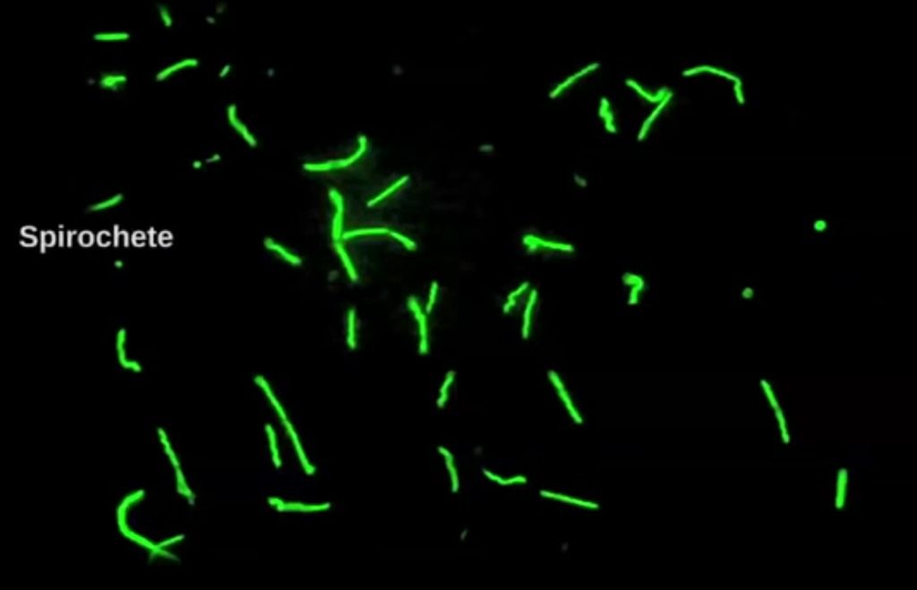
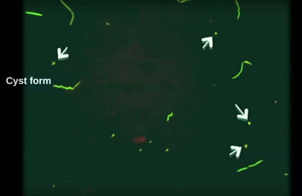
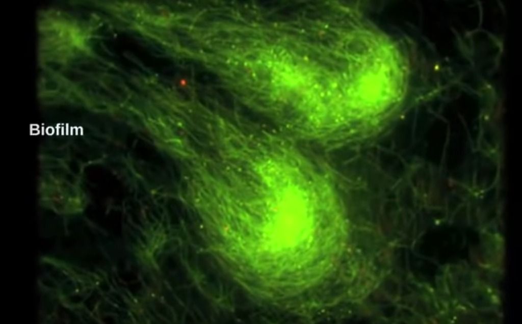
At time 12:40 in this French documentary, they show us video of Borrelia burgdorferi. Shortly after that, Dr. Eva Sapi shows Borrelia, cyst, & biofilm pictures.
posted
My plan apo 100x oel with iris is a Zeiss. Certainly a older model, but great.
You can find great deals on eBay for objectives.
There is a significant difference between the stock objectives and others you can upgrade to.
I know Nikon uses their own threading, and Leica might as well.
Most other companies use RMS threading, so any objective will fit your microscope.
Posts: 163 | From USA | Registered: Oct 2015
| IP: Logged |
TNT
Frequent Contributor (1K+ posts)
Member # 42349
posted
Had a sample under darkfield since Friday evening in which I initially saw plenty of ketes (more than I would like to see and more than I have seen in the last couple wet mounts). Now, almost all the free spirochetes have turned into those "granules," the same objects that Dude demonstrated recently, and the ones I previously mentioned that resemble "Everlasting Gobstoppers," (if you've ever watched the movie Charlie and the Chocolate Factory).
I guess the fewer ABX I've been on recently have not been enough. I did not see any granules when the sample first went under the scope. I see a number of perfectly round gemma cysts as well, but the number of gemma cysts appear to have remained the same from when the sample was fresh.
With all the festivities this weekend, I have not been able to do a time-lapse. But, I am determined to capture the conversion of a kete to one of these granules.
Posts: 1308 | From Eastern USA | Registered: Oct 2013
| IP: Logged |
Lymedin2010
Frequent Contributor (1K+ posts)
Member # 34322
posted
Yea, there sure is a difference with a Zeiss lens in general & the APO's in particular.
Seeing the spiros break up into blebs is awesome, I can never get enough of it. Love it when we capture a cyst transformation.
Below are the pictures of Immuno Stained Borrelia directly from Dr. Sapi's labs. Notice how some of the spiros have BULBOUS (rounded) tips.
Cyst forms:
Biofilm: Posts: 2094 | From NY | Registered: Oct 2011
| IP: Logged |
posted
TNT, I see those frequently. Especially during a regimine change in antibiotics.
Those are great photos to observe, lymedin.
Hey, anyone open to doing some Skype conversation? It'd be cool to have a group Skype chat while prepping and looking at slides and whatnot. Talk about methods, etc. Might be an interesting weekly or monthly thing to do.
Posts: 163 | From USA | Registered: Oct 2015
| IP: Logged |
TNT
Frequent Contributor (1K+ posts)
Member # 42349
posted
quote:Originally posted by Lymedin2010:
Cyst forms:
Lymedin, isn't this picture you posted depicting blebs and not cysts? I'm not arguing, just curious since we've been able to demonstrate the conversion from kete to gemma cyst.....
Posts: 1308 | From Eastern USA | Registered: Oct 2013
| IP: Logged |
Lymedin2010
Frequent Contributor (1K+ posts)
Member # 34322
posted
Dude, you have my email...email me & yea skype would be awesome. It depends on day & energy, as it is unpredictable with me if/when I have enough stamina. But I love the idea.
TNT, I was going to ask what you guys thought, because to me they look like blebs for sure, as they are too small to be cysts from what I know. I could see the very first pic I posted, top right being a cyst with it's size, but even that seems to be a bleb from a thicker spirochete probably.
I posed the same question to the other guys & we will see what the consensus is.
Posts: 2094 | From NY | Registered: Oct 2011
| IP: Logged |
bluelyme
Frequent Contributor (1K+ posts)
Member # 47170
posted
quote:Originally posted by TNT: Had a sample under darkfield since Friday evening in which I initially saw plenty of ketes (more than I would like to see and more than I have seen in the last couple wet mounts). Now, almost all the free spirochetes have turned into those "granules," the same objects that Dude demonstrated recently, and the ones I previously mentioned that resemble "Everlasting Gobstoppers," (if you've ever watched the movie Charlie and the Chocolate Factory).
I guess the fewer ABX I've been on recently have not been enough. I did not see any granules when the sample first went under the scope. I see a number of perfectly round gemma cysts as well, but the number of gemma cysts appear to have remained the same from when the sample was fresh.
With all the festivities this weekend, I have not been able to do a time-lapse. But, I am determined to capture the conversion of a kete to one of these granules.
Is the bvt showing any evidentiary progress? Skype group could be fun ...
-------------------- Blue Posts: 1539 | From southwest | Registered: Dec 2015
| IP: Logged |
TNT
Frequent Contributor (1K+ posts)
Member # 42349
posted
quote:Originally posted by bluelyme:
quote:Originally posted by TNT: Had a sample under darkfield since Friday evening in which I initially saw plenty of ketes (more than I would like to see and more than I have seen in the last couple wet mounts). Now, almost all the free spirochetes have turned into those "granules," the same objects that Dude demonstrated recently, and the ones I previously mentioned that resemble "Everlasting Gobstoppers," (if you've ever watched the movie Charlie and the Chocolate Factory).
I guess the fewer ABX I've been on recently have not been enough. I did not see any granules when the sample first went under the scope. I see a number of perfectly round gemma cysts as well, but the number of gemma cysts appear to have remained the same from when the sample was fresh.
With all the festivities this weekend, I have not been able to do a time-lapse. But, I am determined to capture the conversion of a kete to one of these granules.
Is the bvt showing any evidentiary progress?
Yes, it's definitely helping! But, I seem to be in a bit of a slump at the moment. It might be time to switch things up a bit....
Posts: 1308 | From Eastern USA | Registered: Oct 2013
| IP: Logged |
TNT
Frequent Contributor (1K+ posts)
Member # 42349
posted
Lymedin, your new video today is interesting. The few spirochetes I can see look like SOME of the ones we see in our blood..... straight and a little rigid. I don't notice much undulation.
So, in your opinion, would you consider these true spirochetes?? I'm sure they are, but those in our blood we generally are not as quick to claim they are with absolute certainty.
Sorry for the wording of that last sentence....(under the influence of Lyme brain at the moment).
Lymedin2010
Frequent Contributor (1K+ posts)
Member # 34322
posted
TNT, thanks.
The reason I can say they are spiros, is because when I first observe right after preparation, I do not see them. More likely the RODI shocked them & forced them into cysts. Then after a while the solution balances out in the tick juice + RODI & they start to emerge from the cysts.
More & more come out over time. Sometimes they have a bit of undulation & I have some video & I will post in the future. I already posted a past tick video, where I do a negative of the screen (bluish screen) & you can notice the wave-like spiraling motions in that particular lengthy one.
I do not see these in many ticks that do not have any spiros. So there were many ticks that did not have these rigid spiros. I also do not see any of the very small & very active zig-zagging spiros & I imagine they might be a different species. Those smaller & zaggers were also dividing with a thin spiro strip between two adult segments & that was interesting to see that in the past.
I also see L-shaped forms & sometimes "Tennis Rackets" as well. I am uploading 002 in the next series of tick juice time lapse & you will see a nice sized tennis racket form there (this will take some time to upload).
Posts: 2094 | From NY | Registered: Oct 2011
| IP: Logged |
Lymedin2010
Frequent Contributor (1K+ posts)
Member # 34322
posted
TNT, here is a video with a spirochete with a bit more motility. It comes from the same prep as the previous 2 & there are many other examples too & perhaps even better ones...just too much to go through all to find right now.
On another note has anyone seen Borrelia with a stain yet?
" The cells are gram negative and stain well with Giemsa and Warthin-Starry stains. Unstained cells are not visible by bright-field microscopy but are visible by dark-field or phase-contrast microscopy. " "A review of reports on the genetic and phenotypic characteristics of strains of the spirochete which causes Lyme disease revealed that these organisms are representative of a new species of Borrelia. We propose the name Borrelia burgdorferi for this species. The type strain of B. burgdorferi is strain B31 ( ATCC 35210). "
I have not seen them stain with Giemsa, but of course the blood dries within minutes for a Giemsa stain, which doesn't allow enough time for them to exit the RBCs or WBCs.
On some of my first (failed) stains I used a dyed methanol that did pick them up, although the color was not typical. Those pics are on page 5.
The microbiologyresearch link you posted above says "the page requested is not available."
I'm pretty sure Giemsa does not pick up Bb spirochetes like it does Relapsing Fever spirochetes. Another thing I wouldn't agree with from that quote you posted above is that you cannot see Bb spirochetes with brightfield, because you can, although we all know they're more difficult to see with brightfield than with darkfield or phase contrast.
Posts: 1308 | From Eastern USA | Registered: Oct 2013
| IP: Logged |
TNT
Frequent Contributor (1K+ posts)
Member # 42349
It has a Zeiss 100x Plan objective with an iris that you could sell (since you already have a Zeiss 100x Plan Apo with iris) to put towards getting the couple Phase objectives it appears to be missing.
Posts: 1308 | From Eastern USA | Registered: Oct 2013
| IP: Logged |
Lymedin2010
Frequent Contributor (1K+ posts)
Member # 34322
posted
Thanks TNT.
Look up the text within the quote, all of it & it will be the first link. Somehow it just does not seem to work when I post in here.
I got some good news for us. I have seen a confirmed case of Bartonella in the blood back in 2014, but the owner did not want to share that video. I've been dying to share it with you guys, but could not.
Now someone else has posted a video on what they thought are vacuoles. BUT when I looked at it, it reminded me so much of the Bart video & so I looked at that original bart vidoeo again & sure enough it is EXACTLY like that.
So here are some points about it:
1) Round or oval, & all depending on which side is displayed as it moves one can see a slight pointy tip (almost teardrop).
2) It moves with more intent & in a jerky fashion and does not move in mere Brownian motion.
3)The original video I saw had at least 9-10 of them, of various thickness & size WITHIN the rbc & they can be seen soooooo clearly with darkfield. Also there were at least 2-3 barts outside the rbc & one thick one in particular. Adjacent rbc's ~3-4 had between 1-3 barts in them as well.
When the video starts you will see a 2 arrows & one bart zipping across from left to right. What he is describing as vacuoles is probably rbc's that have been whittled down & probably have thin walls. Those objects within them are bart as well & they have that same jerky motion.
I am happy I can share this video with you. This person is confirmed with HIGH levels of bart ( 1/1280) they told me after I told them that I think it is bart.
A great video explaining Bart & they show you cappillaries & blood & biofilm on collagen from Bart (Time 47.00). https://youtu.be/uYEHsRRxrQw?t=2816
[ 12-30-2016, 06:19 PM: Message edited by: Lymedin2010 ]
Posts: 2094 | From NY | Registered: Oct 2011
| IP: Logged |
TNT
Frequent Contributor (1K+ posts)
Member # 42349
posted
I'm not sure, Lymedin. I have seen that kind of stuff pretty often and there's no way to actually verify it's Bart. I think we would need more verification than just that the person has high titers for Bartonella. What if they also have a high load of something else that resembles Bart that they haven't been tested for, or even some type of apicomplexan they haven't been tested for.
If those vacuoles do indeed contain Bart organisms, you should be able to see those RBC vacuoles with organisms inside on a Giemsa or Wright stain.
I'm not saying it's not Bart, just that I personally would feel more comfortable with more evidence. In the second video, I don't see the objects actually enter the RBCs. If they did, that would be incredible to see, but still would not differentiate between Rickettsias or apicomplexans (or anything else) unless something specific to the entry was seen that would give us a clue.
But, definitely interesting. Too bad I can't read German.
Posts: 1308 | From Eastern USA | Registered: Oct 2013
| IP: Logged |
Lymedin2010
Frequent Contributor (1K+ posts)
Member # 34322
posted
Look at the 2nd video & look at the vacuoles WITHIN the rbc. This is Bart!!!
Posts: 2094 | From NY | Registered: Oct 2011
| IP: Logged |
Lymedin2010
Frequent Contributor (1K+ posts)
Member # 34322
Time 4:35 slightly larger vacuole of bart AND you can see the bart within the vacuole. So vacuole within the rbc & bart with in the vacuole....DEAD GIVEAWAY!!!!! https://youtu.be/Bv7dKi4Wcaw?t=275
The Lyme Disease Network is a non-profit organization funded by individual donations. If you would like to support the Network and the LymeNet system of Web services, please send your donations to:
The
Lyme Disease Network of New Jersey 907 Pebble Creek Court,
Pennington,
NJ08534USA http://www.lymenet.org/


 UBBFriend: Email this page to someone!


![[Smile]](smile.gif)


 Printer-friendly view of this topic