posted
Hi! I have had a few problems getting started with microscopy, and am hoping for some help...
First the 20w bulb doesn't seem to be able to illuminate very well at 40x regaurdless of how I adjust the condenser. I put my 320lumen led flashlight under the condenser and get a much better view - any ideas why this is?
I hooked my point and shoot up to my third viewing port, but it doesn't get a very good image - I have to zoom quite a bit to see anything, and still have some trouble. It also doesn't do well through the eyepiece, but my ipad does.
I took this video with my ipad held up to thee yepiece and an LED flashlight held below the condenser. It is about 8 hours after making the slide:
What do you think? Do these things look like spirochetes? Any suggestions about how to use the camera better?
Posts: 30 | From CA | Registered: Oct 2006
| IP: Logged |
TNT
Frequent Contributor (1K+ posts)
Member # 42349
posted
Hi May, welcome to the thread, and thanks for joining!
What kind of scope do you have? Improvising with the flashlight is a good idea, but you might want to look into Lymedin2010's suggestion about the LED (?) bulb from Home Depot. It seems that most microscope lights are less than adequate. Generally, the more light, the better the view.
It sounds like you may need a relay lens for your third port viewing. That would explain why you have to zoom to get an image. I'm not sure why your camera is not getting a good view through the eyepiece lens, though. That's what I do, and it works very well for me.
Those objects do resemble ketes, but I can't say they are for sure since they are not undulating like true spirochetes. Nor do I see the bulbous tips. Yours look more rigid; like the ones I refer to as L-form, or cell-wall-deficient ketes.
What power objective are you using for that video? I would guess it to be 100x (1000x total magnification).
You could purchase a camera adapter that mounts to your eyepiece and see if that helps your camera get a better capture through your eyepiece lens. I gave links for some adapters further up in the thread. That would help to keep your camera steady which might help it focus better.
I hope those suggestions help.
You're doing a great job. That video was nice and clear, and overall very good.
My next step is to get a true eyepiece camera and start doing time-lapse. Also, I need to perfect my staining abilities. I found out that I bought and have been using the wrong stain. So, hopefully once I get the correct one(s) I will have more to share.
Posts: 1308 | From Eastern USA | Registered: Oct 2013
| IP: Logged |
I wonder what else those things could be? I could only find 2 before the blood sat for a while - then there were many.
I'm using an amscope 490t dark field with oil condenser. I could modify my system to use an Mr16 led, but it isn't drop in compatible. What seems strange is that others seem to be doing fine with the stock 20w halogen.
I'll look into a relay lens.
I'm using 40x objective and 20x eyepiece. The 100x shows nothing. I was thinking about getting a 60x or a 100x oil with iris - I wonder if anyone has any experience with the 100x oil/iris?
Posts: 30 | From CA | Registered: Oct 2006
| IP: Logged |
TNT
Frequent Contributor (1K+ posts)
Member # 42349
posted
quote:Originally posted by TNT: Hey thatdudefromkansas,
Here is a nice Olympus scope with phase contrast. All it needs is a 100x Olympus phase objective.
For those wanting to buy a scope, please be informed about your item before purchasing (especially a used scope). I am still a novice, and I take no responsibility for your purchases, but I think I know a good deal when I see it.
Posts: 1308 | From Eastern USA | Registered: Oct 2013
| IP: Logged |
Lymedin2010
Frequent Contributor (1K+ posts)
Member # 34322
posted
"Woman who has live PARASITES 'wiggling around everywhere' in her blood flies to Germany to undergo controversial treatment for Lyme disease
Tahlia Smith, 21, was diagnosed with Lyme disease in January this year
She has travelled to Germany to undergo controversial treatment
A test showed Tahlia's blood contained live Spirochetes bacteria
The treatment involves blood filtering and heating her body to 42 degrees"
Any ideas whether this is a good deal and would allow me to see spiroochetes?
Posts: 696 | From New York | Registered: Aug 2006
| IP: Logged |
TNT
Frequent Contributor (1K+ posts)
Member # 42349
posted
Hey WakeUp, both of those scopes are good deals. But, I don't know if either one is phase contrast.
I don't have time to answer thoroughly right now, but as I mentioned earlier, phase contrast is wonderful for seeing organisms in live blood.
The upright Olympus CH for $300 looks like it may have a darkfield condenser, but I'm not sure.
The inverted looks like a really good scope. If the whole scope is Olympus like its eyepiece lens, then it is a great deal. It does not appear to be phase contrast. And, inverted scopes take up a lot of space and are not as easily moved.
Posts: 1308 | From Eastern USA | Registered: Oct 2013
| IP: Logged |
TNT
Frequent Contributor (1K+ posts)
Member # 42349
posted
WakeUp, that Olympus CH phase contrast scope I linked to above (on Ebay) is almost exactly like the CH you linked to on CL except the Ebay one is almost fully equipped for phase contrast (it's missing the 100x phase objective, but I linked to one for that as well).
The Ebay scope is not currently equipped for darkfield, but if I'm not mistaken, all you would need to do would be to get a darkfield annulus to put in the turret condenser.
This scope has a "Make an offer" option, so they may let it go for considerably less. You never know.
With Plan phase objectives, and all the annuli in the turret, this scope is a lot of scope for several hundred dollars.
TNT
Frequent Contributor (1K+ posts)
Member # 42349
posted
Here is another brand name phase turret scope, but I hesitate to recommend this one because there is no description and I'm not sure what kind of shape it's in.
There are just too many questions about this scope to recommend it, but it is a nice scope for the price. It's a trinocular that appears to have plan objectives (I can discern the word "Plan" on one of the objectives at least). It does NOT appear that they are phase objectives. Nor can one tell which (if any) annuli are installed. Also, the turret looks a little off-center.
Thanks again--- scopes are a new world for me... As a child I read the book the Microbe Hunters--- and I have always been intrigued since then.
Wish I had been able to film my blood before I went on a new herbal regime that has IMPROVED my arthritis in just a few days (Neem(spiros), Grapefruit Seed Extract(spiros and cysts), Burdock root(biofilms), Sarsaparilla(spiros and biofilm), Mangosteen juice(spiros), Pomegranate Juice(biofilm) and NAC(biofilm), Boluoke(biofilm)...Mango juice(biofilm), Cilantro and Chlorella (detox of heavy metals) and Monolaurin (spiros).
Yes--- I am actually walking up and down the stairs, and I was able to string christmas lights without massive pain...! Wish I could see if my blood looks different!!
Posts: 696 | From New York | Registered: Aug 2006
| IP: Logged |
TNT
Frequent Contributor (1K+ posts)
Member # 42349
posted
WakeUp, that's great that you are more mobile!
Yeah, you need to get a scope soon! You don't really want to totally rely on others to do your trials for you, do you? ha ha
I think that it can appear that the blood looks worse with effective treatment...at least until the load is actually lowered. There are times when my treatment is doing something and I'm feeling slightly better that my blood actually looks more infected. I can think of a couple times that happened at least. Though, overall, it seems to be fairly random.
One thing that has really improved under the scope for me is the prevalence of what many live blood microscopists refer to as candida, or perhaps it's byproduct gas bubbles.
I think there is a possibility it could be something different, but I'm not sure what. But whatever it is, I have almost none of it anymore. Whereas before, I was loaded very heavily with it. My gut is getting better, so perhaps it is candida.
Microscopy was a "new world" for me, too. I am so glad I got into it. And, I am so glad I got a phase contrast scope.
I just got more stain, and my slides are much better now. I'm using another ready-to-use solution that is easier to use. It's a Wright-Giemsa stain, and the differentiation is more like what I had originally expected (the first stain I got was plain Giemsa with less differentiation). The company I got it from actually gives out free samples, so I didn't even have to pay for it!
Posts: 1308 | From Eastern USA | Registered: Oct 2013
| IP: Logged |
posted
Hey guys, a little late to the party but I'm glad I found this thread. Over the past few months I've been using a new scope I have purchased to help try and unravel my families health mysteries.
Hopefully I can get some pics and video up soon 😃
-------------------- Ryan Posts: 10 | From Utah | Registered: Nov 2015
| IP: Logged |
Live blood on Phase from the other night. Not sure if its a spirochete or what. thoughts
-------------------- Ryan Posts: 10 | From Utah | Registered: Nov 2015
| IP: Logged |
TNT
Frequent Contributor (1K+ posts)
Member # 42349
posted
Hi Gerald12,
Welcome to Lymenet and welcome to this thread! It's great you have joined! That's a great view, and a nice capture.
That's an interesting bright phase view. Do you have a color filter? My bright phase lens doesn't give me that amount of color. What kind of scope are you using?
That "string" organism could easily be a spirochete but without obvious bulbous tips it's kind of hard to verify that it is. If it is, you will definitely see more typical-looking ketes eventually.
The other organism is of more interest to me. I can't quite discern from the video, but did it appear to have flagella? It almost appears like there could be flagella at the 5 & 6 o'clock positions (on the organism).
I really like the detail your scope gives the white blood cells.
Keep us updated. We will give you as much help and advice that we can.
Posts: 1308 | From Eastern USA | Registered: Oct 2013
| IP: Logged |
Lymedin2010
Frequent Contributor (1K+ posts)
Member # 34322
posted
There are objects that resemble spirochetes in our blood & the two can thus be confused.
With Oblique Illumination I have been able to see them in normal blood as well. So to differentiate between spiros & those objects, which might be fibrin, we look for the following criteria from my previous post:
"When observing I give higher credibility to when I find the following:
1) BULBOUS OR ROUND TIPS ON BOTH ENDS & MEDIUM LENGTH SPIROCHETES. There are spirochetes that do not have bulbous ends & I have seen video of large quantities of these spirochetes PROPOGATED from the people who have chosen to culture them. So they can exist & I have tons of video on those as well, but I chose to dismiss them. There are also many VERY thin ones which lack the rounded tips, which I also have tons & tons of video but I choose to dismiss.
AND
2) SPIRAL OR UNDULATE WITH SOME AGGRESSION. I prefer to see a bit of life in their mobility. Even Dr. Alan MacDonald makes reference to these relatively docile forms that appear rather lifeless.
AND
3) LARGE QUANTITIES OF ITEM 1 & 2. Large quantities of the 2 items above eliminate the possibility of the chance encounter of any aggregates conforming to this configuration.
If you look at the videos of professionals who release their videos, they fulfill 2-3 out of the aforementioned for a high impact reception.
Furthermore, the morphological transformations of cysts & blebbing is a huge advantage for even more positive identification."
Posts: 2094 | From NY | Registered: Oct 2011
| IP: Logged |
Lymedin2010
Frequent Contributor (1K+ posts)
Member # 34322
posted
A new DNA test that takes blood & various other samples (such as knee fluid).
"The test offers a highly sensitive and reliable molecular diagnosis independent of clinical manifestations and serology test results. Before performing PCR amplification, the borrelial bacteria are concentrated by differential centrifugation from the blood and body fluids for DNA extraction to further increase the sensitivity of the detection method. "
Don't forget they found spiros in semen & vaginal secretions. Has anyone checked their sperm yet (if male)? I did quickly once, but was too symptomatic to do a thorough check.
Lymedin2010
Frequent Contributor (1K+ posts)
Member # 34322
posted
Guys really ANY LABORATORY GRADE MICROSCOPE will allow one to see the spirochetes. The difference will be in how well designed & polished the lenses are and that will translate to the clarity, sharpness, & focus of the image.
It is difficult to know with certainty which one is better than another. An analogy would be the countless lenses for photography cameras & photographers purchasing them only to have mixed reviews on public forums. Some will be satisfied with the image, others will hunt for a sharper lens that may cost more.
Remember what Morten Laane said in his interview in OUS2...one can see the spiros with a 100 year old microscope, but he does not mention anything about how clearly or sharply one can see them.
I would stick with the top guns such as Olympus, Zeiss, Nikon, Reichert...etc. Zeiss is known for its sharp lenses & even on Sony PNS cameras I notice how well they perform. My Zeiss scope lenses have not disappointed as well.
You should go back & check out all the videos that are made of Borrelia & keep in mind the make/model of the scopes & the type of light you want (light, dark, phase, obligue, or DIC)
Posts: 2094 | From NY | Registered: Oct 2011
| IP: Logged |
Live blood on Phase from the other night. Not sure if its a spirochete or what. thoughts
I can see (I could be wrong) what look like at least two spirochetes hanging off your red blood cells--- one is very long with a bulbous end, and the other is short... They seem to be able to hook onto red blood cells very tightly.
Good show!
Posts: 696 | From New York | Registered: Aug 2006
| IP: Logged |
Lymedin2010
Frequent Contributor (1K+ posts)
Member # 34322
posted
Gerald12, that is such a great video & it has enough aggression/movement to be borrelia. You should check out "normal" blood as well to make comparisons. I was not able to see any of these things in normal blood until I used a new oblique illumination microscope, which then brought to light a mesh of fibrin scattered everywhere between the plasma in all people with "normal" & non-LD'd blood.
Some of the fibrin I observed were detached from all the surround fibers & they move & gyrate similarly to spirochetes, albeit with less force/aggression.
I have also now noticed that these fibrin strands sometimes become sticky with WBC's & they drag them along the tail end as footings. For this reason I use the 3 items that I came up with above to increase the chances of a positive ID, which is about all we can do now without DNA/RNA testing. To this date I have never seen in normal blood the 3 items I listed above & have never even seen a medium size one with bulbous tip. I have never seen any bulbous tip ones in normal blood ever, but I have spent considerably more time on blood of those who were bit by ticks & in some of these blood the bulbous tip spirochetes are abundant & easy to identify. I have also seen in normal blood what looks like small dumbbells & I am yet to accept an identification that I am comfortable with.
You can see the bulbous tips on the medusa colony of the My Horrific Blood video. Do you see any bulbous tips spiros in your blood? As I said before, not all spiros will have bulbous tips either & the best way to next come closer to an ID is via time lapse to observe either cysting or blebbing. To me the surrounding activity in your blood, aside from the longer strand, is an added clue to Lyme blood, but it is not definitive.
Posts: 2094 | From NY | Registered: Oct 2011
| IP: Logged |
Lymedin2010
Frequent Contributor (1K+ posts)
Member # 34322
posted
Now take a look at this blood sample that was cultured in medium & allowed to grow for a few days. This person did not see this many spiros in their blood, yet with the right medium the spiros multiplied. Observe how many have bulbous tips & how many do not & how many are in different morphological forms. Even the most steadfast inquisitor will have to succumb to the notion that we are dealing with a living entity AT THE VERY LEAST.
This observation accompanied with your observation of normal blood will provide you with clues as to what you can more safely dismiss vs accept as Borrelia. My series of normal blood vs lyme blood videos is not complete, but should help everyone out. I will also in time give you the recipe how to culture the spiros yourself.
Posts: 2094 | From NY | Registered: Oct 2011
| IP: Logged |
posted
Thank you for all the kind words and compliments. I have ordered a few staining kits so I can hopefully get a little more info on these organisms.
The scope I use actually was less than a 1000.00. But I have spent a lot of time fine tuning my staining techniques .
I have many fascinating images that I have collected since I got my scope a few months ago.
Heres another very interesting organism that has been very hard to identify .
Maybe a basphil or eosinophil that has expanded due to added hydration in the saliva perhaps? Have you tried scraping from under your gums (gingival sulcus)? Just make sure it has some saliva on there. You can check out this guys video, click on his Youtube name & then "videos" to see more.
I have also tried checking tears & sperm & vaginal secretions besides saliva. I have only done this once each & did not find anything, but I did not have enough stamina to thoroughly check as I should have.
Looking forward to more videos.
Posts: 2094 | From NY | Registered: Oct 2011
| IP: Logged |
Lymedin2010
Frequent Contributor (1K+ posts)
Member # 34322
posted
Lyme is hell & I don't always have the stamina or will power for all things Lyme related. I will go back to some of the previous posts & comment, since I missed so much good stuff...sorry.
thatdudefromkansas, you are not getting enough zoom with your recording equipment, can you zoom in more? Despite this it looks like clear & cut spirochetes in this blood sample that fulfill all 3 requirements on the list....awesome work!!!!
Maybe a basphil or eosinophil that has expanded due to added hydration in the saliva perhaps?
Gerald12,
I would also agree that that odd shape is not a parasite but a red blood cell fragment. I honestly feel it could be a RESULT of a parasite (ie babesia-like, or even bart-like organisms) that has ruptured the RBC, leaving a misshapen fragment in it's wake. I have watched RBCs "die" from "normal" circumstances and they either pop and vanish, or burst and ooze out. I think a fragment with a cell wall mostly intact could possibly suggest parasitization.
In the saliva sample the two objects appear to be WBCs. I can see the nuclei (and granules) in both objects.
Those are some great views! Keep up the good work. I would really be interested in seeing some of your stains.
Posts: 1308 | From Eastern USA | Registered: Oct 2013
| IP: Logged |
Lymedin2010
Frequent Contributor (1K+ posts)
Member # 34322
posted
I don't think they are parasitized either, as I also see them in normal blood. You hit yourself & bang yourself & apply pressure in various parts of your body. Extruding blood from the fingers is another form of trauma & in particular smearing across a glass slide. This will deform a few RBC's.
TNT, your ruptured RBC's are a different thing & I think ARE due to a parasite. I was going to get to a response when going through it. I do not see that type of ruptured cells in mine or in normal blood & malaria is known to rupture RBC's. So I wonder if it is babesia? Look for ruptured RBC in malaria & you see a similar bursting of RBC's, almost identical to your images.
Posts: 2094 | From NY | Registered: Oct 2011
| IP: Logged |
Lymedin2010
Frequent Contributor (1K+ posts)
Member # 34322
TNT
Frequent Contributor (1K+ posts)
Member # 42349
posted
quote:Originally posted by Lymedin2010: Look for ruptured RBC in malaria & you see a similar bursting of RBC's, almost identical to your images.
Thanks. That's why I shared those photos.
The shape, position, and proximity of the ruptured RBCs and the shape, size, and proximity of the other objects seriously suggest apicomplexans bursting forth from RBCs.
Posts: 1308 | From Eastern USA | Registered: Oct 2013
| IP: Logged |
Lymedin2010
Frequent Contributor (1K+ posts)
Member # 34322
posted
Have you seen any cruciforms (X or cross forms) or the ring forms?
Posts: 2094 | From NY | Registered: Oct 2011
| IP: Logged |
TNT
Frequent Contributor (1K+ posts)
Member # 42349
posted
Originally posted by Lymedin2010:
quote:Have you seen any cruciforms (X or cross forms) or the ring forms?
Not sure. Earlier on I thought I had, but if I remember correctly, it was the red blood cells that had funny folds and creases-and even holes-and not objects inside of them. I'll have to find the folders that have those pics and videos of that.
[ 11-19-2015, 05:35 PM: Message edited by: TNT ]
Posts: 1308 | From Eastern USA | Registered: Oct 2013
| IP: Logged |
Lymedin2010
Frequent Contributor (1K+ posts)
Member # 34322
posted
I read somewhere that typically only <.01 of RBC's would be affected by babs & for Bart something on the order of <.001. It is nice to see you guys expanding into uncharted territory & against such odds.
thatdudefromkansas has almost as many spiros as I had & it is interesting how he was diagnosed with MS (as well as his friend). What are your total MS symptoms & do the symptoms overlap into LD symptoms partly?
Just an FYI, out of all the countless stuff I have taken only Cowden Protocol along with pulsed ABX has managed to decrease the amount of spiros directly in my blood. I had Doxy dependency & nothing was able to take it away other than just Cowden alone.
At one point Doxy started to relieve many of my symptoms & prevented what appeared like a sure stroke for me & it was a great drug when I needed it. But over time it has done nothing more than continue my LD progression & I deeply regret staying on it. I knew about Dr. Eva Sapi's study on doxy producing 300% more cysts, but I thought it might have been affecting myco & the reason it was helping me so much initially.
After the Cowden/abx pulse, I now have trouble finding spiros in my blood, albeit it does not change the severity & my multitude of symptoms.
Also it is interesting how I observed spiros in my wife's blood in 2011 & predicted she would show signs of LD. I hear her joints crack & snap, every night, yet her symptoms remain minimal. I think besides all that we know about LD (co-infections, supplements, probiotics...etc) the ability for your body to clear toxins is a major factor in producing pain symptoms. When I get exposed to various substances that I was normally fine with before chronic LD, now I develop additional LD symptoms.
Posts: 2094 | From NY | Registered: Oct 2011
| IP: Logged |
Lymedin2010
Frequent Contributor (1K+ posts)
Member # 34322
posted
Peter Kemp's latest Borrelia video & it looks like a pretty damn long one there. He may just hold the record?
That video is interesting because those are some of the laziest ketes I have seen. There's not much undulation.
Posts: 1308 | From Eastern USA | Registered: Oct 2013
| IP: Logged |
TNT
Frequent Contributor (1K+ posts)
Member # 42349
posted
In this video Peter Kemp mentions the presence of l-form spirochetes:
I wonder what he is referring to. Is it what's pictured from 2:02-2:06 (the bright round objects hanging on the outside of the bright RBC)?
Posts: 1308 | From Eastern USA | Registered: Oct 2013
| IP: Logged |
Lymedin2010
Frequent Contributor (1K+ posts)
Member # 34322
posted
I have seen lazier & they appear to be actual ketes with sharp bends at various points & not fibrin. I saw the most of these very stagnant & lifeless ones when I took a combo of doxy & Penicillin VK together.
I had asked this question as well earlier on as I always though that it referred to the fission of a spirochete in the middle, which then forms into what sometimes looks like an L or greater or lesser than sign. But it turns out he is referring to all of the strings, which are atypical (non-textbook spiraling) & L forms. So every string we see in that video that is not a cyst or bleb/spore.
Posts: 2094 | From NY | Registered: Oct 2011
| IP: Logged |
Lymedin2010
Frequent Contributor (1K+ posts)
Member # 34322
posted
Just what we need, more complications with EBV in the mix...over 60 varieties of EBV.
TNT
Frequent Contributor (1K+ posts)
Member # 42349
posted
quote:Originally posted by Lymedin2010: ... it turns out he is referring to all of the strings, which are atypical (non-textbook spiraling) & L forms. So every string we see in that video that is not a cyst or bleb/spore.
I'm still not sure what you mean or are referring to. L-forms are a different morphology; a non-spiraling spirochete would be just that-a dead kete. That would not be a different morphology. Have you been able to ask Peter what he is referring to?
I guess I could post a question under his video.
Posts: 1308 | From Eastern USA | Registered: Oct 2013
| IP: Logged |
Lymedin2010
Frequent Contributor (1K+ posts)
Member # 34322
posted
I have asked the question to friends of his in the same circle in the past & they say that they refer to all the strings that we see in our blood. I too was taken back by the response, as I thought they were the ones with bends within the bodies.
He won't answer the Youtube video. Instead go to FaceBook & search for "Peter Kemp" check his page & he usually likes to post about LD & UK politics and ask him there.
Posts: 2094 | From NY | Registered: Oct 2011
| IP: Logged |
Lymedin2010
Frequent Contributor (1K+ posts)
Member # 34322
posted
Check out his other video, with clues as to the "motile coccoid" stuck in the middle of the L-form spirochete (the same strings we see in our blood).
TNT
Frequent Contributor (1K+ posts)
Member # 42349
posted
L-form, or cell-wall-deficient bacteria are just that, bacteria without a distinct cell wall -but, rather, an outer membrane. So, theoretically, L-form could be any possible shape. But I think they would probably tend to resemble the shape of the cell-wall form.
That's why I am inclined to think that the faint, thicker, spirochete-looking objects that appear to undulate (or at least wave around) are l-form ketes. Especially the ones that have darker tips.
Posts: 1308 | From Eastern USA | Registered: Oct 2013
| IP: Logged |
TNT
Frequent Contributor (1K+ posts)
Member # 42349
posted
quote:Originally posted by Lymedin2010: Check out his other video, with clues as to the "motile coccoid" stuck in the middle of the L-form spirochete (the same strings we see in our blood).
I am not sure I agree with the description about the objects or what is happening in the video.
The "coccoid" appears to be a lyzosome attacking a typical spirochete. It appears to move up and down ON the OUTSIDE of the kete.
I cannot see anything that differentiates that "l-form kete" from a typical spirochete. There is not enough magnification or resolution to discern between a cell wall and a cell membrane.
Posts: 1308 | From Eastern USA | Registered: Oct 2013
| IP: Logged |
TNT
Frequent Contributor (1K+ posts)
Member # 42349
posted
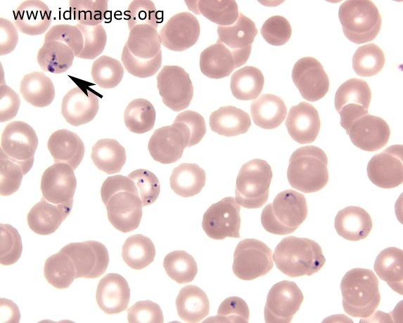
Here are some examples of what I earlier thought was babesia-like organisms, and maltese crosses:
There were a number of stock pictures I could choose from, but here is just one example I thought my pictures resembled:
Posts: 1308 | From Eastern USA | Registered: Oct 2013
| IP: Logged |
Lymedin2010
Frequent Contributor (1K+ posts)
Member # 34322
posted
The coccoid he is referring to is a bleb (aka spore, aka granular form). It is the dots in the string of pearls, which break up at the end & give just the coccoid.
It is a way that Borrelia breaks up into the smallest unit containing just DNA/RNA in one vesicle & designed to spread effectively. So one borrelia undergoes SoP formation & forms 10-20+ coccoids, which can then EACH develop into new spiros & repeat the cycle.
Peter knows this now, since after my video he has done time lapse & see long strings at the beginning & then hours later only COCCOIDS, which is the same name for blebs or spores or granular forms.
Posts: 2094 | From NY | Registered: Oct 2011
| IP: Logged |
Lymedin2010
Frequent Contributor (1K+ posts)
Member # 34322
posted
Those are unstained pics I believe. I see those types in normal blood too & are halos & have to do with the way the RBC is biconcave & bends lights between the sloped points at the center.
The 1st pic is more convincing on your behalf & could be though.
You should however look at normal blood at some point & it will explain a lot.
Posts: 2094 | From NY | Registered: Oct 2011
| IP: Logged |
The Lyme Disease Network is a non-profit organization funded by individual donations. If you would like to support the Network and the LymeNet system of Web services, please send your donations to:
The
Lyme Disease Network of New Jersey 907 Pebble Creek Court,
Pennington,
NJ08534USA http://www.lymenet.org/


 UBBFriend: Email this page to someone!


![[Wink]](wink.gif) , or monsters like Lyme?
, or monsters like Lyme? 


 Printer-friendly view of this topic