TNT
Frequent Contributor (1K+ posts)
Member # 42349
posted
Lymedin,
I guess if he is trying to show that that coccoid is a bleb, then time-lapse would be best. I guess this is the kind of thing we are up against with proving it. Because, honestly, it looks to me like one of the many lyzosomes are attacking the kete.
I think it was S13 that showed time-lapse of SOP to bleb formation?? I remember your one video showed that too.
The thing I find hard to believe is that he is referring to the kete as an "l-form spirochaete." I don't think he can prove that by the video.
Posts: 1308 | From Eastern USA | Registered: Oct 2013
| IP: Logged |
TNT
Frequent Contributor (1K+ posts)
Member # 42349
posted
Here's an early video (just uploaded in response to our conversation on the last page) of my blood showing parasitized RBCs, possible "maltese cross," and ketes with brightfield at 1000x:
TNT
Frequent Contributor (1K+ posts)
Member # 42349
posted
The other thing that makes me speculate that these cells are parasitized by apicomplexans is that babesia and malaria tend to infect the reticulocytes (immature RBCs).
The affected cells appear to be reticulocytes.
Posts: 1308 | From Eastern USA | Registered: Oct 2013
| IP: Logged |
Lymedin2010
Frequent Contributor (1K+ posts)
Member # 34322
posted
You captured some very clear spirochetes in that video, great work. Do you see them all over & scattered in your blood, or are they hard to find?
It could very well be, but the best way to capture them is via staining. This is no easy feat as you may have to do a whole lot of staining to increase your chances of capture.
I have seen normal blood & the RBC's look as if it can be loaded with parasites because of the effect I described previously. I thought they were parasites in my blood too when I first got into microscopy, but after some time with normal blood, I have to easily dismiss them.
Just look at this vid of normal blood & focus on the RBC, which might one mistake as parasitized? And by no means is this the best video of this illusion & I have recorded better in the past.
The other way to capture them is via darkfield & I have seen some very convincing video of parasitized RBC's under darkfield.
For sure you have parasites in your RBC's, as your ruptured RBC's & the pictures you took of the occlusions exiting rbc suggest & that was beautiful work which I have not seen from any other LD person doing microscopy. Congrats!
Consider the possibility of culturing them in chocolate agar, as I had mentioned in a previous post. This will allow you to place a larger amount of blood & again increase chance of seeding the medium.
Posts: 2094 | From NY | Registered: Oct 2011
| IP: Logged |
Lymedin2010
Frequent Contributor (1K+ posts)
Member # 34322
posted
I don't know if I did a good enough job on the My Horrific blood video? One of the major points was to show SoP formation & how it breaks up & scatters in the blood.
So if you wait a few hours you will see the spiros come out more & more into the blood plasma. But if you wait even more hours the plentiful collection of spiros in the plasma just vanish & what is left is a lot of debri & many coccoids/blebs/spores/granular forms (all those words are interchangeable for the same Borrelia structure....the dot).
P. Kemp has done maybe about 4 months ago a before & after video of the same thing that I have. First he shows the spiros in plasma & then a few hours later all one sees is the blebs scattered in the plasma. I suppose he used coccoid as a more general term to be safe & can apply to any object. I know from watching his videos that his culturing produces A LOT of blebs/coccoids in the plasma.
Posts: 2094 | From NY | Registered: Oct 2011
| IP: Logged |
Lymedin2010
Frequent Contributor (1K+ posts)
Member # 34322
posted
It looks like slowly the world is learning...this is recently & directly from Dr. Alan MacDonald...
" Macro Particulate forms of Amyloid (coating living Borrelia bio-films) exist in Circulating Blood from dementia patient. Link: https://www.facebook.com/whyamistillsick
Amyloid ""Globs" up to 100 or more microns in size were captured in photographs by me after I completed a Congo Red stain [ for amyloid } on a peripheral blood smear from a 62 year old woman with dementia, and with previously documented circulating large size Borrelia bio-films in her blood smear. No one, on planet earth ,has ever stained a blood smear with Congo Red Stain to search for amyloid.
No one on earth has ever performed FISH method DNA hybridization for borrelia Miyamotoi DNA. No one on earth has ever photo documented the existence of living Borrelia biofilm communities in the circulating blood.
These discoveries occurred on October 7,2015. One of the Images of a huge Borrelia biofilm Coated with Amyloid is below. Other images are posted through the link above.
This is a Major new insight into the biology of Infectious Alzheimer's Disease.
Respectfully, Alan B. MacDonald MD, FCAP October 9,2015"
TNT
Frequent Contributor (1K+ posts)
Member # 42349
posted
quote:Originally posted by Lymedin2010: You captured some very clear spirochetes in that video, great work. Do you see them all over & scattered in your blood, or are they hard to find?
That last video was from the second slide I ever did, so it was very early on. At that point I was seeing COLONIES of spirochetes, as I pointed out in previous videos. It goes without saying that this was not cultured blood....so yeah, MANY, MANY ketes! Now I do not see that many (most of the time).
Here is another early video of the same area that shows some of these same elements, but gives a better idea of just how many ketes there were:
I apologize for the shaking and the loud noise in the background. Someone was vacuuming as I shot the video. Plus, the slide/coverslip was under the influence of some type of tension that kept pushing the blood around every time I tried to focus. I think I had too much blood for the surface area of the coverslip that allowed it to "float."
Posts: 1308 | From Eastern USA | Registered: Oct 2013
| IP: Logged |
TNT
Frequent Contributor (1K+ posts)
Member # 42349
posted
Alan is doing AWESOME work! I hope it continues. Does anyone know if it is true about his microscope being taken from him? Can someone provide written proof about it?
Posts: 1308 | From Eastern USA | Registered: Oct 2013
| IP: Logged |
TNT
Frequent Contributor (1K+ posts)
Member # 42349
posted
quote:Originally posted by Lymedin2010: I have seen normal blood & the RBC's look as if it can be loaded with parasites because of the effect I described previously. I thought they were parasites in my blood too when I first got into microscopy, but after some time with normal blood, I have to easily dismiss them.
Just look at this vid of normal blood & focus on the RBC, which might one mistake as parasitized? And by no means is this the best video of this illusion & I have recorded better in the past.
I could be mistaken, but the RBCs in your video don't quite look like what I pointed out. But, please don't go searching in your archives for a better resemblance because I realize that what I am showing in my video may completely be an illusion.
I was simply illustrating what COULD look like "maltese crosses," and pointing out a couple things that could lead a person to consider the possibility of apicomplexan parasites. I don't plan to "go to the bank" with it, because it's not hard evidence.
The other thing I consider is how much Zithromax and Artemisinin has helped me in the past number of months.
So, I am just trying to get the big picture on things, and TOTALLY appreciate all of your help & input (Lymedin, and each one of you).
Posts: 1308 | From Eastern USA | Registered: Oct 2013
| IP: Logged |
quote:Originally posted by TNT: Here's an early video (just uploaded in response to our conversation on the last page) of my blood showing parasitized RBCs, possible "maltese cross," and ketes with brightfield at 1000x:
SIGH---- TNT-- OMG your red blood cells look like crap. I'm sooooo sorry for us all because "our life is in the blood.." and these parasites are literally sucking the life out of us.
It takes red blood cells 3 months to die-- so treatment must be a minimum of 3 months.. just to eradicate an infection in red blood cells, not to mention other cells.
SIGH-----
Those "S" like spirochetes inside your cells are SSSatan's personal taunt...LOL
Posts: 696 | From New York | Registered: Aug 2006
| IP: Logged |
quote:Originally posted by TNT: Alan is doing AWESOME work! I hope it continues. Does anyone know if it is true about his microscope being taken from him? Can someone provide written proof about it?
Elena mentioned that the LDA had withdrawn their loan of the cytoviva Microscope, but I will see if I can verify it-- and the reasons for it. They probably came under "pressure" to take the scope away.
The last thing we patients need is MORE tick studies and tick surveys. Tick studies are great---- BUT--- they distract precious resources from research for a CURE, and I, for one do not want to finance more tick studies and surveys when almost no work or money is going into finding a CURE..
I want to finance researchers who want to CURE--- to eradicate Borrelia biofilm Borrelia spirochetes and Borrelia cysts from our bodies. Denialists harm us because they divert attention from these lines of research onto research that will never find us a cure....
If the microscope loan was called in, then a group of concerned Lyme patients need to buy the Cytoviva to rent to MacDonald for $1 a year. This way, actual patients who want a cure will have control over the equipment-- and not an organization like the LDA which is subject to pressure and compromise because of its funding requirements.
Our only hope rests with grass roots funding of dedicated scientists like Sapi, Miklossy, MacDonald and that pathologist is Norway... (forgot his name-- brain freeze)
One key to which scientists are dedicated is: they always get persecuted when they come close to the truth!!
Posts: 696 | From New York | Registered: Aug 2006
| IP: Logged |
TNT
Frequent Contributor (1K+ posts)
Member # 42349
posted
quote:Originally posted by WakeUp: Elena mentioned that the LDA had withdrawn their loan of the cytoviva Microscope, but I will see if I can verify it-- and the reasons for it...
....If the microscope loan was called in, then a group of concerned Lyme patients need to buy the Cytoviva to rent to MacDonald for $1 a year. This way, actual patients who want a cure will have control over the equipment-- and not an organization like the LDA which is subject to pressure and compromise because of its funding requirements.
Thanks WakeUp, I appreciate you verifying that for us.
If this is true, perhaps someone should put a bug in John Caudwell's ear that MacDonald could use another microscope. The best scope made would cost pocket change to him.
It's too bad MacDonald couldn't get his hands on one of the 2 surviving original Rife microscopes. Then again, there might be a learning curve to using one. They were the most powerful microscopes in the world at that time, and maybe still are.
Posts: 1308 | From Eastern USA | Registered: Oct 2013
| IP: Logged |
TNT
Frequent Contributor (1K+ posts)
Member # 42349
posted
quote:Originally posted by Lymedin2010: I don't know if I did a good enough job on the My Horrific blood video? One of the major points was to show SoP formation & how it breaks up & scatters in the blood.
So if you wait a few hours you will see the spiros come out more & more into the blood plasma. But if you wait even more hours the plentiful collection of spiros in the plasma just vanish & what is left is a lot of debri & many coccoids/blebs/spores/granular forms (all those words are interchangeable for the same Borrelia structure....the dot).
I thought you did a great job with that video. I only wish it could have been filmed at 1000x instead of 400x.
I don't have an eyepiece camera yet, but I recently watched a sample over a week's time. At first there were numerous ketes. But as the week progressed, fewer and fewer ketes were visible until there were NONE. The inverse was true concerning cysts. I saw more and MORE and MORE irregularly-rounded, granular type objects approximately .5-2.5 um in diameter. I had seen a few of these objects in blood before, but was not sure what they were until this sample.
Now I know.
Posts: 1308 | From Eastern USA | Registered: Oct 2013
| IP: Logged |
Lymedin2010
Frequent Contributor (1K+ posts)
Member # 34322
posted
Interesting that your last video is not that sharp & very shaky, yet it is very easy to identify as spirochetes, great job on that one.
You guys know that I was thanked by many for my video & that we have influenced other researches into looking at the blood & spirochetes more seriously now. Hopefully the momentum will keep on going & we will all learn new things as time progresses. What we really need to do is to get the LLMD's involved.
1000x produces a VERY narrow field of view & when focusing on only 1 spiro it becomes difficult to keep focus since it easily moves & looses focus. I am doing more 40x with cam zoom now. Good to see that both you & P. Kemp have noticed a very similar pattern to what I have noticed. I think this info is BIG news & insight for all of us. We have shown the researches what actually happens in our blood & how 1x borrelia can easily lead to 20x or 30x Borrelia entities.
TNT, I again asked for verification on L-form of borrelia & this is what was said:
"...our definition of L form is: typically a long thin string like body with a length between 10 and 20 microns and diameter about 0.3 to 0.5 microns.
That’s typical, however we also frequently see and include. Very long forms that can be 100 microns or more, and very short forms that can be 3-10 microns in length. Frequently the short ones have definite round bodies on the ends and look like dumbells. Some of these can be seen in the video you include."
Posts: 2094 | From NY | Registered: Oct 2011
| IP: Logged |
Lymedin2010
Frequent Contributor (1K+ posts)
Member # 34322
posted
Also I have thought about this many nights, are the CDC really this stupid? Could they really not understand that different morphologies exist. Is it too difficult of a concept to accept that in a BLOOD BORNE DISEASE that the pathogen can be in very large quantities in the blood?
In the beginning I thought I had discovered a species of Borrelia that may not be known as pathogenic to humans or maybe a new one all together. I was able to figure out spores, dotted spirochetes & their breakup all within 4-5 months. I knew nothing of Borrelia burgdorferi as it pertained to what I was seeing. The only sure guarantee that I witnessed was that these things were not in my blood during early infection, when I only had a few symptoms & I was guessing I had Lyme. Then as I developed more & more symptoms these things coincided in abundance.
Could they really be this moronic or do they know this already? Perhaps they are already aware & realize that if we cannot kill it with abx early on, then no matter how much antibiotics or combos of abx we administer subsequently the organism will persists.
If people go on life thinking they have fibro or ME/CFS, then why not leave it at that & not scare the public. I bet when they went out testing, they discovered that many people have the infection, yet they show no symptoms or very little symptoms. Collectively the cost of handling this would be astronomical. So if the infection exists & it does not necessarily translate to disease, then how does one prove that they have full blown LD? This becomes a public fiasco.
Big pharma is making money off us Lymies, but the insurance companies are loosing a tremendous amount. Do you think that the gov will favor Big Pharma over insurance companies? Or are they trying to reach a happy middle ground where the gov does not loose much, big Pharma gets its share by way of drugs for labeled diseases, & insurance companies loose minimally.
Posts: 2094 | From NY | Registered: Oct 2011
| IP: Logged |
TNT
Frequent Contributor (1K+ posts)
Member # 42349
posted
quote:Originally posted by Lymedin2010: TNT, I again asked for verification on L-form of borrelia & this is what was said:
"...our definition of L form is: typically a long thin string like body with a length between 10 and 20 microns and diameter about 0.3 to 0.5 microns.
That’s typical, however we also frequently see and include. Very long forms that can be 100 microns or more, and very short forms that can be 3-10 microns in length. Frequently the short ones have definite round bodies on the ends and look like dumbells. Some of these can be seen in the video you include."
So, they are saying the very long strings we have in our blood and which you can see in many of our videos (like in my flagellate video) are the L-form (cell-wall-deficient) spirochetes?
I wish they would tell us how they know that. What are they seeing about those long ketes that define them as cell-wall-deficient? Because they appear no different than the shorter ones. They do not appear to have the qualities consistent with cell-wall-deficient forms from what I can see.
I also wish I could get hold of a copy of Dr. Lida Mattman's book on cell wall deficient forms! If only it wasn't so expensive.
Posts: 1308 | From Eastern USA | Registered: Oct 2013
| IP: Logged |
Lymedin2010
Frequent Contributor (1K+ posts)
Member # 34322
posted
Small dumbbells, medium, long, & extra long ones are all L-form. In fact everything that we see that is not granular (dot) or cyst is L-form in our blood thus far. I don't know of anyone who has captured a typical spirochete in their blood yet. They are also called atypical & they do not conform to the text-book definition of a Borrelia spirochete. I know someone who has cultured them & had a few typical ones in full spiraling Borrelia form. Some L-forms can never revert back, while some can.
Perhaps because it is cell wall-deficient it does not spiral with aggression. Other organisms also have L-forms such as Bacillus subtilis & mycoplasma.
Posts: 2094 | From NY | Registered: Oct 2011
| IP: Logged |
TNT
Frequent Contributor (1K+ posts)
Member # 42349
posted
Here is a good slide show in the public domain that explains l-forms well:
Posts: 1308 | From Eastern USA | Registered: Oct 2013
| IP: Logged |
TNT
Frequent Contributor (1K+ posts)
Member # 42349
posted
Here is a better picture and description of the first pic (on last post):
Phase contrast image of L-form cells from Bacillus subtilis showing a range of sizes. Scale bar is 5 micrometers:
A couple more pics:
Transmission electron micrograph of L-form Bacillus subtilis, showing a range of sizes. Scale bar is 10 micrometers:
Transmission electron micrograph of L-form Bacillus subtilis. The cells lack the electron-dense cell wall of normal bacteria. Scale bar is 500 nanometers:
Does any of this information give us an idea of what L-form spirochetes look like under the scope? I'm not sure. I will continue to search it out.Posts: 1308 | From Eastern USA | Registered: Oct 2013
| IP: Logged |
Lymedin2010
Frequent Contributor (1K+ posts)
Member # 34322
posted
Good links, but I am sure there is more to learn from Borrelia L-forms.
I think it forms when exposed to harsh conditions, such as the abx we take & it does so easily with Penicillin exposure as well. I cannot really prove they are CWD, as we would need an electron microscope. BUT, it looks like we have them in abundance & the abx we take are not very good at killing them, which is a clue in itself as to CWD. Also their motility is impeded by the loss or degradation of the flagella. So the clues are there, but I have to go by what the say since I do not have the insight to prove it any further.
"Antibody status is immaterial in DNA detection [BANR] procedures . Borrelia spirochetes which bind to Species specific DNA probes, directly visualize under the microscope, the borrelia spirochetes in the body of the [BANR] patient."
"The equipment needed for each physician performing such testing after graduation from the tutorial is inexpensive and straightforward. It consists of a microscope. "
"Point of Care Methologies are now routinely available in many physician offices, and this campaign will expand the capabilities of individual physicians to, with a microscope in their private offices , to evaluate their own patients for DNA evidence of borrelia infections. This harkens back to the early 20th century, when every physician was trained in and equipped with a microscope and with the necessary reagents for staining bacteria ( Gram Staining) , to assist in diagnosis of infections their own patients."
Posts: 2094 | From NY | Registered: Oct 2011
| IP: Logged |
Lymedin2010
Frequent Contributor (1K+ posts)
Member # 34322
posted
It looks like they did confiscate the Cytoviva loaner microscope from Dr. Alan MacDonald.
"I realise now I was mistaken. It turns out that LDA has never given any funding to Dr. Alan Macdonald.
The Cytoviva microscope was effectively a loan, not a gift. You recalled it in a great hurry from Dr. MacDonald just as he began to make important strides in detecting Borrelia biofilms with it.Biofilms are, by definition, proof of persistent infection.
Mrs. Smith, who pressured you to demand that Dr. MacDonald return that microscope so hastily? And why did you concede to that pressure?
These matters, and all of the issues raised in my letter, are relevant to the Lyme community throughout the world, and deserve an answer."
Posts: 2094 | From NY | Registered: Oct 2011
| IP: Logged |
TNT
Frequent Contributor (1K+ posts)
Member # 42349
posted
quote:Originally posted by Lymedin2010: Good links, but I am sure there is more to learn from Borrelia L-forms.
I think it forms when exposed to harsh conditions, such as the abx we take & it does so easily with Penicillin exposure as well. I cannot really prove they are CWD, as we would need an electron microscope. BUT, it looks like we have them in abundance & the abx we take are not very good at killing them, which is a clue in itself as to CWD. Also their motility is impeded by the loss or degradation of the flagella. So the clues are there, but I have to go by what the say since I do not have the insight to prove it any further.
I find it interesting that they describe the change from CWD form to active spirochete in these terms: "the cytomorphic change from atypical nonmotile, "rigid" form to motile helical form..." - So, the l-form is "nonmotile" and "rigid."
In reference to the four biological variations of their Bb cultures they said (concerning #3), "the colonies may grow to resemble mycoplasma;" (I think they refer to the "fried-egg" look of the colonies in the agar dish).
They also mention "membrane blebs."
Also, "[previous] studies showed the presence of large bubbles (1.0-1.6 micrometers) and encysted forms of leptospirae and borreliae."
And, the mention of "elongated forms."
I really don't think that an electron microscope would be needed to see CWD borrelia. I reference the pics I posted of the CWD Bacillus as an illustration. The usual form of Bacillus is roughly the same size as it's CWD form. I imagine the same is true for spirochetes. Greater magnification would therefore not be needed.
In response to some of the great material in that PDF, it might be helpful for me to post the slides defining & showing the two basic forms of CWD bacteria, protoplasts and spheroplasts:
Posts: 1308 | From Eastern USA | Registered: Oct 2013
| IP: Logged |
TNT
Frequent Contributor (1K+ posts)
Member # 42349
posted
Pictures of Spheroplasts:
Bacteria that attempt to divide in the presence of penicillin fail to do so and end up shedding their cell walls in the process.
NASA / Univ. of Alabama A fluorescent stain renders adds a green tinge to the corkscrew-shaped Spirochaeta americana from California’s Mono Lake. Green spots are spheroplasts. Reddish areas are dead cells.
Posts: 1308 | From Eastern USA | Registered: Oct 2013
| IP: Logged |
But these appear to be good examples of what we have just been discussing... Cell-wall-deficient forms.
Those little round translucent "bubbles" appear to be spheroplasts of some type of organism. Could be from gram-negative bacteria of some type, because gram-negative bacteria (as well as other things) produce spheroplasts.
Borrelia spheroplasts? Who knows? Could be! But, they could be some other type of bacteria just as easily.
Great job with both videos! I like them.
Posts: 1308 | From Eastern USA | Registered: Oct 2013
| IP: Logged |
As far as the L-form . Im working with a local college in a infectious disease study of these monsters . It is amazing what they can do and how fast they can change. You throw vancomycin or any other cell wall attacking antibiotic at them on the slide, they imediatly drop their cell wall and hop into the nearest RBC to protect themselves .
its nuts !!!
-------------------- Ryan Posts: 10 | From Utah | Registered: Nov 2015
| IP: Logged |
Lymedin2010
Frequent Contributor (1K+ posts)
Member # 34322
posted
Since the L-form lose the cell wall & flagella it then becomes rigid & what they call nonmotile, although they sure as heck still have motility although it is very passive. I believe they need this motility in order to make it in & out of our cells (RBC's....etc). Imagine how awkward it would be for them to burrow in & out of the RBC's lengthwise if they were "NONMOTILE."
Since the information has been fed to us & we know what to look for in the string/spiral form of the spirochete, then no we don't need an electron microscope. But if we really wanted to prove that there is actually no cell wall or flagella, then yes we do need it. What if there was still a cell wall & flagella but that they were only perforated & damaged & thus producing this non-motility.
In fact your spheroplast picture is an electron micro pic & such a pic as I had posted in a previous time would provide very clear detailing....
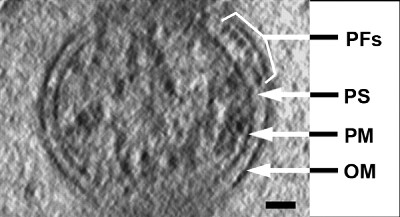
Electron Cryotomography of Borrelia b.
"Figure 1 of Charon et al. Bar, 50 nm. PFs, periplasmic flagella; PS, periplasmic space; PM, plasma (or cytoplasmic) membrane; OM, outer membrane."
I cannot tell whether the blebs are CWD or not & whether they are sphero or proto. They do come in an assortment of shapes & sizes & at times they hold on to part of the Borrelia body.
Posts: 2094 | From NY | Registered: Oct 2011
| IP: Logged |
Lymedin2010
Frequent Contributor (1K+ posts)
Member # 34322
posted
Gerald, such impeccable timing on subject matter for this video.
I really love this video since it shows the physical spirals of a spirochete & this may be the in between a typical & atypical spiro, whereby it is losing the cell wall & flagella. It also has what is reminiscent of the text-book spiral movement, albeit still not as aggressive as the dogmatic naysayers would like to see it in.
Beautiful!!!!
Posts: 2094 | From NY | Registered: Oct 2011
| IP: Logged |
Lymedin2010
Frequent Contributor (1K+ posts)
Member # 34322
posted
Yes, I am very well aware of how RESPONSIVE these things are. Within minutes of taking a particular herb or abx I can feel mass migrations, tremors, vibrations, pins & needles as they scatter & hit nerves, muscle twitches & movement as they form cysts.
When you keep in mind a systemic infection that involves the total blood volume & its incredible rapid response to environmental factors, then one can explain many Lyme symptoms via this model of infection & occupancy.
I liken the spirochetes in the blood as a person being dragged in a river or rapids. The person has some movement, but not total control. Both Borrelia & the person moves intentionally & passively with sticky bolbous tips or sticky hands & feet that grab onto or stick to what is need (rocks vs cell walls).
Also it would be huge for researches to apply a microscope to our capillaries to follow the spiros as abx are administered. I also see a synthetic capillary with a semi-permeable membrane directly to our blood supply via IV & monitored by a microscope. Since our blood can live outside of our bodies for hours & days these blood experiments can be conducted in vitro as well & will be the hallmark of future studies. They will provide insight as to what is actually going on in real time & with each abx application.
Posts: 2094 | From NY | Registered: Oct 2011
| IP: Logged |
TNT
Frequent Contributor (1K+ posts)
Member # 42349
Very interesting!
Posts: 1308 | From Eastern USA | Registered: Oct 2013
| IP: Logged |
TNT
Frequent Contributor (1K+ posts)
Member # 42349
posted
That lecture is GREAT! She knew this stuff like the back of her hand.
At 41:15 she says that in many of these (chronic, neurodegenerative) diseases the organisms react with Borrelia burgdorferi antibody fluorescent stain, but their respective cysts show that the organisms are fundamentally different from one another; (same genus, different species). The slide (pic of blood culture from ALS patient) shows pleomorphic spirochetes in fluorescent borrelia antibody stain.
Very revealing and interesting! It's not much of a surprise to us of course.
Posts: 1308 | From Eastern USA | Registered: Oct 2013
| IP: Logged |
Lymedin2010
Frequent Contributor (1K+ posts)
Member # 34322
posted
Yea, I watched those a long time ago & loved her lectures. I love when she mentions the guy they used to make fun of who used to clean his hands after touching the door knob. Once he had the knowledge of possible contact transmission, who could blame him after all.
There was some other giant secret that Dr. Burgdorferi had left dangling in the wind & I think it may have been to deal with contact transmission as well. He left off in his video professing that he did not tell us everything!
On another note, have you guys checked the blood of a normal person? Do you see any spirochetes in normal blood with the same criteria used for a more positive identification? I personally have not been able to see a single one yet with bulbous tips.
Posts: 2094 | From NY | Registered: Oct 2011
| IP: Logged |
TNT
Frequent Contributor (1K+ posts)
Member # 42349
posted
I have wondered about the strong possibility of contact transmission many times. There is no doubt people have contracted Lyme through body fluids. It's possible it could be with far more casual contact than we would like to believe!
Even Burgdorfer himself contracted it from the urine of an infected rabbit that he was dissecting in the lab. The urine squirted him in the eye and he came down with the tell-tale symptoms. The lab physicians diagnosed him with Borreliosis and immediately put him on ABX. His symptoms resolved.
Later, under pressure, the lab physicians retracted his diagnosis saying it couldn't have been Lyme!!! The politics went against un-refutable evidence with the very discoverer of Lyme disease. Even Willy himself said (with an incredulous chuckle) "What else could it have been?"
Posts: 1308 | From Eastern USA | Registered: Oct 2013
| IP: Logged |
Lymedin2010
Frequent Contributor (1K+ posts)
Member # 34322
posted
From my microscopy studies I can see how some people can harbor the bacteria, yet not show any/many symptoms, but I just don't know why exactly. I will go on a limb & say there are more people who don't know they have Lyme than there are those who have it, as I was one of them too & certain people on my block continued with life after the bite & treatment, but forever changed.
Even I was an exception & I was loaded with spirochetes until it came to a boiling point & then it all changed in severity in an instant. It is that type of change that we see in HIV+ to AIDS, very similar in immunosuppressive nature.
“Dr. Lida Mattman, Ph.D., who died back in 2008, taught microbiology, virology, pathology at major universities. She identified the Lyme disease spirochete in semen, saliva, mosquitoes, biting flies and spiders – in addition to the well-known deer tick. This means (of course) that we’re all exposed to these conditions and since she found them in things like tears, semen and saliva – that, in addition, it’s contagious.”
Posts: 2094 | From NY | Registered: Oct 2011
| IP: Logged |
Lymedin2010
Frequent Contributor (1K+ posts)
Member # 34322
posted
The mortar and pestle tick grinder that Dr. Burgdorferi used at Rocky Mountain Labs to crush ticks & check for Borrelia.
TNT
Frequent Contributor (1K+ posts)
Member # 42349
posted
quote:Originally posted by Lymedin2010: On another note, have you guys checked the blood of a normal person? Do you see any spirochetes in normal blood with the same criteria used for a more positive identification? I personally have not been able to see a single one yet with bulbous tips.
I did.
The friend I've referred to before, who is as healthy and strong as an ox, had 3 ketes in the sample. One of those was irrefutably a lyme spirochete. It was one of the thick spirochetes (not a thin string) with bulbous tips on both ends and approx. 6-8 microns in length. It was a textbook borrelia spirochete.
I didn't know how to explain it to him, so I never told him (and didn't show it to him). Regretfully, I didn't document it with pics or video. I've kicked myself ever since.
Posts: 1308 | From Eastern USA | Registered: Oct 2013
| IP: Logged |
Lymedin2010
Frequent Contributor (1K+ posts)
Member # 34322
posted
Take a look at this beaut coming out of the RBC with some aggression. Probably on the way to becoming a L-form.
Lymedin2010
Frequent Contributor (1K+ posts)
Member # 34322
posted
TNT, if you can take video next time that would be awesome. I have checked quite a few people so far & I don't see what I see in my blood & my wife's blood & a few other people with Lyme.
Like I said before I see small dumbbells & fibrin strings, but nothing that meets the criteria so far. I did find loads of stringy objects (fibrin???) in my father-in-law's blood who has Parkinson's & was bitten by many ticks when he was younger. But I just don't have any proof that they are spirochetes or alive for that matter. I will post that video up eventually.
Posts: 2094 | From NY | Registered: Oct 2011
| IP: Logged |
Lymedin2010
Frequent Contributor (1K+ posts)
Member # 34322
"A simple method has been found that tells people who have become seriously ill after a tick bite once and for all whether they have bacteria in their blood.
Over the past year, two experienced biologists at Oslo University have seen something that very few scientists experience. They have been sought out by a persistent stream of people from all over Norway who are asking for help."
Posts: 2094 | From NY | Registered: Oct 2011
| IP: Logged |
TNT
Frequent Contributor (1K+ posts)
Member # 42349
posted
quote:Originally posted by Lymedin2010: Take a look at this beaut coming out of the RBC with some aggression. Probably on the way to becoming a L-form.
Definitely an aggressive one! I've seen quite a few of these short aggressive ketes in my blood already. Interesting, because many of those I've seen have been "coming out" of RBCs, too (but not all of them).
The bright, granular, jiggling small object to the immediate left of the infected RBC appears to be a borrelia cyst. That's exactly what the objects in my blood looked like in my week-old sample.
Posts: 1308 | From Eastern USA | Registered: Oct 2013
| IP: Logged |
Lymedin2010
Frequent Contributor (1K+ posts)
Member # 34322
posted
Yes, I think it is a cyst too with some granular forms floating around as well, but it is easy to misinterpret them as well & as we already know.
Take a look at the stacked RBC's on the top right with the Maltese Cross, as that could be a sign of babs, but it can also be the illusion I talked about in the past & better to see those when the smear has been stained.
Posts: 2094 | From NY | Registered: Oct 2011
| IP: Logged |
Lymedin2010
Frequent Contributor (1K+ posts)
Member # 34322
" Research into syphilis in the early 1900’s showed that the causative agent, Treponema, has a unique life cycle which, when under attack from antibiotics or the immune system causes the spirochaete to change into a spore form. Recent research has shown that the Lyme disease spirochaete has a similar life cycle producing spore and cyst forms when under threat - which may account for the persistence and chronic nature of the infection.
In fact, much recent research confirms what homeopaths have long maintained which is that many of the diseases which plagued our forefathers were never completely eradicated, but suppressed to be expressed in various forms later in the individual's life or in future generations. "
Posts: 2094 | From NY | Registered: Oct 2011
| IP: Logged |
posted
2010, that new software for that scope is amazing!! im going to research more and see exactly what it can do and what the magnification levels are.
That could be HUGE for folks being run through the medical ringer
-------------------- Ryan Posts: 10 | From Utah | Registered: Nov 2015
| IP: Logged |
Lymedin2010
Frequent Contributor (1K+ posts)
Member # 34322
posted
That scope may help us to reveal spirals in Borrelia, where the spirals have regressed to the benefit of the spirochete & infection.
Posts: 2094 | From NY | Registered: Oct 2011
| IP: Logged |
TNT
Frequent Contributor (1K+ posts)
Member # 42349
posted
I have not done much with my "new" stains since I got them a few weeks ago. Actually I made three slides right afterwards, and stained two of them immediately. I looked at them under 400x for a while and thought "Neat!" but didn't see anything significant, so I didn't mess with 1000x.
The two new stains I got provide results similar to Wright-Giemsa, and the other, similar to plain Wright stain. I did one slide in the "blue" and the other in the "red." ---Those where the ones I previously looked at under 400x but saw nothing outstanding.
Yesterday, after comparing the two types of stains, I decided to stain the 3rd slide (finally) with the "red" (Wright stain).
For this slide I finally pulled around the 100x objective. I looked for hours and took probably 100+ pics. The final product with these stains are pretty typical (finally) and are really neat. But, after looking at hundreds of WBCs and glancing at thousands of RBCs I didn't see much that was remarkable (except one possible kete and a few inclusions...and my platelets stain purpleish-green...which is a little strange perhaps?).
Nothing very remarkable. UNTIL THIS! (I am feeling dramatic)!
I was ready to hang it up until I spotted two distinct, (mostly) crescent-shaped objects that are stained YELLOW! (I know, S13, you're gonna really love me)They are about 1.5 microns in length and about half that in width.
My obvious question is, what type of organism stains YELLOW???!!! A protozoan? Toxo? But, they're supposed to stain blue or purple I think.
Any ideas?
[ 12-03-2015, 10:34 PM: Message edited by: TNT ]
Posts: 1308 | From Eastern USA | Registered: Oct 2013
| IP: Logged |
The Lyme Disease Network is a non-profit organization funded by individual donations. If you would like to support the Network and the LymeNet system of Web services, please send your donations to:
The
Lyme Disease Network of New Jersey 907 Pebble Creek Court,
Pennington,
NJ08534USA http://www.lymenet.org/


 UBBFriend: Email this page to someone!


 Printer-friendly view of this topic