TNT
Frequent Contributor (1K+ posts)
Member # 42349
posted
I know my lighting is still not the greatest. I need to get an LED and try that. Here's some that are not as bright but highlight a little different (better??) pattern of coloring (my platelets and WBCs are more typically-colored for a Wright stain):
Posts: 1308 | From Eastern USA | Registered: Oct 2013
| IP: Logged |
TNT
Frequent Contributor (1K+ posts)
Member # 42349
posted
I will say, some of this is my camera. I can get better, more typical coloring with my newer camera on the stains. But, I cannot get proper field depth with that one since it has wide-angle lens, so almost all my captures are done with my older A540. Which leaves much to be desired unless I'm doing dark phase with live blood. (The A540 does pretty good with that).
Posts: 1308 | From Eastern USA | Registered: Oct 2013
| IP: Logged |
TNT
Frequent Contributor (1K+ posts)
Member # 42349
posted
quote:Originally posted by TNT: My obvious question is, what type of organism stains YELLOW???!!! A protozoan? Toxo? But, they're supposed to stain blue or purple I think.
Any ideas?
Anyone???
Posts: 1308 | From Eastern USA | Registered: Oct 2013
| IP: Logged |
Lymedin2010
Frequent Contributor (1K+ posts)
Member # 34322
posted
Personally I don't have a whole lot of staining experience, but I would have just paid more for the real Wright-Giemsa stain if this is what you indeed wanted to do & not a stain that is "like" WG, which would introduce new variables & uncertainties.
It is very hard to say what that is exactly, as it has the consistency of the rest of the surrounding material. It could very well be fragments of RBC"s...just not sure? I think identification of babs, bart, & BLO's requires a trained eye from one who has seen the bacteria in action & has spent some time with it, which I think none of us here have this experience. It has been very difficult for me to get a professional opinion on any one of those organisms in the past. This coupled with the knowledge that babs typically infects <.01% of RBC's & bart infects <.001 of RBC's means getting a positive stain is like hitting the lottery.
I would be more confident in ID'ing if a purple stain was present showing one of the known morphologies (such as the Maltese Cross), which would also increase the chances of a negative ID.
Posts: 2094 | From NY | Registered: Oct 2011
| IP: Logged |
TNT
Frequent Contributor (1K+ posts)
Member # 42349
posted
I agree. If I would be seeing the typical ring form or maltese cross in the RBCs I could be 100% sure of what I was seeing. So far, I have not seen that in my stains. I seriously doubt they are RBC fragments. They aren't shaped as such, and they are not stained pink.
As for the quality and consistency of my stains, I don't think that is variable or uncertain anymore. I am using a good standard stain. This is what I used:
Even though the identification of these two objects is uncertain, I still feel they are significant. But more personal investigation is needed to discern what they could be.
According to my Fry protozoan multiplex test, I had a very high load of a type of protozoan that was in the babesia/toxo class. Could these be related to that somehow? I don't know. Probably not if all protozoans stain purple with Wright-Giemsa.
So, at this point, my objects are perhaps "mystery bugs." That's why I posted about it. Just maybe someone will have a clue. In the meantime, I will keep learning, and keep treating.
By the way, I did find another flagellate, but in a family member's blood. They are sick, too. I might publish that video sometime.
Posts: 1308 | From Eastern USA | Registered: Oct 2013
| IP: Logged |
TNT
Frequent Contributor (1K+ posts)
Member # 42349
posted
quote:Originally posted by TNT: Probably not if all protozoans stain purple with Wright-Giemsa.
Correction- that smear was stained with the equivalent of a Wright stain, not Wright-Giemsa.
Posts: 1308 | From Eastern USA | Registered: Oct 2013
| IP: Logged |
Lymedin2010
Frequent Contributor (1K+ posts)
Member # 34322
posted
How many stains in total have you done? The more you do the more likely you are to get a hit.
Funny when I saw all these things grow in my blood as I got sicker & as I saw them grow in my wife's blood, I knew there must be sexual & contact transmission as well. When I mentioned it on this website there was a backlash from some members, who indicated that it was not possible since their partners were not getting sick. But they were forgetting that many people were bitten by ticks & did not experience any discernable symptoms for years. Many people, including myself can carry the bacteria for years without ever knowing. Many people receive treatment & get "cured" only to experience relapses.
My wife is finally getting fatigue, migrating joint pain & migrating body pains on a small scale to the point where she is finally giving into the notion that she is a carrier as well.
It is no wonder that the billionaire John Caudwell has 9 infected family members. His 3x kids, ex wife, 5 close family members & himself all have LD.
"Dairy cattle and other food animals can be infected with B. burgdorferi and hence some raw foods of animal origin might be contaminated with the pathogen. Recent findings indicate that the pathogen may be transmitted orally to laboratory animals, without an arthropod vector. Thus, the possibility exists that Lyme disease can be a food infection."
Posts: 2094 | From NY | Registered: Oct 2011
| IP: Logged |
Lymedin2010
Frequent Contributor (1K+ posts)
Member # 34322
posted
TNT, are you vacuum sealing the slide when you do 40x for spirochete viewing? I have been using Vaseline with a Q-Tip to apply it to the slip cover edges ever so gently with success & it is working beautifully.
Posts: 2094 | From NY | Registered: Oct 2011
| IP: Logged |
TNT
Frequent Contributor (1K+ posts)
Member # 42349
posted
quote:Originally posted by Lymedin2010: How many stains in total have you done? The more you do the more likely you are to get a hit.
About 4 slides now, not including the ones that flopped. Yes, I plan to keep doing them. If I get well again, I plan to work during the day, and moonlight as a pathologist/microbiologist, LOL!
quote:Originally posted by Lymedin2010: Funny when I saw all these things grow in my blood as I got sicker & as I saw them grow in my wife's blood, I knew there must be sexual & contact transmission as well. When I mentioned it on this website there was a backlash from some members, who indicated that it was not possible since their partners were not getting sick. But they were forgetting that many people were bitten by ticks & did not experience any discernable symptoms for years. Many people, including myself can carry the bacteria for years without ever knowing. Many people receive treatment & get "cured" only to experience relapses.
My wife is finally getting fatigue, migrating joint pain & migrating body pains on a small scale to the point where she is finally giving into the notion that she is a carrier as well.
It is no wonder that the billionaire John Caudwell has 9 infected family members. His 3x kids, ex wife, 5 close family members & himself all have LD.
"Dairy cattle and other food animals can be infected with B. burgdorferi and hence some raw foods of animal origin might be contaminated with the pathogen. Recent findings indicate that the pathogen may be transmitted orally to laboratory animals, without an arthropod vector. Thus, the possibility exists that Lyme disease can be a food infection."
I completely agree! Definitely, there is sexual transmission! And, I feel there is casual contact transmission to a certain degree as well. Specifically how that is, is a good question. I have my own ideas. As for it being passed orally between animals (and humans), I don't doubt that at all.
Posts: 1308 | From Eastern USA | Registered: Oct 2013
| IP: Logged |
TNT
Frequent Contributor (1K+ posts)
Member # 42349
posted
quote:Originally posted by Lymedin2010: TNT, are you vacuum sealing the slide when you do 40x for spirochete viewing? I have been using Vaseline with a Q-Tip to apply it to the slip cover edges ever so gently with success & it is working beautifully.
I seal my wet mount samples only about half the time. When I do, I use the immersion oil method.
What is the advantage of using vaseline? I wonder how even that minute amount of manipulation of the coverslip will affect the sample environment.
Posts: 1308 | From Eastern USA | Registered: Oct 2013
| IP: Logged |
Lymedin2010
Frequent Contributor (1K+ posts)
Member # 34322
posted
When I do oil at 100x then I use the oil immersion method, but when I do 40x I use the Vaseline method since the oil is not needed & I don't have to waste it unnecessarily.
Posts: 2094 | From NY | Registered: Oct 2011
| IP: Logged |
Lymedin2010
Frequent Contributor (1K+ posts)
Member # 34322
posted
I wonder if it is possible to exploit this feature to at least clear the spirochetes from the joints of the arms & legs with limited blood flow restriction & exposure to temperatures higher than 106 to the appendages? Continued high temperature exposure in this region might also clear them from the blood. External blood collection & high temperature treatment might work wonders for us & relieve many circulatory symptoms/issues?
"In vitro cultivation of B. burgdorferi at various temperatures demonstrates that the spirochete replicates most quickly at 37°C." (98.6F normal human body temp)
" An increase in temperature to 39°C retards growth significantly, while a 24 hour exposure at 41°C (105.8F) kills all spirochetes in the culture. " It is dangerous to push the body to 106 & not all areas (brain, heart, & organs) may reach this temp because the body protects them through vaso-dilation/restriction throughout).
"Therefore, the optimal growth temperature of B. burgdorferi is only 4°C below the upper lethal limit. The low tolerance of spirochetes for high temperatures is well known and may explain in part the restricted distribution of B. burgdorferi to temperate latitudes and its absence in the tropics, where infected ticks may be exposed to high temperatures detrimental to spirochete survival. Interestingly, the thermal sensitivity of T. pallidum was exploited in the early 1900s prior to the discovery of penicillin by using fever therapy with malaria or relapsing fever infection to treat patients with general paresis (31)."
"During the natural transmission cycle, OspA is produced by the spirochete only in ticks and not in mammals (11, 24, 25), suggesting that growth at higher temperatures downregulates production of this protein. Supporting this concept are observations that the amount of OspA expressed by the bacteria declines when the spirochetes are cocultivated at mammalian host temperatures with tick cells (26), or when they are present in attached, infected ticks feeding on mice (undergoing warming to near 37°C) (24, 25). "
Posts: 2094 | From NY | Registered: Oct 2011
| IP: Logged |
Lymedin2010
Frequent Contributor (1K+ posts)
Member # 34322
Big news in the LD arena as they found a new spirochete (B. bissettii) that causes LD & Bb in people who do not necessarily show classical LD symptoms.
Posts: 2094 | From NY | Registered: Oct 2011
| IP: Logged |
Lymedin2010
Frequent Contributor (1K+ posts)
Member # 34322
posted
Peter Kemp is sick like us with LD & has done his own microscopy & went further out to culture & stain his the spirochetes.
"The researchers successfully cultivated Borrelia burgdorferi and Borrelia bissettii-like spirochete from these individuals. This is the “first recovery of live Borrelia burgdorferi sensu stricto from residents of southeastern United States,” writes Rudenko. “And, the first successful cultivation of live Borrelia bissettii-like strain from a resident of North America.”
A modified Kelly-Pettenkofer medium was used to culture the specimens, rather than a Barbour-Stoenner-Kelly-H (BSK-H) medium. The positive cultures were further characterized by DNA purification, PCR amplification, sequencing, sequence analysis and multilocus sequence analysis (MLSA) followed by transmission electron microscopy."
Posts: 2094 | From NY | Registered: Oct 2011
| IP: Logged |
Lymedin2010
Frequent Contributor (1K+ posts)
Member # 34322
posted
So which microscope do you need to see the Borrelia spirochetes in your Lyme Disease infected blood?
Any "Lab Grade" microscope can be used & even a 100 year old microscope as Morten Laane has said publically in Under Our Skin 2: Emergence.
Below are some links to microscopes being used by some of the investigators who dare to look & to help you in your selection. Darkfield gives the best contrast between the background blood plasma & the spirochetes, but you can use lightfield, phase contrast or DIC just as well & easily see the spirochetes as demonstrated by some of the video links below.
_____________________________________________________________ User/group: Lymedin2010 on lymenet.org
1) The Amscope type cameras are very useful when it comes to time lapse & I adore them for their software & time lapse feature.The newer USB 3.0 are better for on demand viewing on the screen, as lag time has been drastically reduced between the time you change focus on a slide to the time it is actually displayed on the screen & it is practically real time. The older USB 2.0 cams are a pain for real-time viewing, but still powerful for recording & time lapse.
The settings on the Amscope software are a PITA to set & one thing I do not like about the whole Amscope/software setup, but at least they give you presets to save the settings & recall them if needed on the left bottom parameter pane. PNS & phone cameras are so much easier, you just point the camera & mostly everything can be set to auto unless you wish to tinker with some manual settings but not necessary.
To counter this pain nothing beats direct USB connection & the ability to record directly on a PC while viewing on a monitor & quickly doing time lapse & reviewing video on demand. You also avoid having to hit record on the PNS or phone & destabilize the image, as opposed to hitting record on a PC.
2) Option two is to use any PNS camera (or DSLR or video camcorder) that has a video output & add a video card to your PC. Then you download a free software called VirtualDub & I am sure there might be others out there.
Keep in mind that the screens will be small & you may not see what you want to record necessarily & you may have to find it with the binocular first & then switch over to record. Some cameras have a video output & you can connect a TV or monitor & see the live image in real-time.
3) Option 3 is to use your phones with one of the adapters I have linked & download an app like LapseIt Pro.
Some phones also have the ability to attach a mini-HDMI cable & you can see the video in real time on your TV screen.
################################################## 1000x OIL VS 400x, WHICH TO USE?:
Human blood spirochetes by 400x & the naked eye are difficult to see, but not impossible. Thicker spiros will be visible. The smaller ones, thinner ones, and details can easily be missed & therefore it is beneficial to use a camera (to a TV monitor or USB PC) to see them with the compounding magnification of the camera zoom.
A successful culture of spiros in BSK, because of the many available spiros to view, is easier to see than a direct blood smear with the naked eye @400x, but can still drastically benefit by camera zoom & thus a camera is highly recommended.
Personally, I now prefer to view them under 400x + cam zoom, since this provides a deeper depth of field & more details can be viewed & recorded in one instant. This is in direct contrast to the 1000x Oil view, which intrinsically provides a much narrower field of view & forces one to constantly refocus the fine adjustment to keep up with the motility of the spirochete. So there is a trade-off for the 1000x between greater magnification and detail VS depth of field & the annoyance of constantly have to refocus to catch the details of the ever moving spirochete. The latter will also be affected on the position of the spirochete to the surrounding and its orientation, thickness of the spirochete & its relative speed of motility and can be different for each spirochete on the same slide.
Here is the trade-off summary to help better understand:
400x + cam zoom: Greater depth of field, but loss of magnification & detail. But one spends less time constantly refocusing.
1000x: Greater magnification & detail, but at a loss of depth of field (i.e. the field of view is VERY narrow & as a result one is forced to constantly refocus (depending on spiro size & movement).
What good is having all that extra mag & detail if ones is doing time lapse & your spiro moves out of the narrow field of view & what you record is just blurry detail with really nothing to show for. This is why I prefer 400x when doing time lapse & overall. When I want detail & mag, but I am willing to put in the energies to refocus many times over again, then I use 1000x.
################################################## BORROW/USE A MICROSCOPE IF YOU DON'T OWN ONE:
If you do not own a microscope you can always do the following:
1) Check to borrow or use friends & families.
2) Ask to use one in a local high school or college. You can prepare a 1 day old & 2 day old slide before ever walking into the facility & simultaneously make a fresh smear right before going on site. You can then swiftly make observations of multiple time framed slides, allowing spirochetes ample time (before hand) to burrow out of the rbc's.
3) Seek out a local Microscopy Blood Analysis & they typically charge $100-$150, the monies of which could have been used toward the purchase of your own microscope.
Always keep in mind that if you buy a used microscope, that it will retain an intrinsic value & you can resell your used microscope at a similar price for which you bought it for. Be wise & selective in your purchase & monitor prices. Know that prices for microscope start to go way up around Oct-Dec for the holiday season & then gradually spike back down again into spring & summer time, when most people are in vacation mode & microscope sales are sluggish.
[ 02-09-2017, 03:20 PM: Message edited by: Lymedin2010 ]
Posts: 2094 | From NY | Registered: Oct 2011
| IP: Logged |
Lymedin2010
Frequent Contributor (1K+ posts)
Member # 34322
posted
Facebook microscopy thread started for those that wish to contribute.
Lymedin2010
Frequent Contributor (1K+ posts)
Member # 34322
posted
What???
One of the biggest new optical microscopy technology companies has put out videos stating that these dancing strings are spirochetes (Borrelia burgdorferi) in human blood, but the stupid, moronic, idiots at the CDC can't?
" This video illustrates spirochetes (Borrelia burgdorferi) associated with Lyme Disease in a live red blood cell culture. This video was captured using a research grade optical microscope equipped with CytoViva's patented enhanced darkfield optical illumination system." https://www.youtube.com/watch?v=XWR7AFcgKSc
Lymedin2010
Frequent Contributor (1K+ posts)
Member # 34322
posted
For the novice & those that are new....just because one sees strings in their blood, it does not necessarily mean they are spirochetes.
1) For instance one can confuse the glass slide or slip cover glass streaks from the glass cutting process as spirochetes, but they will be very straight, sharp, rigid & non-moving.
2) Then there are artifacts from the RBC, either from the proteins on the RBC's surface or from phospholipid layers of the RBC wall than can break off into a string & move similarly to spirochetes, but with much less aggression. These will not have bulbous tips or bulbous tips in BOTH ends. Sometimes a cocci can stick to one end & give a false illusion.
3) Then there are stand-alone fibrin particles that do not attach or become connected to other fibrin particles & as a result can gyrate similarly to spirochetes, but again they will move with less force & intent and again no bulbous tips.
Because of the above realities I have come up with the below criteria (which I have posted elsewhere before) that will place you into the hump where you can say that you are more confident that these are spirochetes & not artifacts & say to yourself it would be great to ultimately prove this via PCR or DNA Probing.
"When observing I give higher credibility to when I find the following:
1) BULBOUS OR ROUND TIPS ON BOTH ENDS & MEDIUM LENGTH SPIROCHETES. There are spirochetes that do not have bulbous ends & I have seen video of large quantities of these spirochetes PROPOGATED from the people who have chosen to culture them. So they can exist & I have tons of video on those as well, but I chose to dismiss them. There are also many VERY thin ones which lack the rounded tips, which I also have tons & tons of video but I choose to dismiss.
AND
2) SPIRAL OR UNDULATE WITH SOME AGGRESSION. I prefer to see a bit of life in their mobility. Even Dr. Alan MacDonald makes reference to these relatively docile forms that appear rather lifeless.
AND
3) LARGE QUANTITIES OF ITEM 1 & 2. Large quantities of the 2 items above eliminate the possibility of the chance encounter of any aggregates conforming to this configuration.
If you look at the videos of professionals who release their videos, they fulfill 2-3 out of the aforementioned for a high impact reception.
Furthermore, the morphological transformations of cysts & blebbing is a huge advantage for even more positive identification."
################################################ Here are two great resources for many experts in the microscopy field. Furthermore, if anyone has any questions you can always give a shout to microscope dealers locally or online, as they are always willing to help & make a sale.
Peter your work is amazing & just to clarify for the new comers & for those that may not necessarily understand, your culturing work on these spirochetes means the following?
1) You find a smaller amount of these spirochetes in human blood (some peoples blood shows more than others), but when you culture them you are able to see even larger quantities?. Which then means they are living organisms & not mere human cellular debris or artifacts? With this information we can pretty much dispel anyone who says these things are just artifacts?
2) Borrelia produces proteins or antigens on their surfaces & can release them in the surrounding environment. Humans can produce antibodies that are specific to these Borrelia proteins & is the basis for current Lyme Disease testing. The FITC antibodies fluorescence you use are specific for Borrelia antigens & act like a lock and key. Once the key (the FITC fluorescent antibodies) attach to the lock (Borrelia antigens) then they give off a fluorescence. So this not only allows us to say they are living things from item #1 above, but now we can say they truly come from Borrelia & that FITC antibodies fluorescence is commonly used in peer reviewed research papers where there is a need to identify specific organism?
_________________________________________________
Peter's response:
"...that is essentially correct. There is a problem that many of the spirochaetes like the ones that people see on these pages do not bind borrelia antibodies and therefore do not fluoresce with FITC staining. My guess is that this is because most of these are L-forms with unusual or hidden surface proteins. What does stain, is some infected white blood cells, biofilms, some very tiny coccoid forms."
Posts: 2094 | From NY | Registered: Oct 2011
| IP: Logged |
Lymedin2010
Frequent Contributor (1K+ posts)
Member # 34322
posted
All of those objects can be found in normal human blood & it is hard to say for sure. I don't see any clear signs of Borrelia.
Perhaps keep on searching.
I am curious what make/model of microscope you are using, since the video is rather blurry & not very sharp and detailed? It is a sign of the objective lenses not being well made & relatively cheap. Yet this should not stop you from seeing spirochetes if there was spiros in the blood.
Posts: 2094 | From NY | Registered: Oct 2011
| IP: Logged |
quote:Originally posted by Lymedin2010: All of those objects can be found in normal human blood & it is hard to say for sure. I don't see any clear signs of Borrelia.
Perhaps keep on searching.
I am curious what make/model of microscope you are using, since the video is rather blurry & not very sharp and detailed? It is a sign of the objective lenses not being well made & relatively cheap. Yet this should not stop you from seeing spirochetes if there was spiros in the blood.
It is a very cheap setup. I am looking to get a better video camera.
Thanks for looking.
I was Elisa and Western Blot positive 5 years ago and have been on the antibiotic roller coaster ever since.
Feel like crap....go on Doxy for a month...start to feel better.
A few weeks later the cycle starts again.
Finding a Doc willing to put me on something for 6 months or a year has been almost impossible.
Due to HMO restrictions I have not been to a LLMD yet but I think I am going to have to open the wallet and do it on my own.
Posts: 37 | From Midwest | Registered: Jan 2014
| IP: Logged |
Lymedin2010
Frequent Contributor (1K+ posts)
Member # 34322
posted
If Doxy worked for you then the Cowden Protocol was a surprise to me that it was able to take away my doxy dependency. It is expensive though & just like the abx can wear off.
Buhner's protocol has helped others & is much cheaper & I would try that. Maybe with some GSE, garlic extract, & CBD oil.
Sorry, crazy stuff we are dealing with & can feel like the twilight zone.
Posts: 2094 | From NY | Registered: Oct 2011
| IP: Logged |
Lymedin2010
Frequent Contributor (1K+ posts)
Member # 34322
posted
Lida Mattman knows that our blood & RBC's can contain spirochetes, so why doesn't the CDC???
At time 24:28 Lida Mattman shows RBC's parasitized with Borrelia infection. Then she states that "lots & lots of intracellular growth. So you understand how they get hemolyzed cells & are going to feel miserable."
Yea, no kidding!!! Feels like torturous unimaginable hell on earth & as I said before this can explain some of the LD symptoms (circulatory issues, exercise intolerance, feeling of burning & O2 depravation and shortness of breath and heart palps). The RBC's flow randomly throughout the body & at times one gets a mass of heavily concentrated group of highly infected RBC's to parts of the brain or heart & all of a sudden more heavier shortness of breath or the heart starts to palpitate to compensate for the lack of O2 to that organ/area. Then as more randomness comes into play & the higher localized concentration disperses and evens out & balances and the deep shortness of breath & heart palps go away for that moment (or day) only to come back again when it happens again. All this will depend on how heavily infected the blood is AND the infection of the organ/area in question. The hemolytic activity of many RBC's simultaneously can also produce these two latter symptoms.
Lida Mattman goes on to say.... "This is merely blood that was shipped so it had a chance to grow in the shipping temperature & we often find shipped blood will give us a good picture without anymore incubation."
She shows a few more slides & goes on to say that ear pricked blood is a place of stagnation & good for Borrelia detection.
This is basically what I observed & made widely known in my "Horrific Lyme Blood" video, in that spirochetes detect the changes in blood parameters & start burrowing out into the plasma. Over 6-24 hours more & more will come out to the plasma. Exiting will depend on infection load, plasma environment & external temp. Shipping will provide the added benefit of mixing the blood & perhaps favoring some Borrelia growth because of this?
In an ironic twist of fate the simplest of organisms works so efficiently with co-infections and tertiary infections, yet they have gone against the human race & against innocent women & children.
[ 12-30-2015, 04:11 AM: Message edited by: Lymedin2010 ]
Posts: 2094 | From NY | Registered: Oct 2011
| IP: Logged |
Lymedin2010
Frequent Contributor (1K+ posts)
Member # 34322
posted
Who is Lida Mattman you ask?
"Lida Holmes Mattman Ph.D. (1912–2008) Graduated with a M.S. in Virology from the University of Kansas and a Ph.D. in Immunology from Yale University.
Mattman has taught Immunology, Microbiology, Bacteriology, Virology and Pathology. She worked for 35 years in these fields at various schools and institutions including Harvard University, Howard Hughes Institute, Oakland University and Wayne State University.
She was a Professor Emeritus in the Department of Biological Sciences at Wayne State University in Detroit where she was engaged in research and lecturing.
She has served as President of the Michigan Branch of the American Society for Microbiology, as Chairman of the Medical Division of the Michigan Academy of Sciences, and held various offices in the local chapter of Sigma Xi."
Posts: 37 | From Midwest | Registered: Jan 2014
| IP: Logged |
Lymedin2010
Frequent Contributor (1K+ posts)
Member # 34322
posted
Are they stained or just a result of camera settings or pc color editing?
They look similar to what I have seen with babs & bart, but can be fibrin too & so it is difficult to say. It is even better if you can catch these things moving in a video, as I have seen a video of very similar but larger (~1 micron) organisms. We were debating whether they were babs or bart & we thought for sure babs. An expert resolved our question by telling us it was bart.
Locomotion is important in identification when staining is not used. Overall it is more difficult to ID babs & bart without stain & recordings of movement, especially with minimal exposure to such organisms in fresh smears (as is the case for myself & most on here).
Borrelia is so much more easier to identify.
Posts: 2094 | From NY | Registered: Oct 2011
| IP: Logged |
I was PCR tested for Bart, Babs, and Erlichia two years ago and all were negative.
Have never had a FISH test.
My current GP is fairly open minded and if I show him these slide he will probably have a ID look at them and maybe order another round of tests.
I am not overly symptomatic right now but after 5 years of riding the roller coaster I am ready to get off.
Posts: 37 | From Midwest | Registered: Jan 2014
| IP: Logged |
Lymedin2010
Frequent Contributor (1K+ posts)
Member # 34322
posted
The platelet stains are usually 1 micron in diameter & typically stain the same color as your blood, which is usually pink, light purple, or red (depending on quality of stain).
The bartonella video that I have seen are very dark without stain, but they too were around 1 micron.
Yours look smaller than 1 micron & it is possible it is babesia microti, but I cannot tell for certain. It would have been nice to capture ring or maltese cross forms in your RBC's & then be more confident.
b. microti pic: Posts: 2094 | From NY | Registered: Oct 2011
| IP: Logged |
posted
I have looked pretty hard for clear signs of Babs but have not found any.
My symptoms do seem to line up pretty well with Bart including frontal headaches and eye issues(crusty). Also have had a funky rash on my chest since the lymes started and gets a bit better at times but has stuck around since 2011.
Posts: 37 | From Midwest | Registered: Jan 2014
| IP: Logged |
TNT
Frequent Contributor (1K+ posts)
Member # 42349
posted
Great pic above, Lymedin!
These last couple posts are a good lead up to what I have to share.
This is a good article, but I beg to differ that Babesiosis is mainly an animal disease. Also, because of this belief, they seem to believe that there have only been approx. 1000 human cases of B. microti in the U.S. since 1988... not to mention the number of cases of B. duncani they quote...
Below: Infection with Babesia. Giemsa stained thin smears. Note the tetrad on the left side of the image, a dividing form pathognomonic for Babesia.
Below: Babesia sp. in a thin blood smear stained with Giemsa. Note the clumped extracellular forms indicative of Babesia.
Below: Babesia life cycle. I would like to note that even though ticks are specified as the vectors, I believe it's quite obvious that mosquitoes transmit this as well (just like with Malaria).
Notice the pics of the fatal case from Finland near the bottom of the page of the aforementioned link. Notice the rashes on his arms and legs. Of course, this was a very complicated case that involved lyme and Aspergillosis as well. Dare we suspect Bartonellosis as well? Or, is it possible Babesiosis can cause "Bart" streaks as well? It's very possible he was co-infected with bartonella without it even being considered.
More on this topic later!
Posts: 1308 | From Eastern USA | Registered: Oct 2013
| IP: Logged |
Lymedin2010
Frequent Contributor (1K+ posts)
Member # 34322
posted
Great work TNT & you will probably be the local expert here on babs & bart
I would love one of you guys to get a stain with a ring or cross, would be great to see & share.
Tick-borne relapsing fever is found primarily in Africa, Spain, Saudi Arabia, Asia, and certain areas of Canada and the western United States. Other relapsing infections are acquired from other Borrelia species, which can be spread from rodents, and serve as a reservoir for the infection, by a tick vector.
Borrelia crocidurae – occurs in Egypt, Mali, Senegal, Tunisia; vectors – Ornithodoros erraticus, Ornithodoros sonrai; animal host – shrew (Crocidura stampflii) Borrelia duttoni, transmitted by the soft-bodied African tick Ornithodoros moubata, is responsible for the relapsing fever found in central, eastern, and southern Africa. Borrelia hermsii Borrelia hispanica Borrelia miyamotoi [6] Borrelia parkeri Borrelia turicatae
B. hermsii and B. recurrentis cause very similar diseases. However, one or two relapses are common with the disease associated with B. hermsii, which is also the most common cause of relapsing disease in the United States. (Three or four relapses are common with the disease caused by B. recurrentis, which has longer febrile and afebrile intervals and a longer incubation period than B. hermsii.)"
[ 01-02-2016, 02:28 PM: Message edited by: Lymedin2010 ]
Posts: 2094 | From NY | Registered: Oct 2011
| IP: Logged |
TNT
Frequent Contributor (1K+ posts)
Member # 42349
posted
Wow, Lymedin, I NEVER would have thought that the CDC would have endorsed microscopy for the definitive diagnosis of borrelia!!!!!
They don't mention burgdorferi in the list.... something seems pretty suspicious about that. Because that is a pretty big disconnect! So, it definitely seems intended.
BUT, the fact they say this about borrelia in general is something that EVERY Lyme patient can "go to the bank" with. That's exactly what Morten Laane has been saying, and exactly what "they" have been discrediting him about!!
quote:Originally posted by Lymedin2010: I would love one of you guys to get a stain with a ring or cross, would be great to see & share.
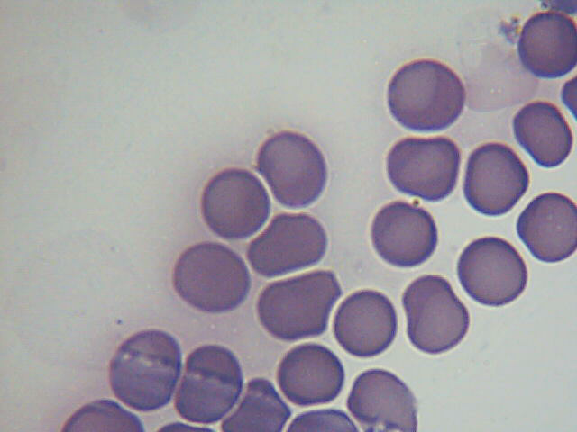
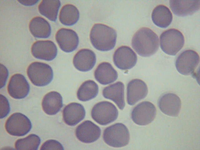
Well, HERE YOU GO!!!! Some of these with my newly-improvised LED makeshift lighting....(the way Lymedin has encouraged us to do). I'm glad I tried this, and should have done it sooner! The colors in my stains are so much truer!
Notice the "signet," or "ring-form" in this neutrophil. Notice also the color difference between the "ring" and the platelets just above the neutrophil-
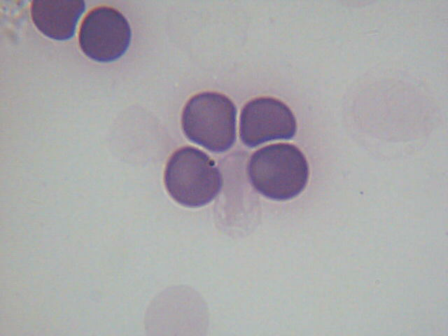
And, the diagnostic "maltese cross"-
Notice also the exo-erythrocytic ring-form on the left-
Since most of my babesia pics right now are with my stock lighting, I will post more "halogen" babesia pics in the next post since I ran out of space in this post (Lymenet gives room for a maximum of 8 pics per entry).
Posts: 1308 | From Eastern USA | Registered: Oct 2013
| IP: Logged |
TNT
Frequent Contributor (1K+ posts)
Member # 42349
posted
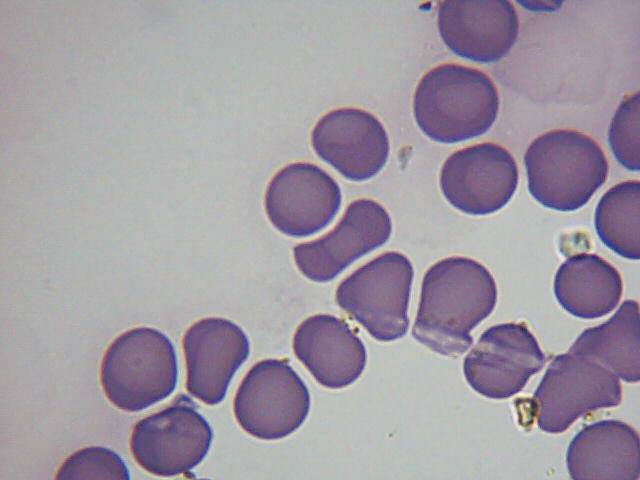
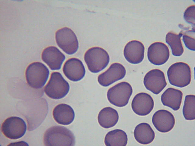
Here's perhaps a better version of my last pic. Sometimes zooming in makes it more "tiresome" to look at.
I'm not sure what form this is, but it is either an exo-erythrocytic maltese cross, or a couple clumped exo-erythrocytic merozoites-
A form of the tetrad/maltese cross?-
An early stage of the tetrad?-
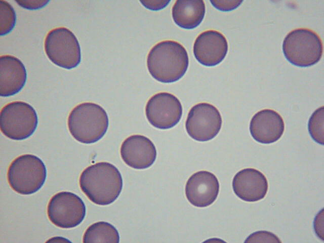
Some more interesting forms-
Posts: 1308 | From Eastern USA | Registered: Oct 2013
| IP: Logged |
TNT
Frequent Contributor (1K+ posts)
Member # 42349
posted
This is almost identical to the stock photo I posted a couple entries ago of the exo-erythrocytic clump of merozoites-
Some more forms-
A "mass" I suspect is/are a form/forms of babesia-
And, a halogen lighting version of the ring in the neutrophil-
Posts: 1308 | From Eastern USA | Registered: Oct 2013
| IP: Logged |
TNT
Frequent Contributor (1K+ posts)
Member # 42349
"Babesia enters erythrocytes at the sporozoite stage. Within the red blood cell, the protozoa become cyclical and develop into a trophozoite ring seen in the second cell [of the above picture].
The trophozoites morph into merozoites, which have a tetrad structure coined a Maltese-cross form.
The tetrad morphology, which can be seen with Geimsa staining of a thin blood smear, is unique to Babesia and serves as a distinguishing feature from Plasmodium falciparum, a protozoan of similar morphology that causes Malaria.
Trophozoite and merozoite growth ruptures the host erythrocyte leading to the release of vermicules, the infectious parasitic bodies, which rapidly spread the protozoa throughout the blood."
(breaking up the paragraph for easier reading for many here)
[ 01-02-2016, 03:38 AM: Message edited by: Robin123 ]
Posts: 1308 | From Eastern USA | Registered: Oct 2013
| IP: Logged |
Lymedin2010
Frequent Contributor (1K+ posts)
Member # 34322
posted
I think you finally got it, congrats & they look beautiful!!!
I think you are the first to find them via stain!
Posts: 2094 | From NY | Registered: Oct 2011
| IP: Logged |
TNT
Frequent Contributor (1K+ posts)
Member # 42349
posted
Here is an interesting page with some info pertaining to the recent posts:
I picked up an oil immersion dark field condenser to use on my microscope.
However, I cannot achieve dark field with it at 100x I have oil on both the condenser and the objective. I only get dark field on 40x and below.
The condenser is 1.36-1.25 The objective is 100x/1.25.
Is there a way to adjust the condenser? It should work since it is 1.36-1.25. Is there some way to adjust between 1.36 and .125 on the condenser that I just don't understand?
Posts: 163 | From USA | Registered: Oct 2015
| IP: Logged |
TNT
Frequent Contributor (1K+ posts)
Member # 42349
posted
I think you will need to use a 100x objective that is less than 1.25. According to this video you can get a drop in plug for your objective that restricts the light value to make it possible to do 100x oil darkfield. It's a very good tutorial that explains darkfield apparatus very well.
posted
Well, I figured out the issue. I'm formally trained in microscopy, so it shouldn't have been such a simple issue. I am not formally trained in any nature in Darkfield, but who really is these days?
The dark field condenser has an aperture of 1.36-1.25. The 100X oil objective has an aperture of 1.25. I know that the condenser must have a higher aperture then the objective lens to attain dark field. I thought the 1.36-1.25 was equivalent to a higher aperture, but it wasn't.
The only fix was purchasing a 100X oil objective with a variable aperture, so I can adjust it so that it is smaller than 1.25.
So, for anyone else curious, if you have a 100X objective with a 1.25 aperture, it is too large of an aperture to achieve dark field with a 1.36-1.25 condenser.
I will report back with, potentially, some new video at 1000X, once I get the new lens in.
Posts: 163 | From USA | Registered: Oct 2015
| IP: Logged |
posted
Also, I may very well have a report, as scientific as i can make, available for anyone to read, including video, and perhaps powerpoint, in a few months.
I am seeking more patients locally (I am one myself).
MS patients. That is my diagnosis. I have another coworker with an MS diagnosis. We both have a demonstrable spirochaetosis (I don't have the equipment, supplies, training to do PCR's or DNA type staining), but it is interesting that any controls I have done (individuals with no illness/disease) do not have the same findings. So I am trying to increase my sample size, gather and organize any and all lab findings/diagnostics used for the diagnosis of MS.
I am currently working to have a diagnosis for a co-worker changed, potentially.
Posts: 163 | From USA | Registered: Oct 2015
| IP: Logged |
Lymedin2010
Frequent Contributor (1K+ posts)
Member # 34322
posted
I am finding the same, as I see the spirochetes in my blood & the family members that have LD, but not in healthy blood.
I was teasing my neighbors cat outside while I was recording, haha. She would meow and I would respond.
Posts: 163 | From USA | Registered: Oct 2015
| IP: Logged |
The Lyme Disease Network is a non-profit organization funded by individual donations. If you would like to support the Network and the LymeNet system of Web services, please send your donations to:
The
Lyme Disease Network of New Jersey 907 Pebble Creek Court,
Pennington,
NJ08534USA http://www.lymenet.org/


 UBBFriend: Email this page to someone!


![[Big Grin]](biggrin.gif)


![[Smile]](smile.gif)


![[dizzy]](graemlins/dizzy.gif)


 Printer-friendly view of this topic