TNT
Frequent Contributor (1K+ posts)
Member # 42349
posted
quote:Originally posted by mustardseed2: Question for the experts:
Are the string of pearl forms and dumbbell looking ones considered cysts or not?
I'm trying to figure out if conventional antibiotics have any activity against these forms. Or, if they're considered cystic forms, then they would require different antibiotics.
I don't really know the physical difference between the 2 forms, or why some antibiotics work on one but not the other, so I'm just curious as to which form these "in-between" forms fall under?
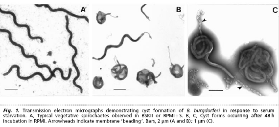
The string of pearls and dumbbell ketes are definitely not cysts.
I don't know if anyone knows what kills the string of pearls, but the short dumbbell ketes would probably be susceptible to the regular borrelia drugs.
There still seems to be some uncertainty (perhaps only in my mind) about whether the spirochetes we are seeing in our blood are l-form or not. If they are, then they would be more susceptible to the tetracyclines and macrolides, but less susceptible to the cell-wall drugs like penicillins and cephalosporins.
Posts: 1308 | From Eastern USA | Registered: Oct 2013
| IP: Logged |
Lymedin2010
Frequent Contributor (1K+ posts)
Member # 34322
posted
Does this family member have night or day sweats? Any shortness of breath episodes? And when you say such "trouble as pain", do you mean wide spread pain or only with the legs?
Any numbness, tremors, vibrations, muscle twitches, joint pain....etc?
Posts: 2094 | From NY | Registered: Oct 2011
| IP: Logged |
TNT
Frequent Contributor (1K+ posts)
Member # 42349
posted
quote:Originally posted by Lymedin2010: Does this family member have night or day sweats? Any shortness of breath episodes?
No sweats, but occasionally mild breathlessness.
quote:Originally posted by Lymedin2010: And when you say such "trouble as pain", do you mean wide spread pain or only with the legs?
Mostly with the legs, but, at it's worse, it can be widespread pain.
quote:Originally posted by Lymedin2010: Any numbness, tremors, vibrations, muscle twitches, joint pain....etc?
Maybe some vibrations at times; and yes to some minimal joint pain.
Posts: 1308 | From Eastern USA | Registered: Oct 2013
| IP: Logged |
quote:Originally posted by mustardseed2: Question for the experts:
Are the string of pearl forms and dumbbell looking ones considered cysts or not?
I'm trying to figure out if conventional antibiotics have any activity against these forms. Or, if they're considered cystic forms, then they would require different antibiotics.
I don't really know the physical difference between the 2 forms, or why some antibiotics work on one but not the other, so I'm just curious as to which form these "in-between" forms fall under?
No, those would not be considered cystic forms.
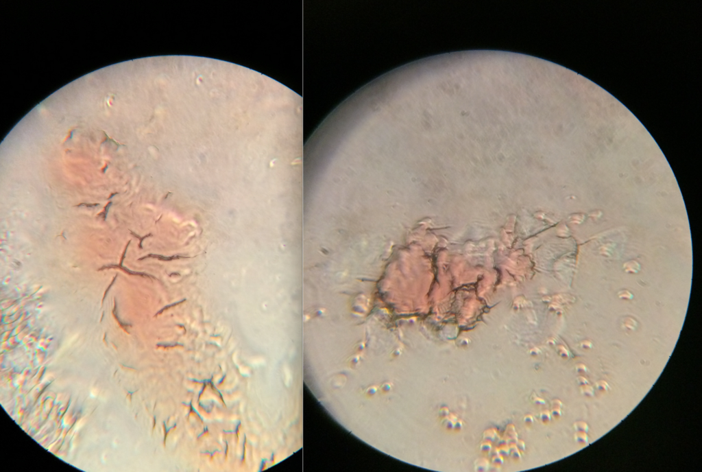
I believe lymedin has some good videos showing the transition from the atypical (non-spiral) form to the cystic form, in a complete cycle.
I posted a video capture of one in the process of going from atypical to cystic, but I cut the capture short.
Here are a couple images you can reference. I'll have to look for some with different staining methods, and something closer to what you would see under your microscope, and not a super high resolution microscope.
Posts: 163 | From USA | Registered: Oct 2015
| IP: Logged |
TNT
Frequent Contributor (1K+ posts)
Member # 42349
posted
quote:Originally posted by thatdudefromkansas:
That is an awesome find and I've actually wondered if this kind of an experiment could be done to demonstrate that the objects we call cysts are indeed dormant borrelia spirochetes.
I considered trying something like that myself, but wasn't sure how to do it with my old cyst-ridden samples without inevitably destroying them in the process.
It's even better that they were able to demonstrate this with the exact same field of view! I'd like to know how they did it with such precision without destroying their sample. I mean, how did they introduce rabbit serum under the coverslip?
I also find it amazing that the cyst to spirochete conversion process took less than a minute!!! That must have been a Eureka moment!
Posts: 1308 | From Eastern USA | Registered: Oct 2013
| IP: Logged |
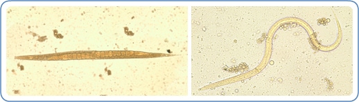
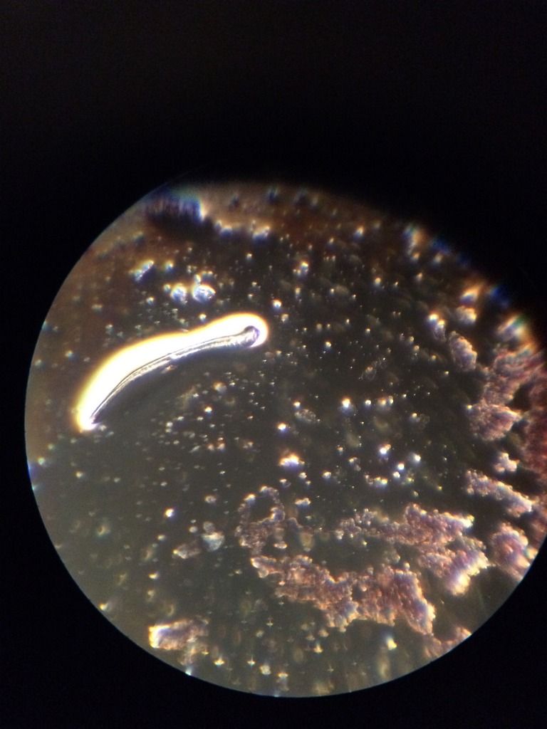
quote:Originally posted by mustardseed2: Check out this worm I found. Took the picture with my cell phone straight through the eyepiece.
10x objective 10x eyepiece
Hi mustardseed--- excellent photo! Its definitely not a piece of lint or an artifact!! Looks like a filarial worm, similar to this one (wucheria bancrofti), but the tips are not as pointed in yours:
I'll see if I can find a photo that matches what you have photographed--- there are many herbs that treat filarial worms( sterilize, kill or disable the worms), but you have to know which species your worm is. Yours might be similar to onchocerca-- and onchocerca-like filarial worms were found by Dr. Eva Sapi in ticks.: Posts: 696 | From New York | Registered: Aug 2006
| IP: Logged |
posted
I've honestly gone back and forth over whether that thing I found was an actual worm or not. It's so hard to tell.
What would be amazing is if someone captures some images of paper fibers and posted them, that would help haha.
Posts: 71 | From Canada | Registered: May 2016
| IP: Logged |
Lymedin2010
Frequent Contributor (1K+ posts)
Member # 34322
posted
You can easily do that. Just leave a slide out in the open for a day or 2 & then add a drop of water & a cover slip on there.
You can also 1 micron filter your own blood & add just the plasma to the dust particles if it so pleases you.
Posts: 2094 | From NY | Registered: Oct 2011
| IP: Logged |
bluelyme
Frequent Contributor (1K+ posts)
Member # 47170
posted
Great vids...rainbow....There is doc here who sends it to african university for darkfield id... its about 300.. idk if these guys do blood as it seems the stain it first... http://www.parasitetesting.com/tests.cfm
Have you done the iver,albendazole, bitricide ,route? Clove,wormwood,papayaseed, rife?
-------------------- Blue Posts: 1539 | From southwest | Registered: Dec 2015
| IP: Logged |
TNT
Frequent Contributor (1K+ posts)
Member # 42349
posted
Hey rainboworiver! Thanks for dropping in. I'm glad to see another person doing microscopy! It's a lot of fun, isn't it? It's the only cool thing about being sick with this disease.
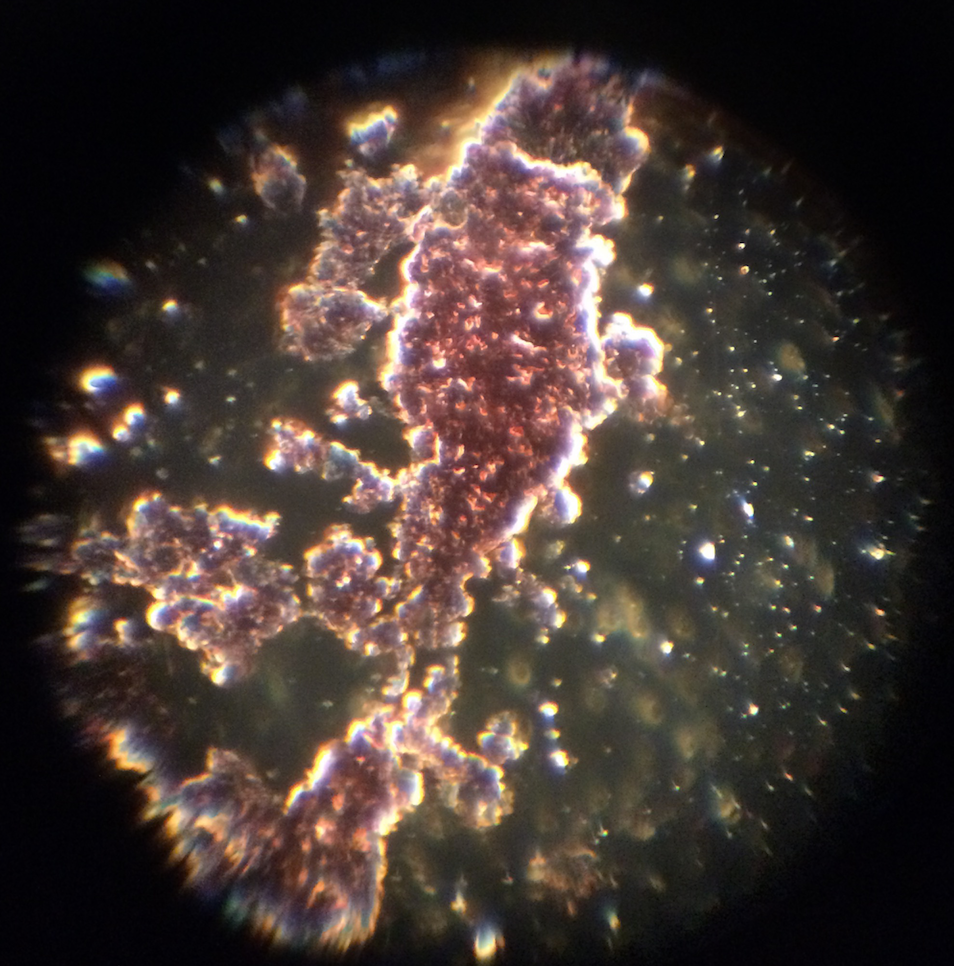
I would have to say I think most of what you are seeing is normal and can be explained fairly easily. Other than possibly the first couple pics, those worm-like objects are almost certainly hair and fiber contaminants. I'm not as sure about the rounder worm-like object in the first two pics.
The misshapen and lemon-shaped red blood cells with legs are most likely RBCs stuck on some fibrin spicules. That one RBC appears to possibly have a string of pearl kete hanging on/out of it.
219jpg and 220jpg are possible (clump(s) of ) platelets snagged on some fibrin spicules.
And, the "huge colony" video and the pic of something similar, could be a clump of platelets or RBCs.
I really doubt anything you showed was related to parasites except the string of pearls on that RBC (18, 19, & 20jpg) and the possible helminth in pics 1 & 2 (2 & 3jpg).
Those parasites, cysts, and eggs (slide #40) are going to be found mainly in fecal matter. The scale of measurement of those cysts were not included unfortunately, but are probably much larger than a red blood cell.
These statements reflect my own experience & opinions, so don't let me keep you from investigating it further if you feel it's necessary.
But, you got some great footage! What type of scope and camera equipment are you using?
Posts: 1308 | From Eastern USA | Registered: Oct 2013
| IP: Logged |
Jordana
Frequent Contributor (1K+ posts)
Member # 45305
posted
Hey there:
I'm sending out a thin blood smear to a lab so they can identify what's in it. So far I've had no luck getting a positive babesia test -- either serology or FISH.
So my question is, since they're looking at ONE DROP of blood, is there a better or worse place to take the draw? Finger? Arm? Torso? Vein in the head? Near lymph nodes?
Thank you!
Posts: 2057 | From Florida | Registered: Feb 2015
| IP: Logged |
TNT
Frequent Contributor (1K+ posts)
Member # 42349
posted
I've heard an ear draw is best for detecting Babesia. That's what is used for testing in dogs.
Babesia is difficult to detect. I have looked and looked for hours before I've found it in a stained smear! When only 1% of your RBCs need to be infected to cause symptoms, it makes it really hard to find.
Posts: 1308 | From Eastern USA | Registered: Oct 2013
| IP: Logged |
Jordana
Frequent Contributor (1K+ posts)
Member # 45305
posted
Yeah, I'm sending in three, just want to increase my chances it shows up. They're also looking for other blood parasites and whatever else, but if they don't find anything I'm just going to buy a microscope .
Posts: 2057 | From Florida | Registered: Feb 2015
| IP: Logged |
TNT
Frequent Contributor (1K+ posts)
Member # 42349
posted
Good luck Jordana, I hope they can help you.
I would encourage you to get a microscope... how many labs or paid technicians can look at a blood slide for hours? None. It just isn't possible for them to look that long and carefully.
But many of us are sick, at home, and have that kind of time to spend on the microscopy (unless we aren't feeling well enough to get out the microscope...which happens too often).
I would like to encourage every chronically ill person to get a microscope! It is one of the BEST expenditures one will make...especially those with known tick-borne illness. I personally believe most chronic illness is caused by a pathogen of some kind. It's just that the establishment is incentivized to NOT find it!
To the mainstream medical INDUSTRY, "cure" is a bad four-letter word.
Posts: 1308 | From Eastern USA | Registered: Oct 2013
| IP: Logged |
Jordana
Frequent Contributor (1K+ posts)
Member # 45305
posted
I believe all illnesses are caused by pathogens; nothing else makes sense to me either.
The thing that's stopped me is mostly I'm just too sick to do anything; learning a whole new thing feels hard. The other thing is, I know I'll find stuff in there and have no idea what it is.
I could drive myself crazy squinting at all those oddly shaped things in blood cells...
Posts: 2057 | From Florida | Registered: Feb 2015
| IP: Logged |
posted
TNT, thank you for your input. I also see a lot of spirochetes in my blood in various forms. I am currently on Nutramedix.
What is the most effective in treatment you have seen to reduce load in blood? After load reduction, does it correlate to feeling better?
I have been on stevia for one month, haven't seen any improvement in spirochete load in blood. I have been feeling worse.
Thanks!
Posts: 99 | From NJ | Registered: Mar 2010
| IP: Logged |
TNT
Frequent Contributor (1K+ posts)
Member # 42349
posted
quote:Originally posted by rainbowriver: TNT, thank you for your input. I also see a lot of spirochetes in my blood in various forms. I am currently on Nutramedix.
What is the most effective in treatment you have seen to reduce load in blood? After load reduction, does it correlate to feeling better?
I have been on stevia for one month, haven't seen any improvement in spirochete load in blood. I have been feeling worse.
Thanks!
I never did the complete Cowden protocol, but did do the main work-horse products for months. It didn't help me. I hear of very few people who get results with Cowden. The Buhner protocol has a better track record.
Lower pathogen burden definitely correlates with feeling better. I personally have seen at least some difference with BVT. My blood used to look horrendous. I am also doing ABX with BVT, and there seems to be synergy with them combined (for me).
Stevia is known to dissolve biofilm. And, may also be spirocheticidal. But, if the stevia is dissolving biofilm faster than you are killing what is being released from the dissolved biofilm, you will only make yourself sicker.
Posts: 1308 | From Eastern USA | Registered: Oct 2013
| IP: Logged |
Lymedin2010
Frequent Contributor (1K+ posts)
Member # 34322
posted
For those interested in growing filarial nematodes from your blood, here is the recipe.
posted
where do you buy staining chemicals/tool kit? I contacted americanmastertech.com and they claim they have high quality staining kit. But it costs $150.
they recommended Wright-Giemsa: it is geared towards a smear and stain procedure. it is good for tick-born infections including parasites.
I may require absolute alcohol, clearing agents (Xylene, or alternative), mounting medium,
Could someone recommend a price to get the complete kit for staining? I am already doing live blood analysis with a phase contast scope.
posted
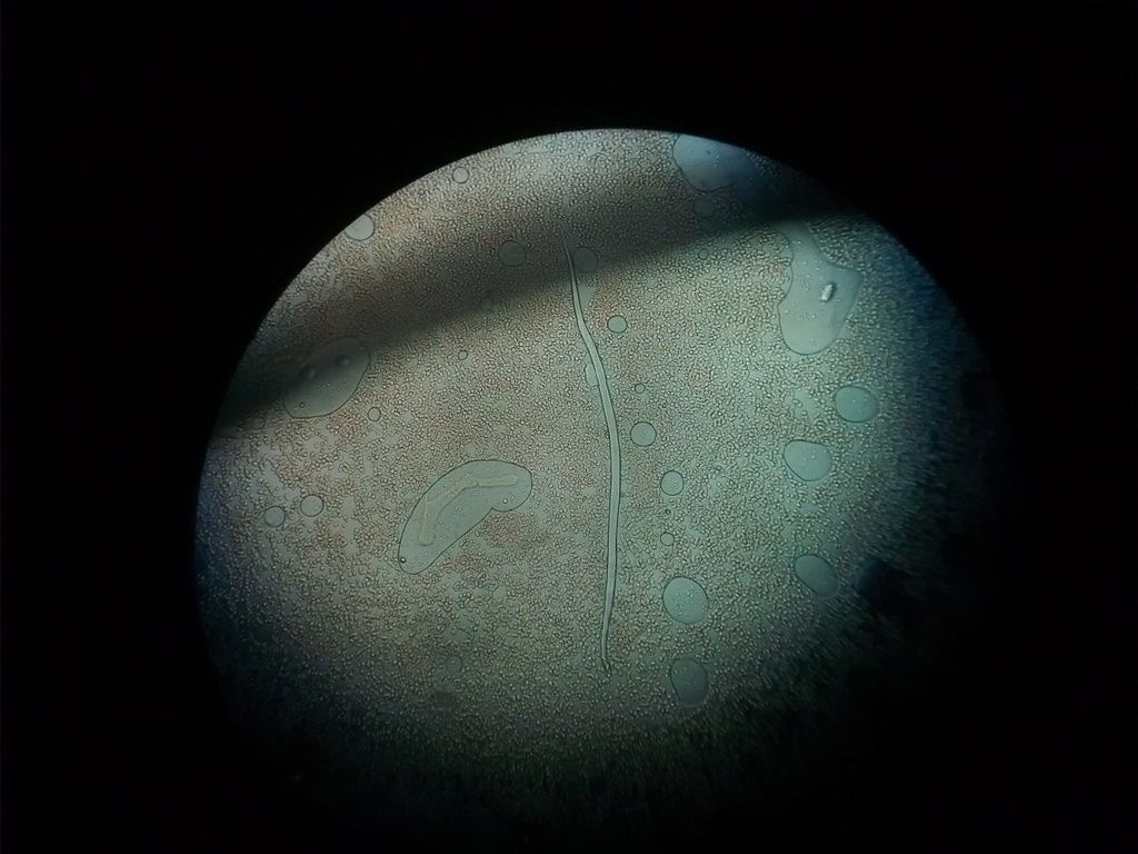
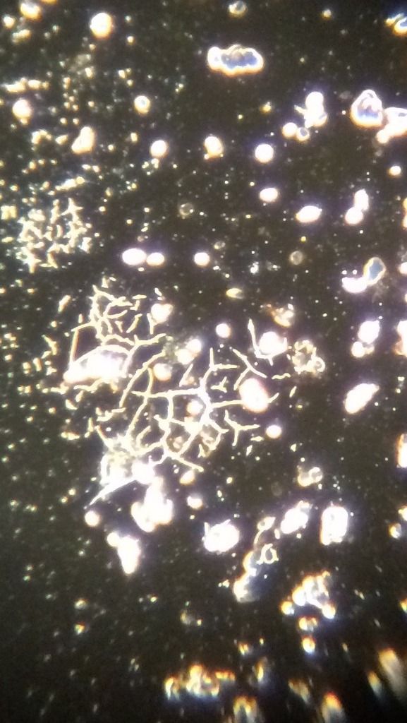
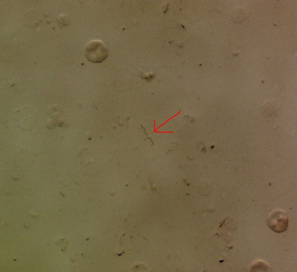
Hi, I'm a new member here. I am just starting to use microscopy to analyze blood. I have a dark field microscope but I can also use it for bright field. I have not dyed slides. I have a lot of questions as to what I'm looking at, if anyone would be able to help! I know I will see RBCs, platelets, WBCs and spirochete forms, but I'm not sure what else I could be seeing. I'm attaching photos. I think I see many forms of spirochetes, but these line-like things could be something else? Is it possible to break RBCs during the blood smear and see broken fragments of their cell membranes?
The following are my specific questions for each image. These are images from 3 different blood smears. Also if anyone who is familiar with microscopy could message me, I would really appreciate it since I'm new at this (I am familiar with microscopes from my university's lab time but not dark field/oil immersion and not looking at blood)
Thanks for any input!
1: Is that a spirochete form coming out of a red blood cell and one in the corner?
2. Here are images of my blood smears. Are the lines seen throughout possible spirochetes?
3. Is that a possible biofilm?
4. What could those black lines be? I did not dye the slide.
5. Is that a group of spirochetes in the middle? Why are the RBCs so clumped?
6. Is that a different parasite on the top left? (possible worm?)
7. Is that just a bad smear (clumped RBCs) or is it due to antibiotic treatment - am I seeing clumping due to antibody work? Posts: 21 | From NJ | Registered: Aug 2016
| IP: Logged |
Lymedin2010
Frequent Contributor (1K+ posts)
Member # 34322
posted
Hi & welcome. It is best that we see videos, as sometimes it is very difficult to tell by just pictures. There are spiro like objects from our blood, which are really fibrin strands & sheddings from the rbc cell wall. From the time you drew the blood to the time you took the picture, how much time elapsed? In other words how old is the blood sample? Did you seal the edge of the slip cover with either Vaseline or some type of oil?
1- dimiss as it is too hard to tell. 2- Is your best set of images & can potentially be spirochetes. Were they wiggling/gyrating around with some aggression? 3- This could be growth from a contamination source, your finger or the air possibly. More likely to be the drying of blood, same as #4 as that one shows it clearly. 4- Dismiss this, as thsoe lines can appear when blood dried up. 5- Clumped, because your blood is drying out & dying, as you probably did not seal the slip cover. If you do not put a large drop of blood & add one that is too small, then when you smear it, it will dry out quicker and look clumped. 6- Could be Strongyloides, but can also too easily be a contamination fiber. Do you find more of these, if so it can buy further clues. https://lookaside.fbsbx.com/file/HorseNematode.PDF?token=AWwl_vuWNqJri2JyO9QO95phUjfuqsNolL4nhOIMPcOZmYVVsX_gwn1CEOcUcmNKtlyQSdFmLHOY8uwAQStzjZbeXLSNUUeWBDb0R_sDpOmtHk_bPmkG0f3rqKD Q_Zfudb2TOOdsO1XUK6r5ijc24VmHC2a6WalGIRsu3ZdceYxIrA 7- Same clumping as I mentioned above.
I would do another sample & take video for the string-like objects/spirochetes.
Here is a nice one I captured, but most of the ones I see do not have strong spirals, so this was a lucky catch.
Also if you look at this spirochetes in this video, it looks like it moves with some aggression & is not partaking in mere Brownian motion. I see many string-like objects too, but I never show them & dismiss most of them. It is not until I find some vitality or spiraling that I show the video.
I need to buy a finger pricking tool because I have been bad at doing it and only got very small drops of blood to use. Therefore, the smears were too thin so the cells were not moving, which explains why they were showing signs of drying blood - I did not know what that looked like, but now I understand, thanks. However, I did take these pictures within minutes of doing the blood smear, so just minutes elapsed. I knew I couldn't look at them for long since the smears were not good. I didn't seal the coverslips for those reasons. Since I saw RBCs I tried to analyze the slides, but now I understand they weren't good enough smears to properly analyze. Thanks for explaining the fibrin and RBC cell wall shedding as spiro like objects from blood, because I couldn't find that type of info online.
Thanks for your photo, links, and input, they will help a lot for my future slides.
Posts: 21 | From NJ | Registered: Aug 2016
| IP: Logged |
Lymedin2010
Frequent Contributor (1K+ posts)
Member # 34322
posted
No problem.
TNT, your mailbox is full & I could not respond back.
Posts: 2094 | From NY | Registered: Oct 2011
| IP: Logged |
TNT
Frequent Contributor (1K+ posts)
Member # 42349
posted
Sorry, Lymedin, I cleaned some out for you!
Posts: 1308 | From Eastern USA | Registered: Oct 2013
| IP: Logged |
TNT
Frequent Contributor (1K+ posts)
Member # 42349
posted
Lymedin....looking forward to your reply.
Posts: 1308 | From Eastern USA | Registered: Oct 2013
| IP: Logged |
Apparently I need to add it to a buffer (pH 6.8-7.0) to make a working Giemsa stain solution. Any suggestions?
Posts: 71 | From Canada | Registered: May 2016
| IP: Logged |
TNT
Frequent Contributor (1K+ posts)
Member # 42349
posted
Would something like this be right? I have a ready-to-use Wright-Giemsa stain, so I am not knowledgeable about buffers/buffering.
posted
Thanks for the info... I'll look into that link.
Ok I have a new question for the experts:
I've made a similar observation as the OP: I don't see a ton of spirochetes right after taking my blood sample, but if I leave the sample out for 24 hours, it becomes very easy to find spirochetes swimming around in my blood plasma.
In my mind, one of two things is happening here:
1.) The spirochetes are hiding in red blood cells, and come out of the cells as the cells start to die.
2.) The borrelia are in cyst form, which I cannot detect easily, and as the blood starts to die, they turn into spirochetes to look for more hospitable territory.
So which of these two scenarios is more likely?
The reason I ask is because I'm on several antibiotics (and have been for a while), but none of which target the cystic form of the bacteria. I'm wondering if they're all just hanging out as cysts, or if there are spirochetes hanging out in my blood cells that I'm not detecting?
Thanks!
Posts: 71 | From Canada | Registered: May 2016
| IP: Logged |
bluelyme
Frequent Contributor (1K+ posts)
Member # 47170
posted
I think they are in rbcs..if i crush them then same thing happens ...maybe why cycle therapy works...let them come ouy then bammmm
dude ks-any luck on your new rig? Pics vid?
-------------------- Blue Posts: 1539 | From southwest | Registered: Dec 2015
| IP: Logged |
Lymedin2010
Frequent Contributor (1K+ posts)
Member # 34322
posted
"New bacteria discovery at biological research lab at DSU could prove revolutionary"
Answer fro above 2 questions: More likely both are happening in the RBC. Lets not forget the rbc's are designed to hold HEMOGLOBIN, so as to oxygenate the rest of the body. What goes into the RBC's & what comes out is VERY particular & controlled. When they measure the concentration of abx in your blood, it does not mean that it profuses to that level in your RBC's & your other tissues. Some cells/tissues are very controlled & more so than others.
Imagine if just anything was let into your rbc's & your body could not oxygenate properly. This would be very detrimental.
So whole spirochetes can be inside rbc's & cysts as well. Also the body becomes more efficient at getting rid of toxins such as abx with time, just like with any drug or alcohol out there. Each dose of the drug or alcohol needs to be more & more, because the body has become efficient at eliminating the toxin. I think this is also a huge reason why we don't get better, since our bodies become accustomed to the new drugs relatively quickly & they wear off effectiveness.
Posts: 2094 | From NY | Registered: Oct 2011
| IP: Logged |
Lymedin2010
Frequent Contributor (1K+ posts)
Member # 34322
posted
Great video of spirochetes in the blood capillaries. Keep in mind this is EARLY infection. Imagine what happens in late disseminated disease & when the spirochetes become more atypical, as they are in our blood.
Lymedin2010
Frequent Contributor (1K+ posts)
Member # 34322
posted
B. mayonii
"These patients have HIGH levels of bacteria in their blood & they seem to have more severe symptoms."
So high that even PCR seems to be the preferred way of testing for Bm, where as Bb does not always show up positive in PCR even though it may be in the body (too low levels in the blood during early infection).
_________________________________________________ Even Wormser can't escape the notion of Borrelia being in the blood in enough quantities to test at times.
"CONCLUSION: Culturing blood for B. burgdorferi may be useful in confirming the diagnosis of Lyme disease in selected patients. Use of spirochete blood cultures may facilitate a better understanding of the pathogenesis and natural history of Lyme disease."
"CONCLUSION: Culturing blood for B. burgdorferi may be useful in confirming the diagnosis of Lyme disease in selected patients. Use of spirochete blood cultures may facilitate a better understanding of the pathogenesis and natural history of Lyme disease."
Wow, for a moment there I wondered how he could say something like that when "they've" bashed the Advanced Lab culture test.... but this is something he said in 1990.
Still relevant though, for sure!
I'm hoping Dude has some updates with the cultures he's been doing!
Posts: 1308 | From Eastern USA | Registered: Oct 2013
| IP: Logged |
Lymedin2010
Frequent Contributor (1K+ posts)
Member # 34322
posted
Sorry TNT, I am always not up to par to elaborating on questions that may need detailed explanations & so I am getting back to you really late on your blood stains.
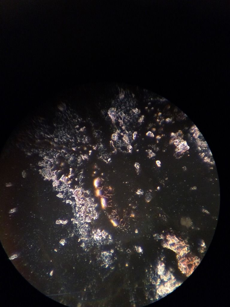
Yes, I pretty much concluded that the plentiful supply of the fake looking large rings are your stain precipitates. BUT, I thought you were questioning some of your rings that really came out looking like possible babesia ring-forms or Theileria that I posted a pic of, as I know I had questioned them. It is possible to have captured a true ring form in the bunch, but I would dismiss them all because of too many artifacts.
This means we need to dismiss the dots on the peripheral of the rbc as well & within the rbc, as they are probably artifacts as well. They are scattered & not in enough quantities to definitely indicate babesia microti.
The mesh artifacts in your plasma I would dismiss as well, and is probably an oddity of the precipitate.
Rickettsia are usually found in endothelial cells & can come in cocci, rod, & string-like forms. Your wbc's stains are interesting, but can easily be granules or even degraded bacteria of sorts that are picked up by the wbc's along the way.
Take a look at this "normal" blood stain, as they too have stained objects in the cytoplasma.
Even this one stain of yours here is most likely stain precipitate on the peripheral of the rbc & not myco. The pink objects in the plasma, which almost look like babesia forms, are probably just platelets.
So the above is my opinion, but here is feedback from the only expert that has anything to say about your stains. I don't agree with the babesia in the stains, but then again I am far from any stain identifying expert:
"very definately looks like babesia RBC inclusions, I would confirm with PCR, also the monocytes might indicate EBV virus. I would check EBV antibody titers. Looks like a round of Mepron might tell the tale through diagnosis by therapeutic response. Does the patient have gasping for air, night sweats, shakes, fevers? Also bears similarity to feline distemper, take a look on google of RBC inclusions flash-cards and make some comparisons. I think treatment is going to tell you a lot. Also might be seeing nucleated RBCs which should not happen in adults and can mean a bone marrow breach, cancers, or most likely an abnormal demand for red blood cells because of babesia, "
"When I compare them to a RBC monograph atlas they look pretty competent. Has the patient ever been to a malaria area? Hydroxyquinone Plaquinil might help with pain and the parasite. Often combined with clindamycin. My guess when therapy for babesia is given he will sweat profusely, have more pain, gasp for breath and then start to improve. Best to take 5000 mcg sublingual B-12 or shots and to take iron to replace iron loss. trial and error gives us more data."
Posts: 2094 | From NY | Registered: Oct 2011
| IP: Logged |
Lymedin2010
Frequent Contributor (1K+ posts)
Member # 34322
posted
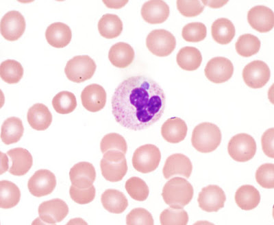
Not that I see this in your stains, but if you are doing stains you should know that the most common Neutrophil pathogen is Anaplasmosis (Rickettsia) & typically has the Morulae bodies adjacent to the wbc membrane, such as this picture. You can check online for additional pictures.
TNT
Frequent Contributor (1K+ posts)
Member # 42349
posted
Thanks for the input, Lymedin.
1. Babesia FISH test on this person was negative. So, I would not only disagree with his opinion based on serology, none of what I showed in the preceding pics really resemble Babesia, except the stain precipitate that looks like Theileria. So, Babesia is practically ruled out.
2. As for the inclusions in the cytoplasm and the dots on the RBCs being possible precipitate, it's possible, except the difference between precipitate and bacteria is pretty discernable, and I did not post any pics in which I saw any "dot" precipitate. I realize that the 2nd-hand evaluation of images of stained blood slides can be tough to tell the difference. 1st hand viewing through the scope is much more reliable since the focus can be varied (which aids in the discernment).
Compare my "mycoplasma" pics with the examples I posted from reliable sources. Not everything is artifacts.
I have looked quite a bit at Anaplasma blood slide pics (from reputable sources), and these objects don't have those characteristics.
As for Distemper, it's true, the inclusions in the cytoplasm have a slight resemblance of distemper, but distemper (a viral infection) appears more like the morulae of Anaplasma in the WBCs and the elemental bodies of Anaplasma in the RBCs.
I have personally seen blood smears of a person with active EBV (backed by serology), and the monocytes in particular were "reactive," and had vacuoles in the cytoplasm. The nuclei of the WBCs also had basophilic inclusions very similar to Distemper. So, this is not EBV.
quote:Originally posted by Lymedin2010: Rickettsia are usually found in endothelial cells & can come in cocci, rod, & string-like forms. Your wbc's stains are interesting, but can easily be granules or even degraded bacteria of sorts that are picked up by the wbc's along the way.
Exactly to all that in your quote above.
And, they could be WBC granules, but they don't appear like typical granules. They appear more like the Rickettsia bacteria in the pics above (from the reputable sources).
All considered, I still lean very heavily towards Rickettsia and Mycoplasma.
Posts: 1308 | From Eastern USA | Registered: Oct 2013
| IP: Logged |
Lymedin2010
Frequent Contributor (1K+ posts)
Member # 34322
posted
PCR for Rickettsia & Mycoplasma tieters and EBV tieters might confirm them, especially if they can be easily picked up in the blood sample.
Posts: 2094 | From NY | Registered: Oct 2011
| IP: Logged |
TNT
Frequent Contributor (1K+ posts)
Member # 42349
posted
Yes, PCR might be helpful, though we all know how dependable it is for Borrelia.
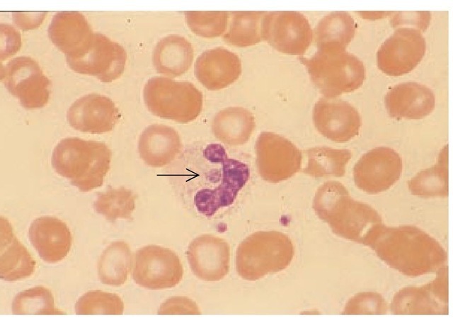
I do feel that the smaller organisms on the RBCs (and possibly the small organisms in the WBCs) are very possibly Mycoplasmas, but I found another possibility for the "Rickettsias" in the shown lymphocytes.
According to this histology page these "large granular" lymphocytes may be:
("T-lymphocytes and B-lymphocytes form the vast majority of lymphocytes in the blood stream, but they do not add up to 100%, and they usually are small lymphocytes. The much less frequent medium-sized or large lymphocytes may represent e.g.
* natural killer (Nk-) cells which belong to the group of large granular lymphocytes, or
* haemopoietic stem cells of which a very few will be circulating in the blood stream.")
Posts: 1308 | From Eastern USA | Registered: Oct 2013
| IP: Logged |
TNT
Frequent Contributor (1K+ posts)
Member # 42349
posted
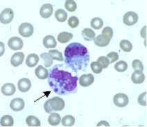
My pic for comparison:
Posts: 1308 | From Eastern USA | Registered: Oct 2013
| IP: Logged |
Lymedin2010
Frequent Contributor (1K+ posts)
Member # 34322
posted
Yup, that looks like a good match & explanation.
Most of what I see in live microscopy are artifacts & I see tons of them, before I find something of interest & decide to post a video and so I have learned from experience.
Posts: 2094 | From NY | Registered: Oct 2011
| IP: Logged |
posted
I have been busy and things have come up in life, so I have not been around.
Also, the microscope came damaged or otherwise in a condition that was different than described, damaged in shipping or otherwise.
So I do not have my new microscope.
So I will have to pursue my objective of staining samples with more specialized staining methods in the future.
I have been following this thread, but have been busy. Hope everyones research is going well and learning and discovering new things that will help them.
Posts: 163 | From USA | Registered: Oct 2015
| IP: Logged |
bluelyme
Frequent Contributor (1K+ posts)
Member # 47170
posted
We understand dude ..keep us updated ..your bsk cultures are at the frontlines .
mustard -any new pics of filarial?
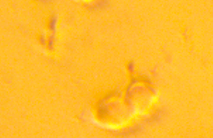
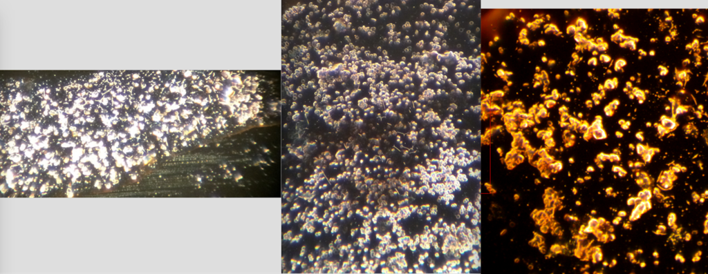
so being inpatient i got a really old wesco and a godawful amscope eyepiece with a bad circular abhorration but that aside i think i caught a sop or a fazed kete under the influence of venom ..
not sure if it is many, or one big un with swelling and bubbled body (as its dying i hope ) forgive the focusing it was the only way to outline it visually..
gosh i want a phase or a dark to see if venom is really doing this to ketes ..most often have to bust open rbc . anyway let me know what you think, thank you all
posted
Nice vid. No I haven't taken any more pics with my camera, but I have seen a few more of these parasites (I think they are).
I also looked at dust particles and tissue particles to see if that what this could be... they were totally different... larger, not smooth, and they don't come to a point at one end. I'm very convinced this thing was... something.
Posts: 71 | From Canada | Registered: May 2016
| IP: Logged |
posted
Without treading through 12 pages, would someone be able to show me a video of an undisputed bleb?
I'm curious to know if some of these dancing round "things" I'm seeing in my blood are blebs.
Posts: 71 | From Canada | Registered: May 2016
| IP: Logged |
posted
Hello. I am new here, just got my lab results with borreliosis with few side infections.
While googling around I bumped into this place and then in this thread and became immediately very interested in trying this myself. So I thought about buying a microscope.
Are any of the USB microscopes any good?
Posts: 31 | From Finland | Registered: Sep 2016
| IP: Logged |
The Lyme Disease Network is a non-profit organization funded by individual donations. If you would like to support the Network and the LymeNet system of Web services, please send your donations to:
The
Lyme Disease Network of New Jersey 907 Pebble Creek Court,
Pennington,
NJ08534USA http://www.lymenet.org/


 UBBFriend: Email this page to someone!


![[Smile]](smile.gif) .
.


 Printer-friendly view of this topic